http://dx.doi.org/10.5534/wjmh.2013.31.1.53
Original Article
Received: Dec 24, 2012; Revised: Feb 4, 2013; Accepted: Feb 4, 2013 Correspondence to: Dong W an Sohn
Department of Urology, The Catholic University of Korea, Yeouido St. Mary’s Hospital, 10 63-ro, Yeongdeungpo-gu, Seoul 150-713, Korea.
Tel: +82-2-3779-1038, Fax: +82-2-761-1626, E-mail: [email protected] Copyright © 2013 Korean Society for Sexual Medicine and Andrology
This is an Open Access article distributed under the terms of the Creative Commons Attribution Non-Commercial License (http://creativecommons.
org/licenses/by-nc/3.0) which permits unrestricted non-commercial use, distribution, and reproduction in any medium, provided the original work is properly cited.
The Relationship between Clinical Symptoms and Urine Culture in Adult Patients with Acute Epididymitis
Sung Dae Kim1, Sun Wook Kim2, Byung Il Yoon2, U-Syn Ha2, Sae Woong Kim2, Yong-Hyun Cho2, Dong Wan Sohn2
Department of Urology, 1Jeju National University School of Medicine, Jeju, 2College of Medicine, The Catholic University of Korea, Seoul, Korea
Purpose: We evaluated adult patients with acute epididymitis to identify the frequency of actual sexual contacts and the causative organism, and compared clinical examinations, degrees of manifested symptoms, and radiological test results.
Materials and Methods: We reviewed the medical records of 65 patients older than 18 years presenting with acute epididymitis who had been treated between 2002 and 2011. Scrotal ultrasonography, urinalysis, and urine culture were performed to diagnose the acute epididymitis. Patients were divided into negative (n=45) and positive (n=20) urine culture groups. Then the latter groups were subdivided into a sexually transmitted organism (STO) culture group (n=13) and a non-STO (n=7) culture group. Data on any history of sexual contact, scrotal pain and tenderness, symptoms of urethritis (discharge, dysuria, urethral burning, or irritation), and lower urinary tract symptoms (dysuria, frequency, and urgency of urination) were obtained from all of the subjects.
Results: Patients in the positive urine culture group were significantly younger than those in the other group (p=0.224) and were more likely to have a history of sexual contact at least two weeks prior to onset of epididymitis (p=0.012). They had also a significantly enlarged epididymal head and significantly more severe complaints of pain or tenderness than those of latter group (p=0.348, p=0.288). However, the difference in these measures between the STO and non-STO group was not significant, except in the case of age (p=0.044).
Conclusions: Patients of the positive urine culture group with acute epididymitis were significantly younger and more sexually active than the others. They also had severe pain or tenderness and an enlarged epididymal head. There was a close association between clinical symptoms, a positive urine culture, and ultrasonographic findings.
Key Words: Epididymis; Infection; Urine; Culture
INTRODUCTION
Acute epididymitis is a common inflammatory disease in men. Many studies have demonstrated that most cases of epididymitis in men aged younger than 35 years are
caused by sexually transmitted organisms.1,2 Chlamydia trachomatis infection is the most common cause of epi- didymo-orchitis in sexually active young men.3 In older men with prostatic hypertrophy and reflux of urine, en- teric organisms are a common cause of epididymitis. This
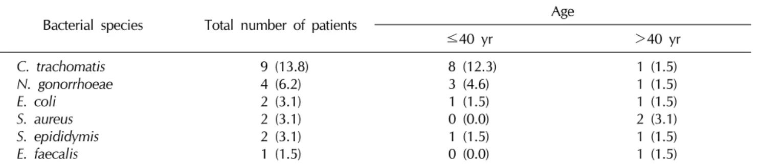
Table 1. Bacterial species detected in men with acute epididymitis
Bacterial species Total number of patients Age
≤40 yr >40 yr
C. trachomatis N. gonorrhoeae E. coli
S. aureus S. epididymis E. faecalis
9 (13.8) 4 (6.2) 2 (3.1) 2 (3.1) 2 (3.1) 1 (1.5)
8 (12.3) 3 (4.6) 1 (1.5) 0 (0.0) 1 (1.5) 0 (0.0)
1 (1.5) 1 (1.5) 1 (1.5) 2 (3.1) 1 (1.5) 1 (1.5) Values are presented as number (%).
C. trachomatis: Chlamydia trachomatis, N. gonorrhoeae: Neisseria gonorrhoeae, E. coli: Escherichia coli, S. aureus:
Staphylococcus aureus, S. epididymis: Streptococcus epididymis, E. faecalis: Enterococcus faecalis.
condition is the result of an infection ascending from the bladder or urethra and is associated with urethritis.4 It should be treated with a course of antibiotics effective against typical urinary pathogens. Studies in other coun- tries have identified an association between sexual con- tact and acute epididymitis, but no such studies have been performed in Korea as yet. Nor has the potential associa- tion between urine culture results and clinical symptoms been investigated. In fact, when examining patients with acute epididymitis in a real clinical setting, it is sometimes possible to encounter patients who report no sexual con- tact at all, or who present with negative culture results from urinalysis. The severity of expressed symptoms also varies among patients. Therefore, we performed this study to analyze the medical examination records of adult pa- tients presenting with acute epididymitis over the past 10 years. We aimed to identify the frequency of actual prior sexual contact and the causative organisms, and to relate these findings to clinical examinations, to the degree of se- verity of symptoms, and to radiological investigations.
MATERIALS AND METHODS
We reviewed the medical records of 65 adult patients older than 18 years presenting with acute epididymitis who had been treated between 2002 and 2011 at St.
Mary’s Hospital. Scrotal ultrasonography, urinalysis, and urine culture were performed to diagnose acute epididy- mitis.
All subjects underwent a genital examination. Their urethral smears were examined for the existence of Gram-negative intracellular diplococci, and their urethral
swabs were cultured for Neisseria gonorrhoeae. An en- dourethral cotton-tipped swab was then inserted 1∼2 cm into the urethra to take a specimen. A midstream urine sample was also obtained and examined microscopically and by routine bacteriological procedures. The urine specimens were tested for C. trachomatis by a PanplexTM HPV/STD4 screening test (Seegene, Seoul, Korea).
Urinalysis was considered positive if the dipstick showed any leukocyte esterase. Urine cultures with greater than 10,000 colony-forming units of a pure strain of bacteria were considered positive. Cases with a positive urethral swab but negative urinalysis and urine culture ≥105 were considered to have had a false positive response and were not counted as cases.
Patients were divided into negative and positive urine culture groups. The positive urine group was further sub- divided into a sexually transmitted organism (STO) group (n=13) and non-STO (n=7) group. Data on any history of sexual contacts, scrotal pain and tenderness, urethritis symptoms (discharge, dysuria, urethral burning, or irrita- tion) and lower urinary tract symptoms (dysuria, fre- quency, and urgency of urination) were obtained from all of the subjects. To quantify scrotal pain and tenderness, the patients reported their pain state as mild or severe.
Statistical analyses were performed using SPSS, version 12.0 (SPSS Inc., Chicago, IL, USA) using a Mann-Whitney nonparametric U test and Fisher’s exact test. p<0.05 was considered statistically significant.
This study protocal was first approved by a Central Ethic Committee (The Catholic University of Korea, Yeouido St.
Mary's Hospital; No. SC12RISI0216).
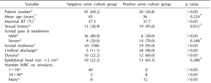
Table 2. Comparison of negative and positive urine culture groups’ patient characteristics
Variable Negative urine culture group Positive urine culture group p value Patient number*
Mean age (years)† Maximal BT (oC)† Sexual history*
Scrotal pain & tenderness Mild*
Severe*
Scrotal erythema*
Urethral discharge*
Dysuria*
Epididymal head size ≥2 cm*
Number WBC on urinalysis 1∼10*
10∼40*
Many*
45 (69.2) 45 37.3 13 (28.9) 36 (80.0) 9 (20.0) 45 (100) 5 (11.1) 10 (22.2) 10 (22.2)
40 5 0
20 (30.8) 36 37.7 19 (95.0)
6 (30.0) 14 (70.0) 19 (95.0) 18 (90.0) 12 (60.0) 13 (65.5)
0 8 12
>0.05 0.224‡
>0.05 0.012‡
>0.05 0.348‡
>0.05
>0.05
>0.05 0.288‡
>0.05
>0.05
>0.05 Values are presented as number (%) or number.
BT: body temperature, WBC: white blood cells.
*Fischer's exact test. †Mann Whitney U test. ‡Statistically significant difference.
Table 3. Comparison ofcharacteristics of positive urine culture patients between STO culture group and non-STO culture group (N=20)
Variable STO culture group Non-STO culture group p value
Number of patient*
Mean age (years)† Maximal BT (oC)† Sexual history*
Scrotal pain & tenderness Mild*
Severe*
Scrotal erythema*
Urethral discharge*
Dysuria*
Epididymal head size ≥2 cm*
Number of WBC on urinalysis 1∼10*
10∼40*
Many*
13 (65.0) 34 37.7 11 (84.6)
3 (23.1) 10 (76.9) 13 (100) 12 (92.3)
8 (61.5) 9 (69.2)
0 5 6
7 (45.0) 41 37.6 5 (71.4) 3 (42.8) 4 (57.1) 6 (85.7) 6 (85.7) 4 (57.1) 4 (57.1)
0 3 6
>0.05 0.044‡
>0.05
>0.05
>0.05
>0.05
>0.05
>0.05
>0.05
>0.05
>0.05
>0.05
>0.05 Values are presented as number (%) or number.
STO: sexually transmitted organism, BT: body temperature, WBC: white blood cells.
*Fischer's exact test. †Mann Whitney U test. ‡Statistically significant difference.
RESULTS
In this series, 65 patients (age range, 18∼80 years) pre- sented with a painful, swollen, and tender epididymis, or with testicular or scrotal pain, and their clinical notes were reviewed retrospectively. Most patients presented with scrotal pain or tenderness (98%), scrotal erythema
(100%), and scrotal swelling (83%). Thirty-two (49%) pa- tients had a history of sexual contact at least two weeks pri- or to the onset of epididymitis.
Twenty (30.8%) of the 65 patients with acute epi- didymitis had a positive urine culture or urethral swab.
Thirteen (65.0%) of these patients had a STO such as C. tra- chomatis or N. Gonorrhoeae, while the rest of them
(45.0%) had a gram negative enteric bacteria (non-STO) like Escherichia coli, Staphylococcus aureus, Streptococ- cus epididymis, or Enterococcus faecalis (Table 1).
The positive urine culture group was significantly younger than the negative culture group (p=0.224) and had a higher incidence of a history of sexual contact at least two weeks prior to onset of epididymitis (p=0.012).
In addition, the patients of the positive urine culture group with epididymitis also had a significantly enlarged epi- didymal head (p=0.348) and significantly more severe complaints of pain or tenderness than those of negative urine culture group (p=0.288). There were 10 patients (22.2%) with an epididymal head diameter ≥2 cm as shown by testicular ultrasonography versus 13 such pa- tients (65.5%) in the positive urine culture group (Table 2).
The STO culture group was younger than the non-STO group, but the differences in these measures between the two groups were not statistically significant, except age (Table 3).
DISCUSSION
Acute epididymitis is one of the most common causes of acute scrotal pain and is found in 74.5% of patients who visit hospital emergency rooms for this complaint.5 Because there are differences in the causes of acute epi- didymitis depending on age, this is an important factor for diagnosis.6 Thus, Melekos and Asbach7 reported that many men aged 40 years and older had general infectious agents of the Chlamydia infections diagnosed as the cause of acute epididymitis. De Jong et al2 reported that 10 out of 12 adults aged 35 years and older had been found with Gram-negative infections, whereas adults aged less than 35 years old had a high rate of infection with Chlamydia.
In this study, Chlamydia was detected in the urethra of 14% of the patients diagnosed with acute epididymitis and 6% of the patients were confirmed with urethritis caused by gonococcal infections. Such epididymiis oc- curs when the bacterial infection starts in the lower urinary tract and ascends to the epididymis. In such cases, if the patient showed pyuria or a positive bacterial culture, anti- biotic treatment was administered.
Hawkins et al8 reported that acute epididymitis caused by Chlamydia tended to advance to severe disease, and
that these patients complained of more severe pain than in cases of epididymitis caused by more general urinary tract infective agents. Hoosen et al9 also reported that epi- didymitis accompanying infections with Chlamydia was more severe than that caused by E. coli, and such cases were characterized by periductal and intraepithelial inflammation. Here we found that of 20 patients with identification of causative microorganisms, nine had uri- nary tract infections caused by Chlamydia and they had a greater diameter of the epididymal head identified from ultrasonography. Moreover, their complaints of pain or tenderness were significantly more severe compared with the patients in whom causative microorganisms were not identified.
Urethral and ejaculatory duct reflux has been suggested as another cause of acute epididymitis besides urinary tract infections and is unrelated to any sexual activity or urethral inflammation caused by occlusion of the lower urinary tract. In this situation, pressure in the prostatic ure- thra is increased during micturition so that urine refluxes to the ejaculatory duct and epididymis, resulting in chem- ical inflammation. Such cases are characterized by the ab- sence of pyuria and microorganisms upon urinalysis.10-12 Lewis et al13 reported that 38% of patients with acute epi- didymitis showed abnormalities in the urethral system and 64% of these patients complained of voiding problems.
Höppner et al14 reported that among patients with acute epididymitis aged 60 years and older, 53% had a lower urinary tract obstruction caused by benign prostatic hyper- plasia, prostate cancer, or a urethral stricture. In the pres- ent study, the positive urine culture group was younger than the others and also had a much higher incidence of recent sexual history. Thus, practically, this means that in sexually active men younger than 40 years of age, epi- didymitis is most often caused by sexually transmitted pathogens such as C. trachomatis and N. gonorrhoeae and in men older than 40 years of age is most often caused by non-sexually transmitted Gram-negative enteric organ- isms causing urinary tract infection. There is also a possi- bility that acute epididymitis could be caused in patients over 40 years of age by chemical inflammation, regardless of sexual activity or urethral infection.
Our study had several limitations. The age of the pa- tients was bimodally distributed. The mean age of the neg-
ative urine culture groups was over 40, while that of the positive urine culture groups was under 40. This could re- flect selection bias in our study. However, acute epi- didymitis is so infrequent that a prospective study design is very difficult. Moreover, studies on acute epididymitis are rare, and this is the first to define the association be- tween urine culture results and clinical symptoms.
Therefore, we believe this is a meaningful study.
Because one cause of acute epididymitis is believed to be an ascending urethral infection with general bacteria, the first-line treatment has involved antibiotics. Neverthe- less, according to recent reports, treatment other than anti- biotics obtained good results in patients with acute epi- didymitis who had no evidence of urethral infection.15,16 According to our national guidelines in Korea,17 if there is no evidence of inflammation, acute epididymitis can be treated sufficiently by bed rest, scrotal elevation, and analgesics. If infection is suspected-depending on the pa- tient’s age, and a history of sexual contact, recent in- strumentation, or catheterization-patients younger than 35 years should be administered doxycycline for 14 days.
If there is a suspected infection from urinary pathogens, it is possible to administer quinolone. The most important treatments for patients with acute epididymitis are scrotal elevation and absolute rest. As the incidence of anti- biotic-resistant microorganisms is increasing rapidly, the uniformed use of antibiotics should be reconsidered among patients with acute epididymitis who have no evi- dence of urethral infection.
CONCLUSIONS
In patients with acute epididymitis, there was a close as- sociation between clinical symptoms, a positive urine cul- ture, and ultrasonographic findings. Patients with a pos- itive urine culture tended to be sexually active and young- er than those others with acute epididymitis. They also had a significantly enlarged epididymal head shown by ul- trasonography and their complaints of pain or tenderness were significantly more severe than in those patients for whom no causative microorganisms were identified.
Therefore, these factors would be worth considering when managing patients with acute epididymitis.
REFERENCES
1. Berger RE, Alexander ER, Monda GD, Ansell J, McCormick G, Holmes KK. Chlamydia trachomatis as a cause of acute
"idiopathic" epididymitis. N Engl J Med 1978;298:301-4 2. De Jong Z, Pontonnier F, Plante P, Gautier JR, Ioualalen A,
Archambaud M, et al. The frequency of Chlamydia trachomatis in acute epididymitis. Br J Urol 1988;62:76-8 3. Anthonisz M. Assessing the impact: the National Chlamydia
Screening Programme. Br J Nurs 2009;18:246-51
4. Geisler WM, Krieger JN. Epididymitis. In: Holmes KK, Sparling PF, Stamm WE, editors. Sexually transmitted diseases. New York: McGraw-Hill Medical; 2008;1127-45 5. Oh DK, Kim SJ, Ahn HS. Experiences of 313 cases of acute
scrotum: properties of acute epididymitis and differential diagnosis of testicular torsion. Korean J Urol 2002;43:
624-30
6. Park CH, Im JK, Kim KK, Park HW. Clinical characteristics of acute epididymitis in children and adults. Korean J Urol 1999;40:674-6
7. Melekos MD, Asbach HW. Epididymitis: aspects concer- ning etiology and treatment. J Urol 1987;138:83-6
8. Hawkins DA, Taylor-Robinson D, Thomas BJ, Harris JR.
Microbiological survey of acute epididymitis. Genitourin Med 1986;62:342-4
9. Hoosen AA, O'Farrell N, van den Ende J. Microbiology of acute epididymitis in a developing community. Genitourin Med 1993;69:361-3
10. Siegel A, Snyder H, Duckett JW. Epididymitis in infants and boys: underlying urogenital anomalies and efficacy of imaging modalities. J Urol 1987;138:1100-3
11. Park HC, Park HJ, Park NC. Adenomatoid tumor of the epididymis. Korean J Androl 2003;21:111-3
12. Lee SW, Jung HW, Kim SY, Kim H, Yang DY, Cho SJ. Clear cell papillary cystadenoma of the epididymis associated with multiple renal and pancreatic cysts. Korean J Androl 2007;25:141-4
13. Lewis AG, Bukowski TP, Jarvis PD, Wacksman J, Sheldon CA. Evaluation of acute scrotum in the emergency depart- ment. J Pediatr Surg 1995;30:277-81
14. Höppner W, Strohmeyer T, Hartmann M, Lopez-Gamarra D, Dreikorn K. Surgical treatment of acute epididymitis and its underlying diseases. Eur Urol 1992;22:218-21
15. Hutson JM, Dewan PA. Acute epididymitis in boys: are antibiotics indicated? Br J Urol 1997;80:970-1
16. Ji YH, Park SC, Lee SK, Choo HS, Kim J, Choi HJ, et al.
Adenomatoid tumor between the epididymis and the testis.
Korean J Androl 2008;26:240-3
17. National guideline for the management of epididymo- orchitis. Clinical Effectiveness Group (Association of Geni- tourinary Medicine and the Medical Society for the Study of Venereal Diseases). Sex Transm Infect 1999;75 Suppl 1:
S51-3