Vol. 2, No. 1, pp 1~8, 2008
Corresponding author:Ki-Won Kim, MD
Department of Orthopedic Surgery, St. Mary’s Hospital, College of Medicine, The Catholic University of Korea, 62 Yoido-dong, Youngdeungpo-ku, Seoul, 150-713, Korea
Tel: +82-2-3779-1192, Fax: +82-2-783-0252, E-mail: kiwonk62@yahoo.co.kr
Senescence of Nucleus Pulposus Chondrocytes in Human Intervertebral Discs
Ki-Won Kim, Kee-Yong Ha#, Jun-Seok Lee$, Ki-Ho Na, Young-Yul Kim, and Young-Kyun Woo
Department of Orthopedic Surgery, St. Mary’s Hospital, College of Medicine, The Catholic University of Korea, Seoul, Korea, Department of Orthopedic Surgery, Kang-Nam St. Mary’s Hospital, College of Medicine,
The Catholic University of Korea, Seoul, Korea#, Department of Orthopedic Surgery, Dongshin General Hospital, Seoul, Korea$
S
Sttuuddyy DDeessiiggnn:: Senescence-related markers were assessed in surgically obtained human nucleus pulposus (NP) specimens.
P
Puurrppoossee:: To demonstrate the mechanism and signaling pathway involved in the senescence of NP chondrocytes.
O
Ovveerrvviieeww ooff LLiitteerraattuurree:: The population of senescent disc cells has been shown to be increased in degenerated or herniated discs. However, the mechanism and signaling pathway involved in the senescence of NP chondrocytes are unknown.
M
Meetthhooddss:: We examined cell senescence markers [senescence-associated β-galactosidase (SA-β-gal), telomere length, telom- erase activity, p53, p21, pRB and p16] and the hydrogen peroxide (H2O2) content in human NP specimens.
R
Reessuullttss:: The percentage of SA-β-gal-positive NP chondrocytes increased with age, while the telomere length and telom- erase activity declined. However, there was no significant correlation between age and H2O2 content. The NP specimens with grade III or IV degeneration showed significantly higher percentages of SA-β-gal-positive NP chondrocytes than those with grade II degeneration. Immunohistochemistry showed that senescent NP chondrocytes in all specimens expressed p53, p21, and pRB, while a few NP chondrocytes in only two specimens expressed p16.
C
Coonncclluussiioonnss:: The present study demonstrates that, with increasing age and advancing disc degeneration, senescent NP chondrocytes increase or accumulate in the NP. Furthermore, the telomere-based p53, p21, pRB pathway, rather than the stress-based p16, pRB pathway, plays a more important role in the senescence of NP chondrocytes in in vivo conditions. Our results suggest that prevention or reversal of senescence of NP chondrocytes can be a novel mechanism by which to pre- vent human disc degeneration.
K
Keeyywwoorrddss:: Senescence, Nucleus pulposus, Chondrocytes, Intervertebral disc, Disc degeneration
Introduction
Cellular senescence is a program activated by normal cells in response to various types of stress1. One important mechanism responsible for cellular senescence is progres- sive telomere shortening and eventual telomere dysfunction that occur due to incomplete DNA replication (an end-repli- cation problem) at the telomeres (‘replicative senescence’
or ‘intrinsic senescence)2,3,4,5,6. This end-replication problem
can be resolved by a holoenzyme telomerase, which elon- gates the telomeric DNA in the 5’-to-3’ direction6,7,8,9,10,11. In the absence of telomerase, or when its expression is very low, the telomeric DNA progressively shortens with each round of cell division12. In addition to replicative senes- cence, cellular senescence can also be induced in a rapid manner by a number of stresses that are independent of telomere shortening [‘stress-induced premature senescence’
(SIPS) or ‘stress or aberrant signaling-induced senescence’
(STASIS)]13,14. Such stresses include oxidative stress, DNA
damage, oncogenic activity, and other metabolic perturba- tions15.
Cellular senescence like apoptosis can be viewed as a powerful tumor-suppressor mechanism that withdraws cells with irreparable DNA damages from the cell cycle16,17. Therefore, the senescence signals, that is, a telomere-based one or a stress-based one, trigger a DNA damage response, and this response shares a common signaling pathway that converges on either or both of the well-established two tumor suppressor proteins, p53 (the p53, p21, pRB path- way) and pRB (the p16, -pRB pathway)1,14,15,18,19,20. In the p53, p21, pRB pathway, senescence stimuli activate p53, which can then induce senescence by activating pRB through p21, a transcriptional target of p53. This senescence can be reversed upon subsequent inactivation of p53. In the p16- pRB pathway, senescence stimuli induce p16, which acti- vates pRB. Once the pRB pathway is engaged by p16, the senescence cannot be reversed by subsequent inactivation of p53, silencing of p16, or inactivation of pRB18. Although there appears to be overlap between the two pathways, the emerging consensus is that the p53, p21, pRB pathway mediates the senescence that is primarily due to telomere shortening, and the p16, pRB pathway is thought to mediate premature senescence1,14,20. However, a population of grow- ing cells suffers from a combination of physiologic stresses that act simultaneously, and the relative importance of the p53, p21, pRB or p16, pRB pathway for the senescence response may differ depending on the tissue and the species of origin1,20. Once cells have entered senescence, they are arrested in the G1 phase of the cell cycle, and they display a characteristic morphology (vacuolated, flattened cells) and gene expression, including that for markers such as a senes- cence-associated β-galactosidase (SA-β-gal)21,22.
Degenerative changes in the intervertebral disc (IVD) occur as a natural part of aging23. Gruber et al and Roberts et al recently provided important insights regarding the close link between cellular senescence and disc degenera- tion, based on the observation that SA-β-gal-positive disc cells increased with advancing disc degeneration and increased in herniated discs24,25. However, the mechanism and signaling pathways involved in the senescence of nucle- us pulposus (NP) chondrocytes are unknown. Our present in vivo study demonstrated that, with increasing age and advancing disc degeneration, senescent NP chondrocytes increase or accumulate in the NP. The NP chondrocytes in aging discs exhibited characteristic senescent features, such as increased SA-β-gal expression, shortened telomeres, and
decreased telomerase activity. We further show that the telomere-based p53, p21, pRB pathway, rather than the stress-based p16-pRB pathway, plays a more important role in the senescence of NP chondrocytes in in vivo conditions.
Materials and Methods
Twenty-five patients (14 female patients and 11 male patients) who underwent open discectomy for symptomatic herniated NP were included in this study. After thorough removal of the protruded or extruded NP fragments, the NP specimens remaining in the central part of the IVD were pooled, and then they were immediately preserved at -75℃.
The NP specimens were grouped according to a grading system for IVD degeneration that was based on the preoper- ative magnetic resonance images (MRI)26: there were 3 patients with grade II degeneration, 17 patients with grade III degeneration, and 5 patients with grade IV degeneration.
In practice, no NP specimens with grade I or grade V degeneration were available because there was no case of NP herniation in any individual with grade I degeneration (normal disc), and there were no remnant NP specimens in patients with grade V degeneration (total collapse of the disc space). The mean patient age was 49 years [range and standard deviation (SD): 20~75, ±14 years].
Senescence-associated β-galactosidase Staining
The SA-β-gal activity in the NP chondrocytes was deter- mined using a SA-β-gal staining kit (Cell Signaling Tech- nology). Sections (4 μm thickness) of the frozen NP speci- mens were placed on slides and fixed with 2% formalde- hyde and 0.2% glutaraldehyde in phosphate buffered saline (PBS) for 10 minutes at room temperature. The slides were rinsed with PBS and then incubated overnight at 37℃ with fresh SA-β-gal staining solution containing 40 mM citric acid/sodium phosphate (pH 6.0), 150 mM NaCl, 2 mM MgCl2, 5 mM potassium ferrocyanide, 5 mM potassium fer- ricyanide, and 1 mg/ml of 5-bromo-4-chloro-3-indolyl-β-D- galactopyranoside (X-gal). After being rinsed with distilled water, the slides were counter-stained with Nuclear Fast Red. All NP chondrocytes and SA-β-gal-positive NP chon- drocytes on the whole section were counted (×200), and the percentage of SA-β-gal-positive NP chondrocytes was calculated.
Telomere Length Assay
Genomic DNA was extracted from 40 mg of the NP spec- imens using the Genomic DNA purification kit (Gentra, Minneapolis, MN). Genomic DNA (2~3 μg) was digested with the restriction enzymes Rsa I and Hinf I at 37℃ for 16 hours and then electrophoresed on 0.6% agarose gels at 50 V for 4 hours. After electrophoresis, the gel was depurinat- ed in 0.25 M HCl for 30 minutes and denatured in 0.4 M NaOH and 1.5 M NaCl for 30 minutes. The DNA was then transferred by capillary transfer onto a positively charged nylon membrane (Hybond-N, Roche, Germany). The mem- brane was prehybridized in hybridization buffer (TeloTAGGG Telomere Length Assay, Roche, Germany) for 1 hour at 42℃ and then hybridized with a telomere probe in hybridization buffer for 3 hours at 42℃. The mem- brane was washed twice with washing solution and rinsed with the recommended blocking reagent. The hybridized probe was detected by the chemiluminescence method, according to the manufacturer’s recommendation. The membrane was exposed to Hyperfilm (Amersham Pharma- cia Biotech, England). The mean telomere length was calcu- lated using the formula: L=∑ (ODi) / ∑ (ODi/Li), where ODi is the integrated signal intensity and Li is the DNA length at position i.
Telomerase Activity
The telomerase activity was analyzed with a TeloTAGGG Telomerase PCR ELISA plus kit (Roche, Germany), accord- ing to the manufacturer’s instructions. The frozen NP speci- mens were suspended in 200 μl of ice-cooled lysis reagent and incubated in ice for 30 minutes. The lysate was cen- trifuged at 16,000×g for 20 minutes at 4℃. The super- natant was carefully removed and transferred into a tube for the TRAP assay. Ten μg of protein extract was used for each assay. Each supernatant was divided into two aliquots.
One aliquot was inactivated at 85℃ for 10 minutes and used as a negative control, while the other was used to eval- uate the telomerase activity. For each test sample and con- trol, 25 μl of the reaction mixture and 5 μl of the internal standard (IS) were transferred into a tube suitable for PCR amplification. The extended products were amplified by PCR using Taq polymerase, the P1-TS, P2 primers, and nucleotides. After a 30 minute incubation at 25℃ to allow the telomerase-mediated extension of the TS primer, and 5 minutes at 94℃ to inactivate the telomerase, the reaction
mixture was subjected to 30 PCR cycles at 94℃ for 30 sec- onds, 50℃ for 30 seconds, and 72℃ for 90 seconds, then 72℃ for 10 minutes on a thermocycler. Using the ELISA method, the amplified products were immobilized on strep- tavidin-coated microtiter plates via biotin-streptavidin inter- action. The amplicons were then detected by anti-digoxi- genin antibodies conjugated to peroxidase. After addition of the peroxidase substrate (3, 3’, 5, 5’-tetramethyl benzidine), the amount of TRAP products was determined by measure- ment of absorbance at 450 nm.
Measurement of H2O2Content in the NP Tissue
The NP tissues (20 mg) were homogenized in PBS. The homogenate was centrifuged at 15,000 rpm for 15 minutes at 4℃, and the supernatant was collected and stored at -75℃.
The H2O2 content was measured using the Quantitative Hydrogen Peroxide Assay Kit (OXIS International, Foster city, CA). This assay is based on the oxidation of ferrous ions (Fe2+) to ferric ions (Fe3+) by H2O2under acidic condi- tions. The ferric ion binds with the indicator dye xylenol orange to form a stable colored complex that can be mea- sured at 560 nm.
Immunohistochemistry
To test the immunohistochemical expression of p53, p21, pRB, and p16INK4(Phamingen, San Diego, CA), the fresh- frozen specimens stored at -75℃ were embedded in OCT (Tissue-Tek, Sakura Finetek, Torrance, CA). The embedded specimens were cut at -25℃, mounted on lysine-coated glass slides, and then fixed for 10 minutes at 4℃ in 4%
paraformaldehyde. The endogenous peroxidase was subse- quently blocked by 3% H2O2for 20 minutes and 0.1% Tri- ton X-100 in PBS for 15 minutes. The cryostat sections were incubated in a humid chamber at 4℃ overnight with the primary antibodies for p16, pRB, p21, and p53. The sec- tions were then exposed to a streptavidin-biotin-peroxidase complex (Histostain Plus kit, Zymed, USA), and the color was developed with 3,3-diaminobenzidine tetrahydrochlo- ride (Lab Vision, Fremont, CA). Mayer’s hematoxylin was used for counterstaining.
Statistical Analysis
The normality of variables (age, percentages of SA-β-gal- positive NP chondrocytes, telomere length, telomerase
activity, and H2O2 content) was assessed using the Kol- mogorov-Smirnov test. Depending on the normality, corre- lations among the variables were analyzed using Spear- man’s or Pearson’s test. The differences in the means of the variables among the degeneration grades were analyzed using the Mann-Whitney U test.
Results
Senescence-associated β-galactosidase Stain
The mean percentage of SA-β-gal-positive NP chondro- cytes was 41.82% (range: 15.65~60.80%; SD: ±11.20%) (Fig. 1).
Telomere Length
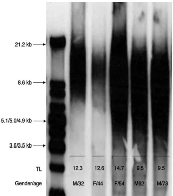
The mean telomere length was 14.42 kb (range: 6.60~
29.80 kb; SD: ±5.73 kb) (Fig. 2).
Telomerase Activity
The mean telomerase activity was 6.41 AUs (range:
0.8~22.3 arbitrary units; SD: ±5.37 arbitrary units).
H2O2Content in the NP Specimens
The H2O2 content in the NP specimens was 0.25 μmol (range: 0.05~0.65 μmol; SD: ±0.14 μmol).
Immunohistochemistry
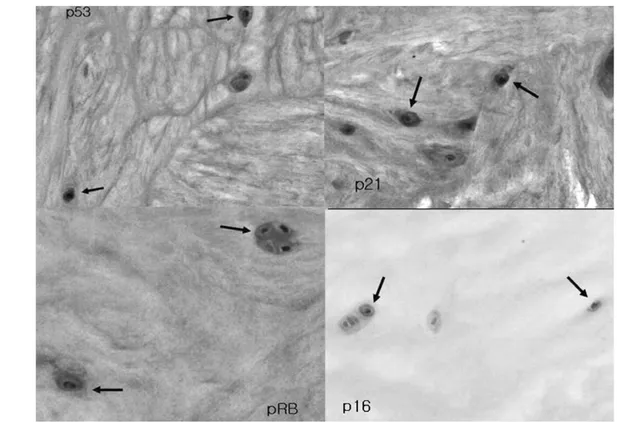
Immunohistochemistry showed that the senescent NP chondrocytes in all 25 specimens expressed p53, p21, and pRB, but only a few NP chondrocytes in two specimens expressed p16 (Fig. 3).
Statistical Analysis
Among the variables (age, percentage of SA-β-gal-posi- tive NP chondrocytes, telomere length, telomerase activity, and H2O2content), age and the percentage of SA-β-gal-pos- itive NP chondrocytes showed a normal distribution. The percentages of SA-β-gal-positive NP chondrocytes increased with age (r=0.82, p<0.001), while the telomere length and telomerase activity declined (r=-0.41, p=0.045 and r=-0.52, p=0.008, respectively) (Fig. 4). However, there was no significant correlation between age and the H2O2
content (p=0.18). The NP specimens with grade III (mean±
SD: 44.11±9.40%) or IV (mean±SD: 46.02±6.11%) degeneration showed significantly higher percentages of SA-β-gal-positive NP chondrocytes than those with grade II (mean±SD: 21.83±6.01%) degeneration (p=0.01 and p=0.025, respectively).
Fig. 1. SA-β-gal-positive NP chondrocytes are shown with arrows.
Fig. 2. The telomere length (TL) of the NP chondrocytes as measured by Southern blotting.
Fig. 3. Expression of p53, p21, pRB, and p16 by NP chondrocytes (arrows).
Fig. 4. Correlation analysis of patient age versus the percent- age of SA-β-gal-positive NP chondrocytes (Pearson’s test), telomere length (Spearman’s test), and telomerase activity (Spearman’s test).
Discussion
In general, senescent cells become unresponsive to mito- genic stimuli, yet they can remain viable for extended peri- ods of time21. They also express elevated levels of extracel- lular matrix (ECM)-degrading proteases, collagenases, and matrix metalloproteinase (MMP) family members20,21. In contrast, they express decreased levels of the MMP inhibitor TIMP1 and decreased levels of ECM components, such as elastin, laminin, and several forms of collagen20,21. These alterations indicate a general shift in the phenotype of the senescent cells from matrix-synthesizing to matrix- degrading20,21,27 .
Such a shift has also been identified in aging and degen- erated IVDs28. The present study showed that the percent- ages of SA-β-gal-positive NP chondrocytes increases with age. This implies that for NP chondrocytes, like primary cells in other organs of the body, senescence with age is unavoidable22. In addition, the NP specimens with grade III or IV degeneration showed significantly higher percentages of SA-β-gal-positive NP chondrocytes than those with grade II degeneration, which supports the previous finding that the senescent disc cell population increases or accumu- lates with advancing disc degeneration24,25. Thus, the increase or accumulation of senescent NP chondrocytes with aging may, at least in part, contribute to IVD degenera- tion, whether it is natural or pathologic.
We further showed that the telomere length and telom- erase activity of NP chondrocytes decline with age, while the H2O2content has no significant correlation with age. In articular cartilage, the chondrocytes were shown to senesce with age via telomere shortening, which was effectively prevented by ectopic telomerase expression29. In contrast, oxidative stress induced by hyperbaric culture conditions caused premature chondrocyte senescence, which was not effectively prevented by ectopic telomerase expression30. Because NP chondrocytes originate from the hyaline carti- lage endplate31,32, which is naturally similar to articular carti- lage, our results imply that the senescence of NP chondro- cytes can be caused by age-dependent telomere shortening and/or age-independent oxidative stress, as occurs in articu- lar chondrocytes. This may explain the increased risk of disc degeneration with age23and the occurrence of unex- pected premature or accelerated disc degeneration in some situations, such as vertebral body fractures33,34.
Although SA-β-gal is a useful senescence marker, its
activity is critically dependent on detection conditions. SA- β-gal is also expressed in non-senescent cells that have a high lysosomal content35,36. Multiple markers of senescence are therefore recommended to demonstrate senescence in vivo. For this, we further determined the expressions of p53, p21, pRB, and p16 by immunohistochemistry. Immunohis- tochemistry showed that the senescent NP chondrocytes in all the specimens expressed p53, p21, and pRB, but only a few NP chondrocytes in two specimens expressed p16.
These results indicate that the telomere-based p53, p21, pRB pathway, rather than the stress-based p16, pRB path- way, plays a more important role in the senescence of NP chondrocytes. However, the co-expression of p16, although uncommon, implies that there is a situation in which both pathways are activated simultaneously. Thus, our results suggest that NP chondrocytes in in vivo conditions are sub- ject to a number of physiological stresses that act simultane- ously, and the total stress magnitude may determine the appropriate senescence response1.
The results of our study, which addressed disc herniation, may not be applicable to other degenerative disc diseases.
In addition, the NP chondrocytes in the herniated NP frag- ments may senesce via different signaling pathways, or they may undergo apoptosis rather than senescence37,38. To mini- mize any potential bias related to disc herniation, we consis- tently obtained NP specimens from the central part of the disc after thorough removal of the protruded or extruded disc fragments. Further studies are required to understand the senescence mechanisms of IVD cells in other degenera- tive disc diseases.
Although cellular senescence helps an organism by sup- pressing life-threatening tumorogenesis16,17, it can also be detrimental to the organism by depleting renewable tissues that contain proliferation-competent progenitor or stem cells. In turn, such depletion can compromise the structure and function of tissues, which is a hallmark of aging. Fur- thermore, senescent cells can persist and acquire altered functions, and so this alters the tissue microenvironments in ways that can promote aging phenotypes20,27,39. The direct relationship between cellular senescence and IVD aging or degeneration is unclear. However, two recent studies24,25, in addition to ours, have provided evidence for the potential role of senescence of disc cells or NP chondrocytes in aging or degenerating IVDs.
In conclusion, the present in vivo study demonstrates that, with increasing age and advancing disc degeneration, senes- cent NP chondrocytes increase or accumulate in the NP.
The senescent NP chondrocytes exhibited characteristic senescent features, such as increased SA-β-gal expression, shortened telomeres, and decreased telomerase activity.
Further, we showed that the telomere-based p53, p21, pRB pathway, rather than the stress-based p16, pRB pathway, plays a more important role in the senescence of NP chon- drocytes in in vivo conditions, although there is probably a scenario in which both pathways are activated simultane- ously. Because the p53, p21, pRB pathway is reversible, prevention or reversal of NP chondrocyte senescence could be a novel mechanism for treating human disc degeneration.
Acknowledgements
We thank Miss Ha-Na Chung for her technical assistant.
This study was supported in part by the Catholic Medical Research Foundation.
REFERENCES
01. Ben-Porath I, Weinberg RA: The signals and pathways activating cellular senescence. Int J Biochem Cell Biol 2005; 37: 961-976.
02. Blackburn EH: Switching and signaling at the telomere.
Cell 2001; 106: 661-673.
03. Hayflick L, Moorhead PS: The serial cultivation of human diploid cell strains. Exp Cell Res 1961; 25: 585- 621.
04. Olovnikov AM: Principle of marginotomy in template synthesis of polynucleotides. Dokl Akad Nauk SSSR 1971;
201: 1496-1499.
05. Olovnikov AM: A theory of marginotomy: The incom- plete copying of template margin in enzymic synthesis of polynucleotides and biological significance of the phenom- enon. J Theor Biol 1973; 41: 181-190.
06. Verdun RE, Karlseder J: Replication and protection of telomeres. Nature 2007; 447: 924-931.
07. Alberts B, Johnson A, Lewis J, et al: DNA Replication, Repair, and Recombination. 4th edition ed. New York, NY:
Garland Science, 2002.
08. Blackburn EH: Structure and function of telomeres.
Nature 1991; 350: 569-573.
09. Chan SR, Blackburn EH: Telomeres and telomerase. Phi- los Trans R Soc Lond B Biol Sci 2004; 359: 109-121.
10. Feng J, Funk WD, Wang SS, et al: The RNA component of human telomerase. Science 1995; 269: 1236-1241.
11. Nakamura TM, Cech TR: Reversing time: origin of telomerase. Cell 1998; 92: 587-590.
12. Harley CB, Futcher AB, Greider CW: Telomeres short- en during ageing of human fibroblasts. Nature 1990; 345:
458-460.
13. Drayton S, Peters G: Immortalisation and transformation revisited. Curr Opin Genet Dev 2002; 12: 98-104.
14. Herbig U, Sedivy JM: Regulation of growth arrest in senescence: telomere damage is not the end of the story.
Mech Ageing Dev 2006; 127: 16-24.
15. Ben-Porath I, Weinberg RA: When cells get stressed: an integrative view of cellular senescence. J Clin Invest 2004;
113: 8-13.
16. Artandi SE, DePinho RA: A critical role for telomeres in suppressing and facilitating carcinogenesis. Curr Opin Genet Dev 2000; 10: 39-46.
17. de Lange T, Jacks T: For better or worse? Telomerase inhibition and cancer. Cell 1999; 98: 273-275.
18. Beausejour CM, Krtolica A, Galimi F, et al: Reversal of human cellular senescence: roles of the p53 and p16 path- ways. Embo J 2003; 22: 4212-4222.
19. Campisi J: Cellular senescence as a tumor-suppressor mechanism. Trends Cell Biol 2001; 11: S27-31.
20. Campisi J: Senescent cells, tumor suppression, and organ- ismal aging: good citizens, bad neighbors. Cell 2005; 120:
513-522.
21. Cristofalo VJ, Lorenzini A, Allen RG, et al: Replicative senescence: a critical review. Mech Ageing Dev 2004; 125:
827-848.
22. Dimri GP, Lee X, Basile G, et al: A biomarker that identi- fies senescent human cells in culture and in aging skin in vivo. Proc Natl Acad Sci USA 1995; 92: 9363-9367.
23. An HS, Anderson PA, Haughton VM, et al: Introduction:
disc degeneration: summary. Spine 2004; 29: 2677-2678.
24. Gruber HE, Ingram JA, Norton HJ, et al: Senescence in cells of the aging and degenerating intervertebral disc:
immunolocalization of senescence-associated beta-galac- tosidase in human and sand rat discs. Spine 2007; 32: 321- 327.
25. Roberts S, Evans EH, Kletsas D, et al: Senescence in human intervertebral discs. Eur Spine J 2006; 15 Suppl 3:
S312-6.
26. Pfirrmann CW, Metzdorf A, Zanetti M, et al: Magnetic resonance classification of lumbar intervertebral disc degeneration. Spine 2001; 26: 1873-1878.
27. Krtolica A, Campisi J: Cancer and aging: a model for the cancer promoting effects of the aging stroma. Int J
Biochem Cell Biol 2002; 34: 1401-1414.
28. Roughley PJ: Biology of intervertebral disc aging and degeneration: involvement of the extracellular matrix.
Spine 2004; 29: 2691-2699.
29. Martin JA, Buckwalter JA: The role of chondrocyte senescence in the pathogenesis of osteoarthritis and in lim- iting cartilage repair. J Bone Joint Surg Am 2003; 85-A Suppl 2: 106-110.
30. Martin JA, Klingelhutz AJ, Moussavi-Harami F, et al:
Effects of oxidative damage and telomerase activity on human articular cartilage chondrocyte senescence. J Geron- tol A Biol Sci Med Sci 2004; 59: 324-337.
31. Kim KW, Ha KY, Park JB, et al: Expressions of mem- brane-type I matrix metalloproteinase, Ki-67 protein, and type II collagen by chondrocytes migrating from cartilage endplate into nucleus pulposus in rat intervertebral discs: a cartilage endplate-fracture model using an intervertebral disc organ culture. Spine 2005; 30: 1373-1378.
32. Kim KW, Lim TH, Kim JG, et al: The origin of chondro- cytes in the nucleus pulposus and histologic findings asso- ciated with the transition of a notochordal nucleus pulposus to a fibrocartilaginous nucleus pulposus in intact rabbit intervertebral discs. Spine 2003; 28: 982-990.
33. Haschtmann D, Stoyanov JV, Gedet P, et al: Vertebral endplate trauma induces disc cell apoptosis and promotes organ degeneration in vitro. Eur Spine J 2007.
34. Kerttula LI, Serlo WS, Tervonen OA, et al: Post-trau- matic findings of the spine after earlier vertebral fracture in young patients: clinical and MRI study. Spine 2000; 25:
1104-1108.
35. Kurz DJ, Decary S, Hong Y, et al: Senescence-associated (beta)-galactosidase reflects an increase in lysosomal mass during replicative ageing of human endothelial cells. J Cell Sci 2000; 113 (Pt 20): 3613-3622.
36. Matthews C, Gorenne I, Scott S, et al: Vascular smooth muscle cells undergo telomere-based senescence in human atherosclerosis: effects of telomerase and oxidative stress.
Circ Res 2006; 99: 156-164.
37. Park JB, Chang H, Kim KW: Expression of Fas ligand and apoptosis of disc cells in herniated lumbar disc tissue.
Spine 2001; 26: 618-621.
38. Park JB, Kim KW, Han CW, et al: Expression of Fas receptor on disc cells in herniated lumbar disc tissue. Spine 2001; 26: 142-146.
39. Rodier F, Campisi J, Bhaumik D: Two faces of p53:
aging and tumor suppression. Nucleic Acids Res 2007.