대한정형외과학회지:제 42 권 제 6 호 2007 J Korean Orthop Assoc 2007; 42: 836-839
836
통신저자:정 영 복
서울시 동작구 흑석동 224-1 중앙대학교병원 정형외과
TEL: 02-6299-1578ㆍFAX: 02-822-1710 E-mail: [email protected]
Address reprint requests to Young Bok Jung, M.D.
Department of Orthopedic Surgery, Chung-Ang University Medical Center, 224-1, Heukseok-dong, Dongjak-gu, Seoul 156-755, Korea
Tel: +82.2-6299-1578, Fax: +82.2-822-1710 E-mail: [email protected]
Heterotopic Bone Formation in Patient who Underwent a Posterior Cruciate Ligament Reconstruction using the Inlay Method and
Posterolateral Corner Sling with a Tibia Tunnel
- A Case Report -
Yong Seuk Lee, M.D.*, Young Bok Jung, M.D., Ho Joong Jung, M.D., Se Jin Park, M.D., and Chang Hyun Nam, M.D.
The Armed Forces Yangju Hospital*, Yangju, Department of Orthopedic Surgery, Chung-Ang University Medical Center, Seoul, Korea
변형된 Inlay 방법을 이용한 후방십자인대 재건술과 경골 터널을 이용한 후외측 재건술을 시행한 환자에서의 이소성 골화
- 1예 보고-
이용석*ㆍ정영복ㆍ정호중ㆍ박세진ㆍ남창현
국군양주병원*, 중앙대학교 의과대학 정형외과학교실
We describe a case of posterolateral capsular heterotopic ossification requiring a surgical excision after a PCL (Posterior Cruciate Ligament) reconstruction using the modified inlay method and PLCS (posterolateral corner sling) with a tibia tunnel. A 21-year-old female patient had suffered a blunt proximal tibial direct trauma 6 months earlier. She did not experience limb ischemia or a pulse deficit before she visited our out patient clinic. She had not suffered any trauma in other sites, and showed a range of motion of 0 to 30o at 4 months after surgery. There was no specific finding on the X-ray images. Arthroscopic adhesiolysis was performed and her range of motion increased to 0 to 120o. However, 6 months after the initial operation, she showed ankylosis and heterotopic ossification at the posterior aspect, which was surgically removed at 12 months postoperatively. After the second surgery, there was no recurrence and she showed a 0 to 140o range of motion at postoperative 42 months.
Key Words: Posterior cruciate ligament reconstruction, Posterolateral corner sling, Adhesiolysis, Heterotopic ossification.
INTRODUCTION
Heterotopic ossification (HO) is defined as the formation of mature lamellar bone in the soft tissue3,7). Ogilvie-Harris and Sekyi-Out described four cases of heterotopic ossification requiring surgical intervention after an arthroscopic anterior cruciate ligament (ACL) reconstruction, and Patton
et al reported 3 cases after knee dislocations, in which the PCL was one of the structures reconstructed7). Most cases did not require a surgical excision, and were due to relatively definite causes. On the other hand, our patient had a vague cause and required a surgical excision. We report this case with a review of the relevant
Heterotopic Bone Formation in Patient who Underwent a PCLR and PLCS 837
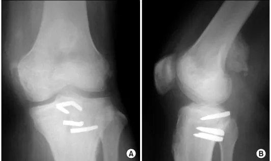
Fig. 2. The roentgenograms 6 months after surgery showing supracondylar heterotopic ossi- fication.
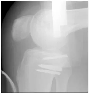
Fig. 1. The stress lateral view of a grade III posterior instability.
literature.
CASE REPORT
A 19-year-old woman visited the out patient clinic with right knee painful instability. She had suffered a direct pretibial trauma when she slipped down 6 months earlier. The clinical examination and MRI results were consistent with a tear of the PCL, a posterolateral corner injury showing PLRI
and an incomplete peroneal nerve injury that recovered completely (Fig. 1). The clinical exami- nation under anesthesia revealed increased pos- terior displacement at 90 degrees flexion with a loss of stepping, a positive reverse Povot-shift test and increased external rotation at 30 and 90o flexion. Arthroscopy revealed pseudolaxity of the anterior cruciate ligament and a drive through sign. The remnant of the PCL bundle was tensioned and anterolateral bundle of PCL was reconstructed using the modified inlay method with an ipsilateral hamstring5,6). The PLRI was restored by PLCS with a tibia tunnel using a contralateral hamstring1). The rehabilitation and follow up were performed as usual5,6). Two weeks after surgery, the patient had an active range of motion of 0 to 70o with no significant discomfort, and she was discharged with PCL brace. However, 6 weeks after surgery, the patient reported significant pain and a range of motion of only 0 to 30o, despite undergoing continued physical therapy. She had persistent pain and a range of motion 3 months after surgery, and the radiographs taken at this time showed no specific abnormality. Under general anesthesia, she showed a similar range of motion. Arthroscopic
838 Yong Seuk Lee, Young Bok Jung, Ho Joong Jung, et al
Fig. 3. The stress lateral view of 30 months after the excision operation showing no recurrence of heterotopic ossification and good stability of the PCL.
adhesiolysis and manipulation under anesthesia were performed, and a passive range of motion of 0 to 120o was gained postoperatively. However, 6 months after the initial surgery, she had a range of motion of 15 to 90o with walking difficulties. The radiographs taken at this time showed heterotopic ossification at the posterior supracondylar aspect (Fig. 2). From that time, the range of motion was not changed and a decision was made to excise the ossification fragment 12 months after surgery.
Arthroscopy revealed a normally looking PCL and no specific intraarticular abnormality. The pati- ent's position was changed and the mass was approached via the posterior aspect.
The mass was palpable, oval shaped and measured 3×5×2 cm. The range of motion improved markedly reaching 0-140o 6 months after the procedure and the patient could walk without any specific difficulty. Thirty months after the excision operation, the patient had no complaints about her knees and there were no changes observed on the radiographs (Fig. 3).
DISCUSSION
Heterotopic ossification is a common complication after a musculoskeletal injury. It is associated with central nervous system damage, burns, trauma, and a hereditary disorder known as fibrodysplasia ossificans progressiva7). Local factors including stasis, hematoma, edema, and prolonged immo- bilization are often cited as contributing factors9). Although heterotopic ossification after an arth- roscopic anterior cruciate ligament reconstruction has been reported, most reports of ligament injuries were of multiligament injuries, which did not require surgical removal7). However, some authors reported medial heterotopic bone requiring a surgical excision in all their patients2,4). We performed more than 150 PCL inlay recon- structions and 100 PLCS procedures but this is the
only case with this complication. Our patient did not suffer a brain injury or other factors known to cause heterotopic ossification. As suggested by other authors7), it is believed that reaming for the graft tunnels particularly PLCS with a femoral tunnel contributed to the process. The link between the posterior surgical bony procedure and posterior capsular ossification has not been recognized, and our report should raise the clinical awareness of such an entity.
For diagnosis, radiographs should be obtained to exclude heterotopic ossification in any patient with pain and a loss of motion, particularly after a knee dislocation, as well as in patients with a history of heterotopic ossification in other anatomic loca- tions. In addition, patients with pain and loss of motion should raise suspicion, even if they have suffered only minor ligament injury or a recon- struction.
The optimal protocol for a resection of heterotopic ossification with regards to the timing and adjunc- tive measures, such as radiation and indomethacin to prevent a recurrence, is controversial10). Most
Heterotopic Bone Formation in Patient who Underwent a PCLR and PLCS 839
authors advocate a minimum wait of 1 year after heterotopic bone formation before the surgical excision8).
In conclusion, reaming of the graft tunnels during PLCS contributes to the process of heterotopic ossification. However, there is the possibility that a capsular injury during the PCL inlay procedure might cause such complications.
REFERENCES
1. Albright JP, Brown AW: Management of chronic postero- lateral rotatory instability of the knee: surgical technique for the posterolateral corner sling procedure. Instr Course Lect, 47:
369-378, 1998.
2. Charnley G, Judet T, Garreau de Loubresse C, Mollaret O: Excision of heterotopic ossification around the knee following brain injury. Injury, 27: 125-128, 1996.
3. Dalury DF, Jiranek WA: The incidence of heterotopic ossification after total knee arthroplasty. J Arthroplasty, 19:
447-452, 2004.
4. Ippolito E, Formisano R, Farsetti P, Caterini R, Penta F:
Excision for the treatment of periarticular ossification of the knee in patients who have a traumatic brain injury. J Bone
Joint Surg Am, 81: 783-789, 1999.
5. Jung YB, Jung HJ, Tae SK, Lee YS, Lee KH:
Reconstruction of the posterior cruciate ligament with a mid-third patellar tendon graft with use of a modified tibial inlay method. J Bone Joint Surg Am, 87(Suppl 1): S247-S263, 2005.
6. Jung YB, Tae SK, Jung HJ, Lee KH: Replacement of the torn posterior cruciate ligament with a mid-third patellar tendon graft with use of a modified tibial inlay method. J Bone Joint Surg Am, 86: 1878-1883, 2004.
7. Patton WC, Tew WM: Periarticular heterotopic ossification after multiple knee ligament reconstructions. A report of three cases. Am J Sports Med, 28: 398-401, 2000.
8. Saito N, Horiuchi H, Takahashi H: Heterotopic ossification in the knee following encephalitis: a case report with a 10-year follow-up. Knee, 11: 63-65, 2004.
9. Stannard JP, Wilson TC, Sheils TM, McGwin G Jr, Volgas DA, Alonso JE: Heterotopic ossification associated with knee dislocation. Arthroscopy, 18: 835-839, 2002.
10. Toyoda T, Matsumoto H, Tsuji T, Kinouchi J, Fujikawa K: Heterotopic ossification after total knee arthroplasty. J Arthroplasty, 18: 760-764, 2003.
= 국문초록=
변형된 inlay 방법을 이용한 후방십자인대 재건 및 후외측 재건을 실시한 환자 중 후외측 관절막에 수술적 제거가 필요했던 이소성 골화가 생긴 1예를 보고하고자 한다. 환자는 근위 경골부 직접 타박상을 6개월 전에 받았으며 내원 당시에 허혈성 소견은 없었고, 다른 부위의 외상도 없었다. 수술적 치료를 시행하였으며 술 후 4개월에 0-30도의 관절 운동 범위를 나타내었다. 방사선 사진에서는 특이 소견이 없었고, 관절경적 유착 박리술 을 시행하였으며 0-120도의 관절 운동범위를 얻을 수 있었다. 술 후 6개월에 후방 관절막에 이소성 골화 소견이 있었으며 술 후 12개월에 성숙된 이소성 골을 제거 하였다. 그후 재발은 없었으며 술 후 42개월째에 0-140도의 관절운동범위를 보였다.