대한정형외과학회지:제 42 권 제 6 호 2007 J Korean Orthop Assoc 2007; 42: 828-831
828
통신저자:박 우 성
제주도 제주시 연동 2030 한라병원 정형외과
TEL: 064-740-5111ㆍFAX: 064-743-3110 E-mail: [email protected]
Address reprint requests to Woo-Sung Park, M.D.
Department of Orthopaedic Surgery, Halla General Hospital, 2030, Yeon-dong, Jeju 690-170, Korea
Tel: +82.64-740-5111, Fax: +82.64-743-3110 E-mail: [email protected]
Lumbar Wedge Resection Osteotomy for Congenital Scoliosis due to a Sacral Malformation
- A Case Report -
Bong-Jin Lee, M.D., Sung-Rak Lee, M.D., Seong-Tae Kim, M.D., Woo-Sung Park, M.D., Kwon-Hee Park, M.D., and Sang Hoon Lee, M.D
Department of Orthopaedic Surgery, Halla General Hospital, Jeju, Korea
천추 기형에 의한 선천성 측만증에 대하여 쐐기형 요추체 절제술을 이용한 치료
- 1예 보고-
이봉진ㆍ이성락ㆍ김성태ㆍ박우성ㆍ박권희ㆍ이상훈 제주 한라병원 정형외과
Congenital scoliosis due to a sacral malformation is quite rare. To the best of our knowledge, most wedge resection osteotomies have been performed to correct a kyphotic deformity in ankylosing spondylitis. However, there is no report of a trapezoidal lumbar wedge resection osteotomy of the vertebral body in the surgical treatment of congenital scoliosis due to a sacral malformation. This paper reports a 41-year-old female with a 25-year history of lower back and buttock pain combined with radiating pain to the lower extremities. The coronal imbalance was 3.8 cm and the scoliosis angle using the Cobb method was 22o. A trapezoidal wedge resection osteotomy of the L5 body was performed, and the scoliosis was corrected. We detail this modification of a vertebral osteotomy technique and show that a fixed coronal deformity could be corrected effectively using this technique.
Key Words: Sacrum, Congenital scoliosis, Wedge resection
A lumbosacral deformity is a fixed spinal defor- mity that cannot be corrected with traction, sus- pension, or side bending. Surgery is often recom- mended because it is difficult to control it with a brace but the method of correcting the deformity is controversial. A trapezoidal wedge resection osteotomy of L5 is generally performed for the treatment of congenital scoliosis due to a sacral malformation. We introduce the surgical technique and show that a wedge resection lumbar osteotomy can correct a coronal and sagittal deformity.
CASE REPORT
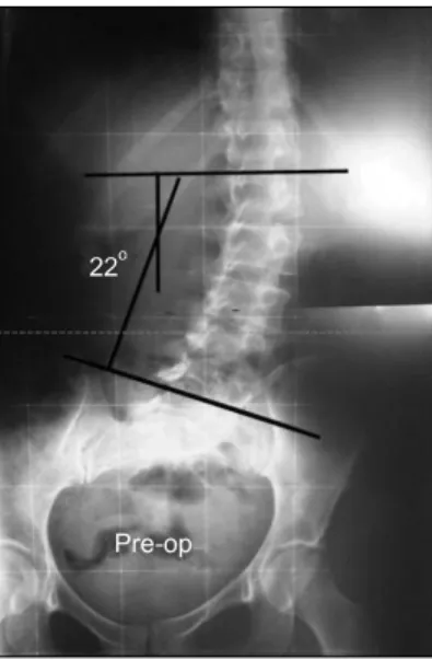
A 41-year-old female complained of a 25-year history of lower back pain and buttock pain ac- companied with intermittent radiating pain to the left leg. The coronal imbalance was 3.8 cm, as determined by the distance between the C7 plumb line and the center sacral line. The scoliosis angle using the Cobb method was 22o (Fig. 1). The sacral malformation was a fixed lumbosacral deformity that could not be corrected by traction or forced side bending. A trapezoidal wedge resection osteo-
Lumbar Wedge Resection Osteotomy for Congenital Scoliosis due to a Sacral Malformation 829
Fig. 1. Pre-operative anteroposterior standing radiograph reveals the malformation of the sacrum and pelvis, and 22 degrees of scoliosis angle between L1 and L5.
Fig. 2. The illustration of the pre-operative planning of the osteotomy. The hatched marks define the extent of the osteotomy.
tomy of the L5 body was planned for the correction of rigid scoliosis.
With the spine exposed posteriorly, the para- spinal muscles were stripped subperiosteally from the spinous processes to the tips of the transverse processes at the L3 to S2 levels. The posterior arch, spinous process, laminae and articular processes of L5 was removed and the dura mater and nerve roots of L5 and S1 on both sides were verified. The pedicles of L5 were removed using a rongeur and the cancellous bone was partially removed through the site of the pedicles by curettage.
The transverse processes were also morcellized with an electrical burr and the normal soft tissues were retracted as far lateral as possible. Pedicular screws were inserted bilaterally into the L3, L4, S1, and S2 levels under fluoroscopic guidance and a temporary rod was inserted at the left side. After meticulous homeostasis had been obtained with bipolar electrocauterization, the dura mater and nerve roots were retracted carefully, and the posterior wall and side walls of the L5 vertebral body were removed.
The lengths of the resection at the pedicle level
were 22 mm on the right side and 9 mm on the left, which were the same as determined in preoperative planning (Fig. 2). A intravertebral trapezoidal wedge resection was performed gradually toward the anterior vertebral body in a so called piecemeal fashion using dissectors, curettes and pituitary forceps The anterior wall was also removed using a Kerrison rongeur, taking particular care to leave the soft tissue anterior to the body intact. This was followed by confirming that the canal was clear of any residual compression.
The temporary rod was removed and the pre- contoured rods were inserted one by one and the osteotomy was closed in a compression manner.
During closure, the nerve roots and dura mater were observed under direct vision of the avoidance of compression. Posterior fusion was performed in a meticulous manner, decorticating all posterior elements and applying autogenous corticocan- cellous bone at all levels instrumented. Before clo- sing the wound, radiographs of the spine were obtained to determine the coronal and sagittal balance.
Postoperatively, the scoliosis angle was corrected
830 Bong-Jin Lee, Sung-Rak Lee, Seong-Tae Kim, et al
Fig. 3. Post-operative anteroposterior standing radiograph re- veals the correction of the scoliosis angle between L1 and L5.
Fig. 4. The illustration of the postoperative status. The scoliosis was corrected with a trapezoidal wedge resection osteotomy of L5.
to 0o and the coronal imbalance had disappeared (Fig. 3 and 4). The patient was allowed to walk 2 weeks after surgery. The well-molded thoraco- lumbosacral orthosis was applied for 4 months when solid fusion was apparent on the radio- graphs. At the 2 year follow-up, the patient returned to normal physical activity without any deformity or low back pain.
DISCUSSION
A posterior closing-wedge vertebral osteotomy has been proposed as corrective surgery for ky- phosis8). The neurological and vascular compli- cations of this technique are much lower than the Smith-Petersen type anterior opening-wedge ost- eotomy, producing a sharp lordotic angle and an elongation of the anterior column. However, most early studies reported the clinical results only in ankylosing spondylitis with kyphotic deformity5,8,9). Since the year 2000, a transpedicular wedge resection osteotomy, or so called pedicle subtrac- tion osteotomy, have been used to treat various fixed sagittal imbalances, including iatrogenic ky- phosis, post-traumatic kyphosis, degenerative ky-
phosis, post-infectious kyphosis, idiopathic sco- liosis and tumors1,3,4).
Congenital scoliosis is most often observed in the thoracic area and least in the lumbosacral area10). Only limited cases of coronal corrections in the treatment of congenital scoliosis have been re- ported1,6,7). Our case was a congenital scoliosis due to a sacral malformation, which was considered to be a unilateral failure of the vertebral formation of S1.
A spinal deformity at the lumbosacral region poses a unique problem. The lack of a mobile spine below the anomaly often results in early truncal decompensation and a long compensatory curve above, which can progresses with time. A lum- bosacral deformity frequently produces functional impairment and neurological involvement. Pain is a common symptom in later years.
There are only a few reports on a vertebral column resection for the treatment of rigid coronal decompensation. Most were accomplished using combined anterior and posterior approaches in one or two stages2). Vertebral column resections through the posterior approach only were reported in the
Lumbar Wedge Resection Osteotomy for Congenital Scoliosis due to a Sacral Malformation 831
= 국문초록=
천추의 기형에 의해 발생한 선천성 척추 측만증은 매우 드물다. 문헌을 고찰한 결과, 대부분의 쐐기형 추체 절제술은 강직성 척추염에 속발한 후만 변형의 치료에 이용되어 왔고, 천추 기형에 의한 선천성 측만증의 치료에 사용한 예는 찾을 수가 없었다. 25년 동안의 허리 통증과 둔부 통증 및 하지로의 방사통이 있고, 제7경추 중심선 과 천추 중심선 사이의 거리가 3.8 cm이었으며, 코브방식으로 측정한 측만각이 22도인 41세 여자 환자에서, 제5요추의 사다리꼴 쐐기형 추체 절제술을 시행하여 측만각이 교정됨을 확인하였다. 이에 관상면에서 고정된 척추의 변형에 유효한 척추 절제술의 한 변법을 소개하고자 한다.
색인 단어: 천추, 선천성 측만증, 쐐기형 절제술
treatment of congenital scoliosis and kypho- scoliosis. However, the cases reported were hemi- vertebrae at the thoracic or lumbar level7). We performed a modification of a vertebral wedge osteotomy in a patient with a rare condition, and confirmed the correction capacity of this procedure, both clinically and radiologically. The characteristics of this vertebral osteotomy were a trapezoidal shaped resection in the coronal plane and a three-column resection in the sagittal plane.It is believed that an intravertebral trapezoidal wedge resection osteotomy is an effective tech- nique for correcting a fixed coronal and a sagittal deformity in various spinal deformities.
REFERENCES
1. Boachie-Adjei O, Ferguson JA, Pigeon RG, Peskin MR:
Transpedicular lumbar wedge resection osteotomy for fixed sagittal imbalance: surgery technique and early results. Spine, 31: 485-492, 2006.
2. Bradford DS, Tribus CB: Vertebral column resection for the treatment of rigid coronal decompensation. Spine, 22: 1590- 1599, 1997.
3. Bridwell KH, Lewis SJ, Lenke LG, Baldus C, Blanke K:
Pedicle subtraction osteotomy for the treatment of fixed sagittal
imbalance. J Bone Joint Surg Am, 85: 454-463, 2003.
4. Cho KJ, Bridwell KH, Lenke LG, Berra A, Baldus C:
Comparison of Smith-Petersen versus pedicle subtraction osteotomy for the correction of fixed sagittal imbalance. Spine, 30: 2030-2037, 2005.
5. Kim KT, Suk KS, Cho YJ, Hong GP, Park BJ: Clinical outcome results of pedicle subtraction osteotomy in ankylosing spondylitis with kyphotic deformity. Spine, 27: 612-618, 2002.
6. Suk SI, Chung ER, Lee SM, Lee JH, Kim SS, Kim JH:
Posterior vertebral column resection in fixed lumbosacral deformity. Spine, 30: E703-E710, 2005.
7. Suk SI, Kim JH, Kim WJ, Lee SM, Chung ER, Nah KH:
Posterior vertebral column resection for severe spinal deformities. Spine, 27: 2374-2382, 2002.
8. Thomasen E: Vertebral osteotomy for correction of kyphosis in ankylosing spondylitis. Clin Orthop Relat Res, 194: 142- 152, 1985.
9. van Royen BJ, Slot GH: Closing-wedge posterior osteotomy for ankylosing spondylitis. Partial corporectomy and tran- spedicular fixation in 22 cases. J Bone Joint Surg Br, 77:
117-121, 1995.
10. Winter RB, Moe JH, Eilers VE: Congenital scoliosis. A study of 234 patients treated and untreated. J Bone Joint Surg Am, 50: 1-47, 1968.