https://doi.org/10.4047/jkap.2017.55.3.258 ORIGINAL ARTICLE
c cc
2017 The Korean Academy of Prosthodontics
This is an Open Access article distributed under the terms of the Creative Commons Attribution Non-Commercial License (http://creativecommons.org/licens- es/by-nc/3.0) which permits unrestricted non-commercial use, distribution, and reproduction in any medium, provided the original work is properly cited.
*Corresponding Author: Hee-Jung Kim
Department of Prosthodontics, School of Dentistry, Chosun University 303 Pulmun-daero, Dong-gu, Gwangju 61452, Republic of Korea +82 (0)62 220 3829: e-mail, [email protected]
Article history: Received April 5, 2017 / Last Revision May 30, 2017 / Accepted June 13, 2017
서론
많은 사람들에게 있어 치아를 상실하는 것은 구강 기능, 전 신 건강, 심미와 삶의 질에 정신 사회적으로 부정적 영향을 준 다.1,2이러한 무치악 및 부분 무치악 환자에 있어 활용될 수 있 는 여러 치료 방법 중에 수술 기법의 발전과 골이식 치료의 질 적 향상에 따라 임플란트 치료가 차지하는 비중이 점차 커져 임플란트는 무치악 및 부분 무치악 환자의 치료에 있어 가장 우선적인 치료법으로 선택된다.3초기 임플란트 연구는 임플 란트의 골유착을 증진할 수 있는 외과적 수술 방법4과 임플란 트 표면 처리 방법5에 대한 연구에 중점을 두었다면, 근래에는
임플란트 상부 보철물의 기능성을 증진할 수 있는 방법에 대 한 연구에 중점을 두고 있다.6
이러한 기능적인 보철물 제작을 위해서 임플란트와 보철물 을 연결하는 상부 구조물인 임플란트 지대주가 강조되고 있 다.7임플란트 고정성 보철물에서 지대주는 치은 조직을 관통 하여 보철물과 임플란트를 연결하는 역할을 한다. 이러한 지 대주는 torque, preload, clamping, thread mechanics, micromovement와 settling과 관련된 생역학 기준들과 연관되어 있다.8또한 임플란 트 주위염은 지대주 - 임플란트 연결구조 주변에서 발생하게 되며, 임플란트 주변 치주조직의 건강과 연관이 되어 있다.9이 에 UCLA 지대주가 환자 맞춤형 지대주로써 난증례에 적용하
CAD/CAM으로 제작된 임플란트 맞춤형 지대주의 방사선학적 평가
윤태경∙이경제∙정재헌∙김희중*
조선대학교 치과대학 치과보철학교실
Radiographic evaluation of computer aided design/computer aided manufacturing (CAD/CAM) customized abutment of implant
Tae-Gyeong Yun, Gyeong-Je Lee, Chae-Heon Chung, Hee-Jung Kim*
Department of Prosthodontics, School of Dentistry, Chosun University, Gwangju, Republic of Korea
Purpose: In this study, the retrospective radiographic study is executed to evaluate amount of bone loss of various conditions in patients using customized abutment for 4 years of follow-up. Materials and methods: The subjects of this study were implant fixed dental prosthesis using CAD/CAM customized abutments. CAD/CAM customized abut- ment and fixed dental prosthesis were manufactured by the Prosthodontics Department of Chosun University Dental Hospital from August 1, 2011 to July 31, 2012. Radiological assessments were performed on the patients who were treated by the fixed prosthodontics. After each treatment, a retrospective study was performed for a total of 4 years at 3 months, 6 months, 1 year, 2 years, 3 years, and 4 years. Results: As a result of the study, the customized abutment using CAD/CAM showed less bone loss than the results of existing research. There was no statistically significant differences at alveolar bone loss between splinting group and non-splinting group (respectively 0.27 mm, 0.5 mm).
Also, there were statistically significant differences at alveolar bone loss in mx. anterior, mx. posterior, mn. anterior and mn. posterior part (respectively 1.37 mm, 0.39 mm, 0.00 mm, 0.30 mm). Conclusion: The customized abutment using CAD/CAM showed less bone loss than the results of existing research, there were statistically significant differences at alveolar bone loss in implant positions. (J Korean Acad Prosthodont 2017;55:258-63)
Keywords: CAD/CAM; Dental implant abutment; Alveolar bone loss; X-ray
※This study was supported by research fund from Chosun University, 2015.
는 방법으로 소개되었다.10그러나, UCLA 지대주는 재료, 비용 및 기공 과정에서의 어려움이 존재하였고, 이러한 단점들을 극복한 Computer Aided Design/Computer Aided Manufacturing (CAD/CAM) 방식의 환자 맞춤형 지대주가 2000년대 초반에 소 개되었다.11
최근 들어 많은 문헌들이 CAD/CAM 지대주 제작의 장점과 정확성을 보고12하고 있으며 이러한 기술이 국내에도 도입이 되었으나, CAD/CAM 지대주를 이용하여 실제로 보철물을 장착 한 환자에 있어서 장기간의 지속적인 경과 관찰에 대한 데이 터가 부족한 실정이다.
이에 본 연구에서는 조선대학교 치과병원에서 제작된 임플 란트 중 CAD/CAM 으로 제작된 맞춤형 지대주의 방사선학적 평가를 시행하여 이에 대한 효용성에 관한 평가를 시행하고자 하였다.
대상 및 방법 1. 대상
1) 환자 선정
이번 CAD/CAM 방식으로 제작된 맞춤형 지대주를 이용한 보철 치료에 대한 후향적 연구는 2011년 8월 1일부터 2012년 7월 31일까지 조선대학교 치과병원 CAD/CAM 센터에서 제작된 임플란트 보철을 대상으로 하였다.
총 200명의 환자가 고정성 임플란트 보철 치료를 진행하였으 며, 4년간의 경과 관찰 기간 중에서 200명의 환자 중 30명의 환 자에서 경과 관찰이 이루어지지 않아 최종적으로 170명의 환 자에서 357개의 임플란트를 대상으로 4년간의 임상적 평가를 시행하였다.
환자 그룹의 성별 구성은 104명의 남성과 66명의 여성으로 이루어졌으며, 환자들의 평균 나이는 54.45 ± 12.40세였다 (Table 1).
2) 임플란트 선정
임플란트 식립 부위와 식립 개수를 나타내었다 (Fig. 1).
식립된 임플란트의 종류는 Astra (Dentsply, York, PA, USA), Biomet 3i (Zimmer-Biomet 3i, Jacksonville, FL, USA), Zimmer (Zimmer Holdings, Warsaw, IN, USA), Dentis (Dentis, Daegu, Korea), Osstem (Osstem Implant, Seoul, Korea)의 총 5개의 회사에서 제작된 internal type의 임플란트를 사용하였다 (Table 2).
CAD/CAM 지대주를 사용한 임플란트 수를 악궁과 연결 고정 여부에 따라 분류하였다 (Table 3).
Table 1. Causes of missing tooth
Group Periodontal disease Caries Trauma Congenital missing Periodontal disease & Caries Total
No. of patient 89 32 18 7 24 170
No. of tooth 207 62 31 10 47 357
Fig. 1. Number of implants placed in each location (eg. 26 maxillary right 1st molar were replaced with implant supported single tooth prostheses).
Table 2. Number of implant classified by implant company
Classification of implant fixture
Astra Biomet 3i Zimmer Dentis Osstem Total
No. of fixture 80 83 34 53 107 357
Table 3. Total number of dental archs, number of fixtures and splinted fixtures
No. of dental arch No. of fixture No. of splinted fixtures Proportion of splinted fixtures
Mx. 85 199 161 81.0%
Mn. 79 158 119 75.3%
2. 방법
환자에 대한 임플란트의 정기적인 검진은 최종 장착 후 3개 월, 6개월, 1년, 2년, 3년, 4년 단위로 검진을 시행하였다. 검진 약 속 이외에 내원을 한 경우에는 가장 근접한 검진 약속일에 평 가를 시행하는 것으로 하였다.
환자에 대한 평가로 방사선 사진을 이용한 임플란트 주위 골 흡수양상을 조사하였다. 위 평가는 일관성과 통일성을 지니기 위해 동일한 기기를 이용하여 1명의 술자가 정기적인 환자에 대한 평가를 시행하였다.
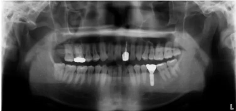
방사선학적인 평가를 위하여 매 내원마다 치근단 방사선 사 진 촬영 (Fig. 2, Focus, Palodex, Tuusula, Finland) 및 파노라마 방사 선 사진 촬영 (Fig. 3, Planmeca Proline XC, Planmeca, Helsinki, Finland) 을 시행하였다. 방사선 사진에 대한 평가는 1명의 술자 가 평가를 시행하였다. 방사선 사진에 대한 평가는 1명의 술자 가 평가를 시행하였다. 골소실 정도는 파노라마 방사선 사진 과 평행 촬영법으로 촬영한 치근단 방사선 사진 상에서 임플 란트 주위 골소실은 수술 시 방사선 사진과 내원 시의 방사선 사진 상 임플란트 나사선과 변연골 수준을 측정한 뒤 실제 임 플란트 나사선과 비교하여 평가하였다.13결과를 각각 연결 고 정 여부와 식립부위에 따라 분류하였다. 통계적 평가 시, 골유 착에 실패한 임플란트는 방사선학적인 평가에서 제외하였다.
3. 통계학적 분석
SPSS ver. 22. 0 (SPSS Inc., Chicago, IL, USA) 프로그램을 이용하 여 각 환자의 지대주 장착 이후 골소실 양을 계산하였다. 연결 고정 여부, 임플란트 식립 부위에 따른 골 평가를 시행하기 위 하여 각각 Independent t-test와 ANOVA test를 시행하였다.
결과
방사선학적인 평가
1) 연결 고정 여부에 따른 골소실 양 간의 관계
이번 연구에서 사용된 임플란트는 모두 고정성 보철물로 제 작되어 각각의 CAD/CAM 맞춤형 지대주는 연결 고정이 되지 않은 그룹과 연결 고정이 된 그룹으로 구분지을 수 있다. 연결 고정이 되지 않은 임플란트 그룹에서의 평균 누적 골소실은 0.27 mm, 연결 고정이 된 임플란트 그룹에서의 평균 누적 골소 실은 0.50 mm를 보였다. 방사선적인 검사를 통해서 도출된 골 소실양을 연결 고정 된 그룹과 연결 고정 되지 않은 그룹 간에 독립 표본 t-test로 분석을 시행한 결과, 각 그룹 간에 검진 시기 및 누적 골소실 양에서 모두 통계적으로 유의한 차이를 보이 지 않았다 (Table 4).
2) 임플란트 식립 부위에 따른 골소실 양 간의 관계
이번 연구에서 사용된 임플란트는 모두 고정성 보철물로 제 작되어 각각의 CAD/CAM 맞춤형 지대주를 식립 부위(상악 전 치, 상악 구치, 하악 전치, 하악 구치)에 따라 분류하였다. 상악 전치부의 평균 누적 골소실은 1.37 mm, 상악 구치부의 평균 누 적 골소실은 0.39 mm, 하악 전치부의 평균 누적 골소실은 0.00 mm, 하악 구치부의 평균 누적 골소실은 0.30 mm이다. 방사선학 적인 검사를 통해서 도출된 골소실양을 상악 전치부, 상악 구 치부, 하악 전치부, 하악 구치부 간에 독립 표본 t-test로 분석을 시행한 결과, 상악 전치부와 다른 그룹 간에 통계적으로 유의 한 차이를 나타내었다 (Table 5).
Table 4. Vertical bone loss of around the implants classified by splinting
Mean of vertical bone loss of around the implants (mm)
3-month recall 6-month recall 1-year recall 2-year recall 3-year recall 4-year recall Total
No Splinting 0.00 0.00 0.00 0.03 0.09 0.15 0.27
Splinting 0.00 0.00 0.00 0.12 0.19 0.19 0.50
P1) .571 .385 .165 .094 .362 .732 .350
1)Statistically significant difference (α= .05) (Independent t-test)
Fig. 2. Periapical X-ray taking at final setting of implant prosthesis. Fig. 3. Panoramic X-ray taking at final setting of implant prosthesis.
고찰
이번 연구에서는 임플란트 고정성 보철물에서 수술적 고려 뿐만 아니라 방사선학적인 고려에 대한 성공 기준으로서 평가 를 시행하였다.14,15
지대주의 제작 방법 및 재료는 다양하나, 1980년대 이후에 컴퓨터 기반의 기술이 발달하면서 현재에는 CAD/CAM 맞춤형 지대주는 1) 비용, 시간, 제작의 편이성, 2) 자연치, 인접치, 대합 치의 형태학적 고려가 용이, 3) 상부 보철물 제작의 용이성, 4) 정확한 제작 과정이라는 장점을 지니고 있다.16,17그러나, 아직 CAD/CAM 지대주의 장기적 경과 관찰에 대한 연구가 부족하 여 본 연구에서는 CAD/CAM 맞춤형 지대주의 경과 관찰을 다 양한 기준으로 시행하여 통계적인 평가를 시행하였다.
기존 논문의 방사선학적인 평가와 이번 논문의 방사선학적 인 평가를 비교하였다. Galindo-Moreno18에 의하면 기성 internal-type 지대주를 사용한 임플란트(Dentsply implant, Waltham, MA, USA) 주변에서 임플란트 변연골의 소실은 6개월 후 검진에서 4.5 mm, 5 mm 직경의 임플란트에서 각각 0.56 mm, 0.55 mm의 골소실 이 일어났으며, 12개월 뒤 검진에서는 4.5 mm, 5 mm 직경의 임플 란트에서 각각 1.09 mm, 1.27 mm의 골소실이 일어난다고 보고하 였다. 이 연구에서는 직경과 기간에 따른 골소실양의 통계학 적 차이는 없다고 보고하였다. CAD/CAM 맞춤형 지대주를 사 용한 이번 연구에서는 연결 고정을 하지 않은 경우와 연결 고 정을 한 경우에 4년 뒤 검진에서 각각 0.27 mm, 0.50 mm로, 기존 연구에 비해 더 적은 골소실양이 관찰되었다.
Cho 등19에 의하면 기공실 지대주를 사용한 경우 상악 전치부 에서 1.32 mm의 골소실을 보고하였다. 이번 연구에서는 상악 전치부에서 1.37 mm로, 기존 연구와 유사한 골소실양이 관찰되 었다.
Vigolo와 Zaccaria 등20에 의하면 기성 external-type 지대주를 사용 한 임플란트(Biomet 3i implant, Zimmer-Biomet 3i, Jacksonville, FL, USA)에서 연결 고정을 하지 않은 경우와 연결 고정을 한 경우 에 5년 뒤 검진에서 각각 0.7 ± 0.2 mm, 0.8 ± 0.2 mm를 보고하였 다. CAD/CAM 맞춤형 지대주를 사용한 이번 연구에서는 연결 고정을 하지 않은 경우와 연결 고정을 한 경우에 4년 뒤 검진에 서 각각 0.27 mm, 0.50 mm로, 기존 연구와 연결방식의 차이는 있 었으나 더 적은 골소실양이 관찰되었다.
Wahlström 등21은 5 년간의 경과 관찰 기간 동안 임플란트 실패 는 46개의 임플란트에서 2개의 임플란트가 실패(8.6%) 한다고
보고 하였다. 이번 연구에서는 골 유착 실패에 따른 임플란트 실패가 2.2%에서 발생하였다. 이번 연구는 연결 고정을 하지 않은 단일치 수복을 기존 연구에 비해 더 많은 비율로 조사하 게 되면서 측방력이 가해질 때 흔하게 발생하는 보철물 탈락 과 지대주 파절이 더 많이 조사된 것으로 사료된다.
그러나 본 실험의 경우 제한된 개체 수와 짧은 경과 관찰을 시행하여 정확한 평가를 평가하기에는 한계가 있을 것으로 사 료되며, 추후 장기적인 연구가 필요할 것으로 보인다.
결론
CAD/CAM 맞춤형 지대주를 이용하여 제작한 보철물에 대 해서 4년간 경과 관찰을 시행한 결과 다음과 같은 결론을 얻을 수 있었다.
1. 연결 고정 여부에 따라 골소실 양을 평가한 경우에, 연결 고정 되지 않은 그룹에서의 누적 골소실은 0.27 mm, 연결 고정 된 그룹은 0.50 mm를 보였다. 이는 검진 일시에 따라 모든 그룹 간에 통계적으로 유의한 차이를 보이지 않았다 (Independent t-test, P > .05).
2. 식립 부위에 따라 골소실 양을 평가한 경우에, 상악 전치부 의 누적 골소실은 1.37 mm, 상악 구치부에서 0.39 mm, 하악 전치부에서 0.00 mm, 하악 구치부에서 0.30 mm를 보였으며 상악 전치부에서의 누적 골소실이 통계적으로 많음을 나 타내었다.
ORCID
Tae-Gyeong Yun https://orcid.org/0000-0002-8374-8115 Hee-Jung Kim https://orcid.org/0000-0002-2015-1530
References
1. Goto T, Nishinaka H, Kashiwabara T, Nagao K, Ichikawa T. Main occluding area in partially edentulous patients: changes before and after implant treatment. J Oral Rehabil 2012;39:677-83.
2. Mericske-Stern RD, Taylor TD, Belser U. Management of the edentulous patient. Clin Oral Implants Res 2000;11:108-25.
3. Brånemark PI, Adell R, Breine U, Hansson BO, Lindström J, Ohlsson A. Intra-osseous anchorage of dental prostheses. I.
Experimental studies. Scand J Plast Reconstr Surg 1969;3:81-100.
Table 5. Vertical bone loss of around the implants classified by location of implant
Mean of total vertical bone loss of around the implants (mm)
Anterior part of maxilla Posterior part of maxilla Anterior part of mandible Posterior part of mandible
Total vertical bone loss 1.37a) 0.39b) 0.00b) 0.30b)
a), b)Statistically significant difference between a) and b) (α= .05) (ANOVA test)
4. Albrektsson T, Brånemark PI, Hansson HA, Lindström J.
Osseointegrated titanium implants. Requirements for ensuring a long-lasting, direct bone-to-implant anchorage in man. Acta Orthop Scand 1981;52:155-70.
5. Le Guéhennec L, Soueidan A, Layrolle P, Amouriq Y. Surface treatments of titanium dental implants for rapid osseointegration.
Dent Mater 2007;23:844-54.
6. Hebel KS, Gajjar RC. Cement-retained versus screw-retained im- plant restorations: achieving optimal occlusion and esthetics in implant dentistry. J Prosthet Dent 1997;77:28-35.
7. Döring K, Eisenmann E, Stiller M. Functional and esthetic considerations for single-tooth Ankylos implant-crowns: 8 years of clinical performance. J Oral Implantol 2004;30:198-209.
8. Brunski JB. Biomaterials and biomechanics in dental implant de- sign. Int J Oral Maxillofac Implants 1988;3:85-97.
9. Gross M, Abramovich I, Weiss EI. Microleakage at the abutment- implant interface of osseointegrated implants: a comparative study.
Int J Oral Maxillofac Implants 1999;14:94-100.
10. Duke ES. The status of CAD/CAM in restorative dentistry.
Compend Contin Educ Dent 2001;22:968-72.
11. Lewis S, Beumer J 3rd, Hornburg W, Moy P. The "UCLA" abut- ment. Int J Oral Maxillofac Implants 1988;3:183-9.
12. Fuster-Torres MA, Albalat-Estela S, Alcañiz-Raya M, Peñarrocha- Diago M. CAD / CAM dental systems in implant dentistry: up- date. Med Oral Patol Oral Cir Bucal 2009;14:E141-5.
13. Yoo HS, Kang SN, Jeong CM, Yun MJ, Huh JB, Jeon YC. Effects of implant collar design on marginal bone and soft tissue. J Korean
Acad Prosthodont 2012;50:21-8.
14. Albrektsson T, Zarb G, Worthington P, Eriksson AR. The long- term efficacy of currently used dental implants: a review and pro- posed criteria of success. Int J Oral Maxillofac Implants 1986;1:11-25.
15. Porter JA, von Fraunhofer JA. Success or failure of dental im- plants? A literature review with treatment considerations. Gen Dent 2005;53:423-32.
16. Priest G. Virtual-designed and computer-milled implant abutments.
J Oral Maxillofac Surg 2005;63:22-32.
17. Miyazaki T, Hotta Y, Kunii J, Kuriyama S, Tamaki Y. A review of dental CAD/CAM: current status and future perspectives from 20 years of experience. Dent Mater J 2009;28:44-56.
18. Galindo-Moreno P, Leo′n-Cano A, Monje A, Ortega-Oller I, O'Valle F, Catena A. Abutment height influences the effect of plat- form switching on peri-implant marginal bone loss. Clin Oral Implants Res 2016;27:167-73.
19. Cho YB, Moon SJ, Chung CH, Kim HJ. Resorption of labial bone in maxillary anterior implant. J Adv Prosthodont 2011;3:85-9.
20. Vigolo P, Zaccaria M. Clinical evaluation of marginal bone level change of multiple adjacent implants restored with splint- ed and nonsplinted restorations: a 5-year prospective study. Int J Oral Maxillofac Implants 2010;25:1189-94.
21. Wahlström M, Sagulin GB, Jansson LE. Clinical follow-up of unilateral, fixed dental prosthesis on maxillary implants. Clin Oral Implants Res 2010;21:1294-300.
CAD/CAM으로 제작된 임플란트 맞춤형 지대주의 방사선학적 평가
윤태경∙이경제∙정재헌∙김희중*
조선대학교 치과대학 치과보철학교실
목적: 이번 연구에서, 다양한 상황에서 골소실양을 평가하기 위하여 맞춤형 지대주를 사용한 환자에서 4년간의 후향적 방사선학적인 연구를 시행 하였다.
대상 및 방법: 이번 연구의 대상은 CAD/CAM 맞춤형 지대주를 이용한 임플란트 고정성 보철물이다. 2011년 8월 1일부터 2012년 7월 31일 까지 조선대 학교 치과병원 보철과에서 제작하여 고정성 보철물로 사용된 CAD/CAM 맞춤형 지대주와 상부 보철물들을 대상으로 하였으며, 고정성 보철물을 제작한 환자에 대하여 방사선학적 평가를 시행하여 보철물 장착 후 3개월, 6개월, 1년, 2년, 3년, 4년 단위로 총 4년 간의 후향적 연구를 시행하였다.
결과: 연구 결과, CAD/CAM을 이용한 맞춤형 지대주는 기존 연구 결과에 비해 골소실 양이 적었다. 연결 고정된 그룹과 연결 고정되지 않은 그룹 간 에 골소실 양(각각 0.27 mm, 0.5 mm)에 통계적으로 유의한 차이가 없었다. 또한 상악 전치부, 상악 구치부, 하악 전치부, 하악 구치부의 부위별 골소실 양(각각 1.37 mm, 0.39 mm, 0.00 mm, 0.30 mm)에 통계적으로 유의한 차이가 있었다.
결론: CAD/CAM을 이용한 맞춤형 지대주는 연결 고정 여부에 따른 골소실 양에서 통계적으로 유의한 차이가 없었으며, 임플란트 식립 부위에 따른 골소실 양에서 통계적으로 유의한 차이가 있었다. (대한치과보철학회지 2017;55:258-63)
주요단어: 캐드캠; 임플란트 지대주; 치조골소실; 방사선 사진
*교신저자: 김희중
61452 광주 동구 필문대로 303 조선대학교 치과대학 치과보철학교실 062 220 3829: e-mail, [email protected]
원고접수일: 2017년 4월 5일 / 원고최종수정일: 2017년 5월 30일 / 원고채택일: 2017년 6월 13일
2017 대한치과보철학회
이 글은 크리에이티브 커먼즈 코리아 저작자표시-비영리 3.0 대한민국 라이선스에 따라 이용하실 수 있습니다.
c cc
※이 논문은 2015학년도 조선대학교 학술연구비의 지원을 받아 연구되었음.