Cephalometric Characteristics of TMD Patients based on RDC/TMD Axis I Diagnosis
Ji-Yeon Ahn, D.D.S.,M.S.D., Yong-Woo Kim, D.D.S.,Ph.D., Young-Ku Kim, D.D.S.,Ph.D., Jeong-Yun Lee, D.D.S.,Ph.D.
Dept. of Oral Medicine and Oral Diagnosis, School of Dentistry & Dental Research Institute, Seoul National University, Seoul, Korea (ROK)
The aims of this study were to investigate whether the facial skeletal patterns previously reported to be related to temporomandibular disorder (TMD) in other studies could be consistently observed in the TMD patients diagnosed according to Research Diagnostic Criteria for Temporomandibular Disorder (RDC/TMD) Axis I and evaluate its usability in the orthodontic clinics to examine the patients with TMD related symptoms.
The clinical records and radiographs of female patients who visited the TMD and Orofacial Pain Clinic of Seoul National University Dental Hospital and were diagnosed as TMD were consecutively filed for this study. Patients were clinically examined and diagnosed according to the revised diagnostic algorithms of RDC/TMD Axis I and the lateral cephalogram, panoramic orthopantomogram, temporomandibular joint (TMJ) orthopantomogram, and transcranial radiograph of each patient were taken and digitalized. The data of patients who were under 18 years of age or had any systemic disease, trauma history involving the TMJ, or skeletal deformity at the time of the first examination were excluded. The remaining data of 96 female patients were finally analyzed. The obtained results were as follows:
1. There are no significant differences of cephalometric measurements between RDC I (muscle disorders) diagnostic groups.
2. Only the articular angle of the RDC group IIc (disk displacement without reduction without limited opening) patients was larger than patients of the no diagnosis of RDC II group (disk displacement).
3. Larger articular angle and smaller facial height ratio were observed in RDC IIIc group (osteoarthrosis) compared to IIIa group (arthralgia). Larger articular angle, larger Björk sum, smaller posterior facial height, and smaller facial height ratio were observed in RDC group IIIc compared to no diagnosis of RDC III group (arthralgia, arthritis, and arthrosis).
4. According to the results of cephalometric analysis in simplified RDC groups, smaller overjet was observed in muscle disorders (MD) group. Facial height ratio and IMPA were smaller and articular angle was larger in disk displacements (DD) group than in no diagnosis of DD group. In arthrosis (AR) group, posterior facial height, and facial height ratio were smaller, and articular angle, gonial angle, facial convexity, FMA, Björk sum, and ANB were larger than in no diagnosis of AR group. In joint pain (JP) group, only posterior facial height was smaller than no diagnosis of JP group.
In conclusion, Facial morphologic patterns showing posterior-rotated mandible and lower posterior facial height is related to RDC group II and III diagnosis of the TMJ in female TMD patients. RDC/TMD Axis I diagnosis can provide a good clinical diagnostic tool for the standardized examination of the TMJ in orthodontic clinics.
Key words: Temporomandibular disorder (TMD), Research Diagnostic Criteria for Temporomandibular Disorder
(RDC/TMD), Lateral cephalogram
1) Ⅰ. INTRODUCTION
Temporomandibular disorder (TMD) is a comprehensive concept that includes various clinical problems such as abnormal morphology of the articular disk and mandibular condyle, displacement or dislocation of the articular disk, inflammation of soft tissue (e.g., synovitis and capsulitis), osteoarthrosis, osteoarthritis, and ankylosis.
Correlation between TMD and dentofacial morphology has been suggested in many studies. It has been reported that a reduction in ramus and posterior facial height and an increase of mandibular plane inclination were related to disk displacement in female adolescents
1-2). It was also suggested in a series of studies that alteration of facial skeletal morphology and facial asymmetry may be related to disk displacement
3-8). Such a relationship between disk displacement and facial morphology was also identified in female orthodontic patients with Class II malocclusion, female patients with osteoarthritis/
osteoarthrosis, and TMD patients divided according to Angle classification on cephalographic analysis
9-11)
. Although, some authors suggested that such data should be cautiously interpreted because a cause and effect relationship could not be derived from the results of these studies, other authors suggested that several skeletal morphologic patterns appeared to be related to TMD and should be considered in the orthodontic diagnosis and treatment process
1-12).
The application of the Research Diagnostic Criteria for Temporomandibular Disorders (RDC/
TMD) has been recommended in the diagnosis of TMD for research purposes to standardize and
Corresponding author: Dr. Jeong-Yun Lee
Assistant Professor, Dept. of Oral Medicine and Oral Diagnosis, School of Dentistry & Dental Research Institute, Seoul National University, Yeongeon-Dong 28, Jongno-Gu Seoul 110-749 Korea (ROK)
Tel: +82-2-2072-0212 Fax: +82-2-744-9135 E-mail: [email protected]
Received: 2011-02-17 Accepted: 2011-03-10enhance reproducibility of the diagnosis process
13). RDC/TMD consists of a dual-axis system. Axis I contains diagnostic criteria for the physical diagnosis of TMD as muscle origin, disk displacement origin, and osteoarthritis/
osteoarthrosis, and Axis II contains psychosocial scales encompassing the behavioral, psychological and physical characteristics of the patient. It has been translated into 19 languages, including Chinese, Spanish, Korean, etc. and has been widely used around the world in clinical research settings for TMD and orofacial pain
14-16). Recently, the reliability and validity of clinical diagnostic algorithms was evaluated and revised algorithms for RDC/TMD Axis I have been proposed to improve the reliability and validity of clinical examination procedures according to RDC/TMD
17-18).
In this study, we investigated whether the facial skeletal patterns previously reported to be related to TMD in other studies could be consistently observed in the TMD patients diagnosed according to RDC/TMD Axis I and evaluated its usability in the orthodontic clinics to examine the patients with TMD related symptoms.
Ⅱ. MATERIALS AND METHODS 1. Materials
The clinical records and radiographs of 114 female
patients who visited the TMD and Orofacial Pain
Clinic of Seoul National University Dental Hospital
and were diagnosed as TMD by a single
experienced examiner specializing in orofacial pain
were consecutively filed for this study. Patients
were clinically examined and diagnosed according to
the revised diagnostic algorithms of RDC/TMD
Axis I
17,18)and the lateral cephalogram, panoramic
orthopantomogram, temporomandibular joint (TMJ)
orthopantomogram, and transcranial radiograph of
each patient were taken and digitalized. The data of
patients who were under 18 years of age or had any
systemic disease, trauma history involving the TMJ,
or skeletal deformity at the time of the first
examination were excluded. The remaining data of 96 female patients whose mean age was 34.5 ± 14.2 years were finally analyzed. The research protocol was approved by the Institutional Review Board of Seoul National University Dental Hospital (#CRI10024).
2. RDC/TMD Axis I Diagnosis
A RDC/TMD Axis I diagnosis was given of each joint after clinical examinations according to the revised diagnostic algorithms and radiographic findings as mentioned in the original version of RDC/TMD for diagnosis of RDC group III (Arthralgia, Arthritis, and Arthrosis) were observed and recorded
13,18). According to the RDC/TMD axis I, multiple diagnoses are allowed but a maximum of one muscle disorder related diagnosis and/or a maximum of one disk displacement diagnosis per joint and/or a maximum of one arthrosis/arthritis diagnosis per joint should be chosen. So when different RDC/TMD axis I diagnosis was given to each side of TMJ because the RDC II (Disk Displacements) and III groups are side specific, the patient was grouped into one of RDC II or III groups, respectively, based on the side with the more advanced diagnosis. Eventually, a separate diagnosis from each RDC/TMD Axis I group was given to every patient. For example, with at least one side of the TMJ diagnosed as RDC IIa (disk displacement with reduction) but neither side was diagnosed as IIb (disk displacement without reduction with limited opening) nor IIc (disk displacement without reduction without limited opening), the paitent was grouped into RDC IIa. With at least one side diagnosed as RDC IIb but not IIc, the patients were grouped into RDC IIb. With any side of the TMJ diagnosed as RDC IIc, the patients were grouped into RDC IIc for the RDC/TMD II group diagnosis.
In case of the RDC group III, RDC IIIb, osteoarthritis, was considered to be a more severe disease state than RDC IIIc, arthrosis. With at least one side of the TMJ diagnosed as RDC IIIa but not IIIb nor IIIc, the paitents were grouped into RDC
IIIa. With at least one side diagnosed as RDC IIIc but not IIIb, the patients were grouped into RDC IIIc.
With any side of the TMJ diagnosed as RDC IIIb, the patients were grouped into RDC IIIb for the RDC/TMD III group diagnosis.
To simplify the relationship between TMD diagnosis and the skeletal facial morphology, the RDC diagnosis was simplified dichotomously as in previous study which tried to evaluate the reliability and validity of diagnostic algorithms of RDC/
TMD
18. The patients with RDC Ia or Ib diagnosis were combined into the muscle disorders (MD) group and the patients with RDC IIa, IIb, or IIc diagnosis were combined into the disk displacements (DD) group. In case of RDC III diagnosis, the patients were divided into two groups in two different ways. First, the patients with RDC IIIa or IIIb were combined into the joint pain (JP) group, and secondly the patients with RDC IIIb or IIIc were combined into the arthrosis (AR) group.
3. Radiographic Examinations
The radiographs were taken into the image plates by Orthopantomograph OP 100 (Instrumentarium Dental, Finland) for panoramic and TMJ orthpantomograms, and by DXG-100N (Listem, Korea) for transcranial radiographs. Images were digitalized by scanning image plates with FCR XG5000 (Fujifilm, Japan). TMJ orthopantomograms were taken according to the optimum standards provided by the manufacturer, with modified focal trough focusing on the TMJ in mouth-open position.
4. Cephalometric Analysis
Cephalometric analysis was also performed by a single experienced examiner specializing in orthodontics based on images appearing on a computer monitor. Linear and angular measurements were calculated with V-ceph software (CyberMed, Korea). Landmarks and measures used in this study are illustrated in Fig.
1, 2, and 3.
Fig. 1. Landmarks used in this study: 1, nasion; 2, sella; 3, orbitale; 4, porion; 5, articulare; 6, A point; 7, B point; 8, pogonion; 9, gnathion;
10, menton; 11, gonion; 12, incisal end of maxillary first incisor; 13, root apex of maxillary first incisor; 14, incisal end of mandibular first incisor; 15, root apex of mandibular first incisor; 16, median point of maxillary and mandibular first molars.
Differences in cephalometic measures were compared among each RDC/TMD Axis I group by ANOVA and independent t-test.
Ⅲ. RESULTS
Out of 96 patients, 63 (65.6%) patients belonged to the RDC group I, 63 (65.6%) to RDC group II, and 50 (52.1%) to RDC group III.
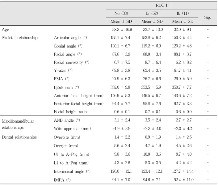
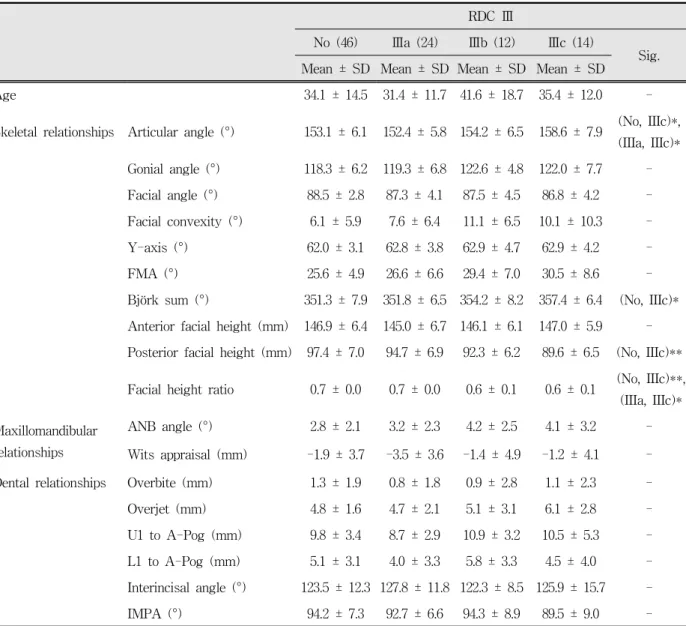
The cephalometric analysis results of each RDC group are shown in table 1. Only the articular angle of the RDC group IIc patients was larger than patients of the no diagnosis of RDC II group. Larger articular angle and smaller facial height ratio were observed in RDC IIIc group compared to IIIa group.
Larger articular angle, larger Björk sum, smaller
Fig. 2. Angular measurements used in this study: 1, articular angle; 2, gonial angle; 3, facial angle; 4, facial convexity; 5, Y-axis; 6, FMA;
7, ANB angle; 8, interincisal angle; 9, IMPA.
Fig. 3. Linear measurements used in this study: 1,
anterior facial height; 2, posterior facial
height; 3, Wits appraisal; 4, overbite; 5,
overjet.
RDC I
No (33) Ia (52) Ib (11)
Sig.
Mean ± SD Mean ± SD Mean ± SD
Age 38.3 ± 16.9 32.7 ± 13.0 32.0 ± 9.1 -
Skeletal relationships Articular angle (°) 155.1 ± 7.4 153.8 ± 6.2 150.3 ± 4.4 - Gonial angle (°) 120.1 ± 6.7 119.2 ± 6.9 120.2 ± 4.8 - Facial angle (°) 87.6 ± 3.9 88.0 ± 3.4 88.1 ± 3.7 - Facial convexity (°) 6.7 ± 7.5 8.7 ± 6.4 6.2 ± 8.2 -
Y-axis (°) 62.8 ± 3.8 62.4 ± 3.5 61.7 ± 4.1 -
FMA (°) 27.9 ± 6.3 26.7 ± 6.6 26.0 ± 5.9 -
Björk sum (°) 352.0 ± 9.8 353.5 ± 5.9 350.7 ± 7.7 -
Anterior facial height (mm) 146.9 ± 5.3 146.5 ± 6.7 143.6 ± 7.2 - Posterior facial height (mm) 94.4 ± 7.7 95.8 ± 7.6 92.7 ± 3.3 - Facial height ratio 0.6 ± 0.1 0.7 ± 0.1 0.6 ± 0.0 - Maxillomandibular
relationships
ANB angle (°) 3.1 ± 2.4 3.5 ± 2.4 2.7 ± 2.7 - Wits appraisal (mm) -1.9 ± 3.9 -2.3 ± 4.0 -2.0 ± 4.2 - Dental relationships Overbite (mm) 1.4 ± 2.2 0.9 ± 1.9 1.4 ± 2.5 - Overjet (mm) 5.6 ± 2.4 4.7 ± 1.9 4.5 ± 2.6 - U1 to A-Pog (mm) 9.8 ± 3.6 10.0 ± 3.6 8.7 ± 4.0 - L1 to A-Pog (mm) 4.3 ± 2.6 5.3 ± 3.5 4.2 ± 4.2 - Interincisal angle (°) 126.0 ± 12.1 123.4 ± 12.1 127.7 ± 14.4 -
IMPA (°) 91.1 ± 7.0 94.6 ± 7.1 92.4 ± 11.0 -
Sig.; Significance by ANOVA, Bonferroni analysis was used for a post hoc test.
-; Not significant SD; Standard deviation
* P<0.05,**P<0.01
Table 1. Cephalometric measurements of RDC/TMD groups. (Continued)
posterior facial height, and smaller facial height ratio were observed in RDC group IIIc compared to no diagnosis of RDC III group.
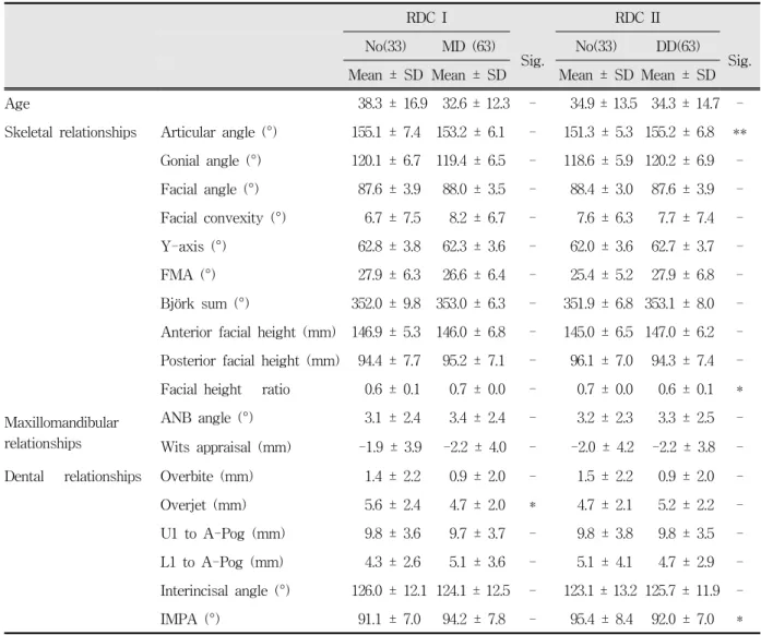
The results of cephalometric analysis in simplified RDC groups are shown in Table 2. Smaller overjet was observed in MD group. Facial height ratio and IMPA were smaller and articular angle was larger
in DD group than in no diagnosis of DD group. In
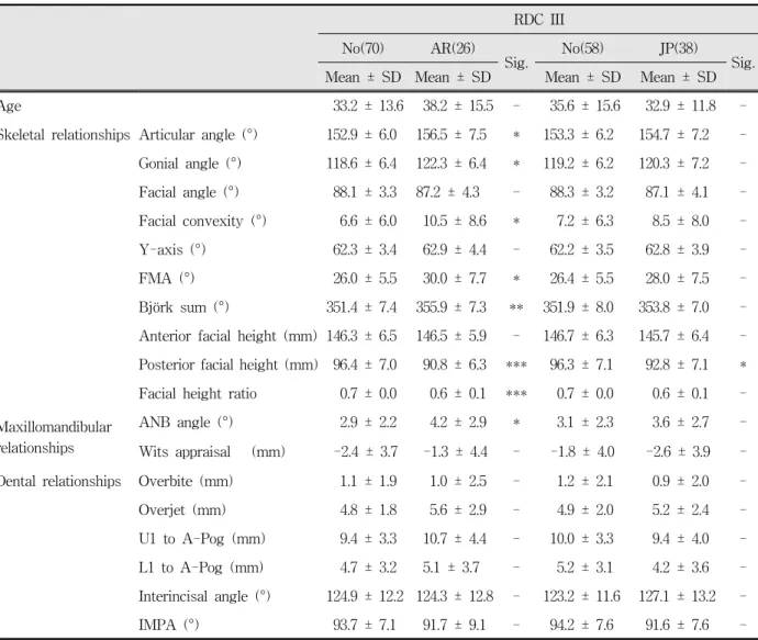
AR group, posterior facial height, and facial height
ratio were smaller, and articular angle, gonial angle,
facial convexity, FMA, Björk sum, and ANB were
larger than in no diagnosis of AR group. In JP group,
only posterior facial height was smaller than no
diagnosis of JP group.
RDC Ⅱ
No (33) Ⅱa (35) Ⅱb (5) Ⅱc (23)
Sig.
Mean ± SD Mean ± SD Mean ± SD Mean ± SD
Age 34.9 ± 13.5 30.0 ± 11.3 46.6 ± 21.0 38.3 ± 15.8 -
Skeletal relationships Articular angle (°) 151.3 ± 5.3 154.2 ± 6.1 158.6 ± 2.4 156.1 ± 8.1 (No, IIc)*
Gonial angle (°) 118.6 ± 5.9 120.3 ± 6.3 118.7 ± 5.3 120.4 ± 8.1 - Facial angle (°) 88.4 ± 3.0 87.2 ± 3.5 87.2 ± 4.1 88.2 ± 4.4 - Facial convexity (°) 7.6 ± 6.3 8.6 ± 6.8 9.7 ± 6.2 6.0 ± 8.4 - Y-axis (°) 62.0 ± 3.6 62.9 ± 3.3 62.8 ± 4.3 62.4 ± 4.2 - FMA (°) 25.4 ± 5.2 27.7 ± 6.2 28.9 ± 6.9 28.1 ± 7.9 - Björk sum (°) 351.9 ± 6.8 353.5 ± 7.2 355.5 ± 7.7 351.9 ± 9.3 - Anterior facial height (mm) 145.0 ± 6.5 146.6 ± 6.3 147.0 ± 7.4 147.7 ± 6.1 - Posterior facial height (mm) 96.1 ± 7.0 94.5 ± 6.9 92.1 ± 8.0 94.5 ± 8.1 - Facial height ratio 0.7 ± 0.0 0.6 ± 0.1 0.6 ± 0.1 0.6 ± 0.1 - Maxillomandibular
relationships
ANB angle (°) 3.2 ± 2.3 3.6 ± 2.5 4.2 ± 1.8 2.7 ± 2.7 - Wits appraisal (mm) -2.0 ± 4.2 -2.0 ± 3.9 -3.2 ± 3.0 -2.4 ± 3.8 - Dental relationships Overbite (mm) 1.5 ± 2.2 1.0 ± 2.1 1.1 ± 2.3 0.8 ± 1.9 - Overjet (mm) 4.7 ± 2.1 5.3 ± 2.4 4.5 ± 0.9 5.2 ± 2.1 - U1 to A-Pog (mm) 9.8 ± 3.8 9.3 ± 3.3 9.8 ± 3.2 10.5 ± 4.0 - L1 to A-Pog (mm) 5.1 ± 4.1 4.2 ± 2.6 5.3 ± 2.8 5.3 ± 3.3 - Interincisal angle (°) 123.1 ± 13.2 128.3 ± 10.7 123.1 ± 9.2 122.1 ± 13.3 -
IMPA (°) 95.4 ± 8.4 91.5 ± 6.2 95.7 ± 10.0 91.9 ± 7.6 -
Sig.; Significance by ANOVA, Bonferroni analysis was used for a post hoc test.
-; Not significant SD; Standard deviation
* P<0.05,**P<0.01
Table 1. (Continued) Cephalometric measurements of RDC/TMD groups. (Continued)
Ⅳ. DISCUSSION
RDC/TMD has been widely used in clinical research settings around the world since it has been introduced in 1992
13). The algorithms of clinical examination of RDC/TMD Axis I have been revised to improve its reliability and validity
17,18). The diagnostic reliability of the revised algorithms ranged from good to excellent. The validity,
however, was acceptable only for the muscle pain
(Ia or Ib), disk displacement without reduction with
limited opening (IIb), and joint pain (IIIa or IIIb)
diagnosis. It has been clearly declared that the
recommendation of the original RDC/TMD
suggesting supplementation of the examination
findings with imaging could not yet be changed
18).
Although it is obvious that MRI and CT guarantee
excellent validity in diagnosis of disk displacement
RDC Ⅲ
No (46) Ⅲa (24) Ⅲb (12) Ⅲc (14)
Mean ± SD Mean ± SD Mean ± SD Mean ± SD Sig.
Age 34.1 ± 14.5 31.4 ± 11.7 41.6 ± 18.7 35.4 ± 12.0 -
Skeletal relationships Articular angle (°) 153.1 ± 6.1 152.4 ± 5.8 154.2 ± 6.5 158.6 ± 7.9 (No, IIIc)*, (IIIa, IIIc)*
Gonial angle (°) 118.3 ± 6.2 119.3 ± 6.8 122.6 ± 4.8 122.0 ± 7.7 - Facial angle (°) 88.5 ± 2.8 87.3 ± 4.1 87.5 ± 4.5 86.8 ± 4.2 - Facial convexity (°) 6.1 ± 5.9 7.6 ± 6.4 11.1 ± 6.5 10.1 ± 10.3 - Y-axis (°) 62.0 ± 3.1 62.8 ± 3.8 62.9 ± 4.7 62.9 ± 4.2 -
FMA (°) 25.6 ± 4.9 26.6 ± 6.6 29.4 ± 7.0 30.5 ± 8.6 -
Björk sum (°) 351.3 ± 7.9 351.8 ± 6.5 354.2 ± 8.2 357.4 ± 6.4 (No, IIIc)*
Anterior facial height (mm) 146.9 ± 6.4 145.0 ± 6.7 146.1 ± 6.1 147.0 ± 5.9 - Posterior facial height (mm) 97.4 ± 7.0 94.7 ± 6.9 92.3 ± 6.2 89.6 ± 6.5 (No, IIIc)**
Facial height ratio 0.7 ± 0.0 0.7 ± 0.0 0.6 ± 0.1 0.6 ± 0.1 (No, IIIc)**, (IIIa, IIIc)*
Maxillomandibular relationships
ANB angle (°) 2.8 ± 2.1 3.2 ± 2.3 4.2 ± 2.5 4.1 ± 3.2 - Wits appraisal (mm) -1.9 ± 3.7 -3.5 ± 3.6 -1.4 ± 4.9 -1.2 ± 4.1 - Dental relationships Overbite (mm) 1.3 ± 1.9 0.8 ± 1.8 0.9 ± 2.8 1.1 ± 2.3 - Overjet (mm) 4.8 ± 1.6 4.7 ± 2.1 5.1 ± 3.1 6.1 ± 2.8 - U1 to A-Pog (mm) 9.8 ± 3.4 8.7 ± 2.9 10.9 ± 3.2 10.5 ± 5.3 - L1 to A-Pog (mm) 5.1 ± 3.1 4.0 ± 3.3 5.8 ± 3.3 4.5 ± 4.0 - Interincisal angle (°) 123.5 ± 12.3 127.8 ± 11.8 122.3 ± 8.5 125.9 ± 15.7 -
IMPA (°) 94.2 ± 7.3 92.7 ± 6.6 94.3 ± 8.9 89.5 ± 9.0 -
Sig.; Significance by ANOVA, Bonferroni analysis was used for a post hoc test.
-; Not significant SD; Standard deviation
* P<0.05,**P<0.01
Table 1. (Continued) Cephalometric measurements of RDC/TMD groups.
and arthritis/arthrosis, respectively, they are not the imaging method of choice because of high cost or radiation exposure problems. In most orthodontic clinics, only clinical examination and plain radio- graphs such as cephalogram and orthopantomo- gram are available. When only limited information is available, a standardized clinical tool for the
diagnosis of TMD is more called for. This study
was performed to evaluate the usability of
RDC/TMD Axis I as a tool for clinical diagnosis of
TMD in the orthodontic diagnosis and treatment
procedure by examining whether the clinical
diagnosis according to RDC/TMD Axis I could
reflect the facial skeletal patterns previously
RDC I RDC II No(33) MD (63)
Sig. No(33) DD(63)
Mean ± SD Mean ± SD Mean ± SD Mean ± SD Sig.
Age 38.3 ± 16.9 32.6 ± 12.3 - 34.9 ± 13.5 34.3 ± 14.7 -
Skeletal relationships Articular angle (°) 155.1 ± 7.4 153.2 ± 6.1 - 151.3 ± 5.3 155.2 ± 6.8 **
Gonial angle (°) 120.1 ± 6.7 119.4 ± 6.5 - 118.6 ± 5.9 120.2 ± 6.9 - Facial angle (°) 87.6 ± 3.9 88.0 ± 3.5 - 88.4 ± 3.0 87.6 ± 3.9 - Facial convexity (°) 6.7 ± 7.5 8.2 ± 6.7 - 7.6 ± 6.3 7.7 ± 7.4 - Y-axis (°) 62.8 ± 3.8 62.3 ± 3.6 - 62.0 ± 3.6 62.7 ± 3.7 - FMA (°) 27.9 ± 6.3 26.6 ± 6.4 - 25.4 ± 5.2 27.9 ± 6.8 - Björk sum (°) 352.0 ± 9.8 353.0 ± 6.3 - 351.9 ± 6.8 353.1 ± 8.0 - Anterior facial height (mm) 146.9 ± 5.3 146.0 ± 6.8 - 145.0 ± 6.5 147.0 ± 6.2 - Posterior facial height (mm) 94.4 ± 7.7 95.2 ± 7.1 - 96.1 ± 7.0 94.3 ± 7.4 - Facial height ratio 0.6 ± 0.1 0.7 ± 0.0 - 0.7 ± 0.0 0.6 ± 0.1 * Maxillomandibular
relationships
ANB angle (°) 3.1 ± 2.4 3.4 ± 2.4 - 3.2 ± 2.3 3.3 ± 2.5 - Wits appraisal (mm) -1.9 ± 3.9 -2.2 ± 4.0 - -2.0 ± 4.2 -2.2 ± 3.8 - Dental relationships Overbite (mm) 1.4 ± 2.2 0.9 ± 2.0 - 1.5 ± 2.2 0.9 ± 2.0 - Overjet (mm) 5.6 ± 2.4 4.7 ± 2.0 * 4.7 ± 2.1 5.2 ± 2.2 - U1 to A-Pog (mm) 9.8 ± 3.6 9.7 ± 3.7 - 9.8 ± 3.8 9.8 ± 3.5 - L1 to A-Pog (mm) 4.3 ± 2.6 5.1 ± 3.6 - 5.1 ± 4.1 4.7 ± 2.9 - Interincisal angle (°) 126.0 ± 12.1 124.1 ± 12.5 - 123.1 ± 13.2 125.7 ± 11.9 -
IMPA (°) 91.1 ± 7.0 94.2 ± 7.8 - 95.4 ± 8.4 92.0 ± 7.0 *
MD; Muscle Disorder, DD; Disk Displacement, AR; Arthrosis/Arthritis, JP; Joint Pain Sig.; Significance by independent t-test
-; Not significant SD; Standard deviation
* P<0.05,**P<0.01
Table 2. Cephalometric measurements of simplified RDC/TMD groups. (Continued)
reported to be related to TMD and investigate which diagnosis group of RDC/TMD was related to such morphologic pattern.
There have been many studies investigating the correlation between specific skeletal patterns and TMD. It has been described that joint shape and bony characteristics are different according to facial type
19). For instance, the articular eminence of brachyfacial pattern is steeper than that of
dolichofacial pattern. A strong positive correlation between the angle of the articular eminence and lingual surface of maxillary central incisor in individuals with good occlusion has been also presented
20). It is assumed that these morphologic features of the maxillofacial skeleton can lead to functional abnormalities of the TMJ.
It has also been reported that disk displacement
influences the morphology of the condyle and
RDC III
No(70) AR(26)
Sig. No(58) JP(38)
Mean ± SD Mean ± SD Mean ± SD Mean ± SD Sig.
Age 33.2 ± 13.6 38.2 ± 15.5 - 35.6 ± 15.6 32.9 ± 11.8 -
Skeletal relationships Articular angle (°) 152.9 ± 6.0 156.5 ± 7.5 * 153.3 ± 6.2 154.7 ± 7.2 - Gonial angle (°) 118.6 ± 6.4 122.3 ± 6.4 * 119.2 ± 6.2 120.3 ± 7.2 - Facial angle (°) 88.1 ± 3.3 87.2 ± 4.3 - 88.3 ± 3.2 87.1 ± 4.1 - Facial convexity (°) 6.6 ± 6.0 10.5 ± 8.6 * 7.2 ± 6.3 8.5 ± 8.0 - Y-axis (°) 62.3 ± 3.4 62.9 ± 4.4 - 62.2 ± 3.5 62.8 ± 3.9 - FMA (°) 26.0 ± 5.5 30.0 ± 7.7 * 26.4 ± 5.5 28.0 ± 7.5 - Björk sum (°) 351.4 ± 7.4 355.9 ± 7.3 ** 351.9 ± 8.0 353.8 ± 7.0 - Anterior facial height (mm) 146.3 ± 6.5 146.5 ± 5.9 - 146.7 ± 6.3 145.7 ± 6.4 - Posterior facial height (mm) 96.4 ± 7.0 90.8 ± 6.3 *** 96.3 ± 7.1 92.8 ± 7.1 * Facial height ratio 0.7 ± 0.0 0.6 ± 0.1 *** 0.7 ± 0.0 0.6 ± 0.1 - Maxillomandibular
relationships
ANB angle (°) 2.9 ± 2.2 4.2 ± 2.9 * 3.1 ± 2.3 3.6 ± 2.7 - Wits appraisal (mm) -2.4 ± 3.7 -1.3 ± 4.4 - -1.8 ± 4.0 -2.6 ± 3.9 - Dental relationships Overbite (mm) 1.1 ± 1.9 1.0 ± 2.5 - 1.2 ± 2.1 0.9 ± 2.0 - Overjet (mm) 4.8 ± 1.8 5.6 ± 2.9 - 4.9 ± 2.0 5.2 ± 2.4 - U1 to A-Pog (mm) 9.4 ± 3.3 10.7 ± 4.4 - 10.0 ± 3.3 9.4 ± 4.0 - L1 to A-Pog (mm) 4.7 ± 3.2 5.1 ± 3.7 - 5.2 ± 3.1 4.2 ± 3.6 - Interincisal angle (°) 124.9 ± 12.2 124.3 ± 12.8 - 123.2 ± 11.6 127.1 ± 13.2 -
IMPA (°) 93.7 ± 7.1 91.7 ± 9.1 - 94.2 ± 7.6 91.6 ± 7.6 -
MD; Muscle Disorder, DD; Disk Displacement, AR; Arthrosis/Arthritis, JP; Joint Pain Sig.; Significance by independent t-test
-; Not significant SD; Standard deviation
* P<0.05,**P<0.01
Table 2. (Continued) Cephalometric measurements of simplified RDC/TMD groups.
mandibular body, and eventually induces alterations in facial morphology. In cases of internal derangement, the backward rotated mandible decreases ramus height, and causes facial asymmetry. We can easily measure the backward rotation of the mandible and actual decrease of ramus height on a lateral cephalogram. It is currently known that these skeletal changes are exacerbated with the progress of internal
derangement
2,3,21-23). The adolescent TMD patients
especially have a short ramus and posterior facial
height, and larger gonial and mandibular plane
angle
24). This is known to be caused by hindered
mandibular growth due to internal derangement or
degenerative osteoarthritis of the TMJ. It was
asserted with certainty that the extent to which
facial form could be affected by TMD is dependent
on both the duration of the disorder and the amount
of growth remaining to be potentially expressed by the individual
25).
The results of this study show that specific clinical morphological patterns similar to what had been reported in previous studies to be related to the presence of TMD
1-12,26)were more likely to be related to arthritis/arthrosis of RDC III group. The larger articular angle, smaller posterior facial height, and smaller facial height ratio of the RDC IIIc group implies that the mandible may have rotated to the posterior. Only a larger articular angle was found in RDC II group. In the simplified RDC groups, such a skeletal pattern was more evident in the DD and AR group, but neither in the MD nor JP group. It can be postulated that such skeletal characteristics are more related to intra-articular changes such as disk displacement and arthrosis than muscular or articular pain. The possible role of disk displacement and arthritis/arthrosis on the facial growth and morphologic changes have been continuously suggested in many studies
2,27-31). On the other hand, a consensus exists that the consistently observed association between intra-articular changes and facial skeletal morphology does not define a cause and effect relationship and its underlying mechanism should be addressed yet
1-8,32).
RDC/TMD Axis I diagnosis is based on clinical examinations which are inevitably less reliable and valid than MRI and CT for the diagnosis of intra-articular disorders such as disk displacement and arthrosis. However, RDC/TMD is a more comprehensive diagnostic tool than imaging techniques since intra-articular disorders cannot sufficiently reflect the disease state of TMD patients. Disk displacement and arthritis/arthrosis is very common in the adult population and in many cases are not accompanied by any clinical signs and symptoms that need medical treatment. Disk displacement was observed in 10-30% of asymptomatic subjects and examination of TMJ specimens through autopsy revealed changes consistent with arthritis/arthrosis in 22% to 40%
33-40)
. While many studies suggested an association between disk displacement and arthritis/arthrosis,
arthritis/arthrosis was also observed in the joints without any evidence of disk displacement
27,41-43). When screening for the presence of TMD before starting orthodontic treatment and monitoring every 6 months are planned as recommended, the diagnosis of TMD should be made comprehensively by all means available including clinical, historical, and radiological examinations than by only intra-articular findings observed in MRI or CT
44). Moreover, it has also been pointed out that the extent of discordant interpretations would suggest that radiograph diagnoses should not be considered to be stand-alone gold standards for TMJ intra- articular disorders
17). Considering such suggestions and the results of this study, RDC/TMD aided by plain radiographs could be a good option for the standardized examination of TMJ state and the diagnosis of TMD in most orthodontic clinics where only plain radiographs are available. Its revised algorithms of clinical examinations have good reliability and validity in diagnosing pain disorders of the TMJ. As shown in our results, it also effectively reflects the facial skeletal pattern known to be associated with TMD in previous studies, and this association was more obvious in RDC III group.
V. CONCLUSIONS
Facial morphologic patterns showing posterior- rotated mandible and lower posterior facial height is related to RDC group II and III diagnosis of the TMJ in female TMD patients. RDC/TMD Axis I diagnosis can provide a good clinical diagnostic tool for the standardized examination of the TMJ in orthodontic clinics.
REFERENCES
1. Nebbe B, Major PW, Prasad NG. Adolescent female craniofacial morphology associated with advanced bilateral TMJ disc displacement. Eur J Orthod.
1998;20:701-712.
2. Nebbe B, Major PW, Prasad N. Female adolescent
facial pattern associated with TMJ disk displacement
and reduction in disk length: part I. Am J Orthod
Dentofacial Orthop. 1999;116:168-176.
3. Gidarakou IK, Tallents RH, Kyrkanides S, Stein S, Moss ME. Comparison of skeletal and dental morphology in asymptomatic volunteers and symptomatic patients with bilateral disk displacement with reduction. Angle Orthod. 2002;72:541-546.
4. Gidarakou IK, Tallents RH, Kyrkanides S, Stein S, Moss M. Comparison of skeletal and dental morphology in asymptomatic volunteers and symptomatic patients with bilateral degenerative joint disease. Angle Orthod. 2003;73:71-78.
5. Gidarakou IK, Tallents RH, Kyrkanides S, Stein S, Moss ME. Comparison of skeletal and dental morphology in asymptomatic volunteers and sympto- matic patients with normal temporomandibular joints.
Angle Orthod. 2003;73:116-120.
6. Gidarakou IK, Tallents RH, Kyrkanides S, Stein S, Moss ME. Comparison of skeletal and dental morphology in asymptomatic volunteers and symptomatic patients with unilateral disk displacement without reduction. Angle Orthod. 2003;
73:121-127.
7. Gidarakou IK, Tallents RH, Stein S, Kyrkanides S, Moss ME. Comparison of skeletal and dental morphology in asymptomatic volunteers and symptomatic patients with unilateral disk displacement with reduction. Angle Orthod. 2004;74:
212-219.
8. Gidarakou IK, Tallents RH, Kyrkanides S, Stein S, Moss ME. Comparison of skeletal and dental morphology in asymptomatic volunteers and symptomatic patients with bilateral disk displacement without reduction. Angle Orthod. 2004;74:684-690.
9. Byun ES, Ahn SJ, Kim TW. Relationship between internal derangement of the temporomandibular joint and dentofacial morphology in women with anterior open bite. Am J Orthod Dentofacial Orthop. 2005 Jul;128:87-95.
10. Ioi H, Matsumoto R, Nishioka M, Goto TK, Nakata S, Nakasima A, Counts AL. Relationship of TMJ osteoarthritis / osteoarthrosis to head posture and dentofacial morphology. Orthod Craniofac Res.
2008;11:8-16.
11. Hwang CJ, Sung SJ, Kim SJ. Lateral cephalometric characteristics of malocclusion patients with temporomandibular joint disorder symptoms. Am J Orthod Dentofacial Orthop. 2006;129:497-503.
12. Ahn SJ, Kim TW, Nahm DS. Cephalometric keys to internal derangement of temporomandibular joint in
women with Class II malocclusions. Am J Orthod Dentofacial Orthop. 2004;126:486-495.
13. Dworkin SF, LeResche L. Research diagnostic criteria for temporomandibular disorders: review, criteria, examinations and specifications, critique. J Craniomandib Disord. 1992;6:301-355.
14. Goulet JP, Lavigne GJ, Lund JP. Jaw pain prevalence among French-speaking Canadians in Qubec and related symptoms of temporomandibular disorders. J Dent Res. 1995;74:1738-1744.
15. List T, Dworkin SF. Comparing TMD diagnoses and clinical findings at Swedish and US TMD centers using research diagnostic criteria for temporomandi- bular disorders. J Orofac Pain. 1996;10:240-253.
16. Lobbezoo-Scholte AM, De Leeuw JR, Steenks MH, Bosman F, Buchner R, Olthoff LW. Diagnostic subgroups of craniomandibular disorders. Part I:
Self-report data and clinical findings. J Orofac Pain.
1995;9:24-36.
17. Look JO, Schiffman EL, Truelove EL, Ahmad M.
Reliability and validity of axis I of the Research Diagnostic Criteria for Temporomandibular Disorders (RDC/TMD) with proposed revisions. J Oral Rehabil.
2010;37:744-759.
18. Schiffman EL, Ohrbach R, Truelove EL, Tai F, Anderson GC, Pan W, Gonzalez YM, John MT, Sommers E, List T, Velly AM, Kang W, Look JO.
The Research Diagnostic Criteria for Temporoman- dibular Disorders. V: methods used to establish and validate revised Axis I diagnostic algorithms. J Orofac Pain. 2010;24:63-78.
19. Grummons D. Orthodontics for the TMJ-TMD Patient. Wright and Co. 1994.
20. Bell DE, Harris EF. Disclusion in mandibular protrusion. Angle Orthod. 1983;53:146-156.
21. Ahn SJ, Baek SH, Kim TW, Nahm DS. Discrimi- nation of internal derangement of temporomandibular joint by lateral cephalometric analysis. Am J Orthod Dentofacial Orthop. 2006;130:331-339.
22. Bsio JA, Burch JG, Tallents RH, Wade DB, Beck FM.
Lateral cephalometric analysis of asymptomatic volunteers and symptomatic patients with and without bilateral temporomandibular joint disk displacement. Am J Orthod Dentofacial Orthop.
1998;114:248-255.
23. Roth RH. Temporomandibular pain-dysfunction and occlusal relationships. Angle Orthod. 1973;43:136-153.
24. Dibbets JM, van der Weele LT, Uildriks AK.
Symptoms of TMJ dysfunction: indicators of growth
patterns? J Pedod. 1985;9:265-284.
25. Dibbets JM, Carlson DS. Implications of temporoman- dibular disorders for facial growth and orthodontic treatment. Semin Orthod. 1995;1:258-272.
26. Tanaka E, Detamore MS, Mercuri LG. Degenerative disorders of the temporomandibular joint: etiology, diagnosis, and treatment. J Dent Res. 2008;87:
296-307.
27. Link JJ, Nickerson JW Jr. Temporomandibular joint internal derangements in an orthognathic surgery population. Int J Adult Orthodon Orthognath Surg.
1992;7:161-169.
28. Trpkova B, Major P, Nebbe B, Prasad N. Craniofacial asymmetry and temporomandibular joint internal derangement in female adolescents: a posteroanterior cephalometric study. Angle Orthod. 2000;70:81-88.
29. Schellhas KP, Pollei SR, Wilkes CH. Pediatric internal derangements of the temporomandibular joint: effect on facial development. Am J Orthod Dentofacial Orthop. 1993;104:51-59.
30. Schellhas KP, Piper MA, Omlie MR. Facial skeleton remodeling due to temporomandibular joint degeneration: an imaging study of 100 patients.
AJNR Am J Neuroradiol. 1990;11:541-551.
31. Stewart A, Harris M. Acquired anterior open bite and facial arthromyalgia: possible aetiology. Br J Oral Maxillofac Surg. 1996;34:174-180.
32. Hall HD. Intra-articular disc displacement Part II: Its significant role in temporomandibular joint pathology.
J Oral Maxillofac Surg. 1995;53:1073-1079.
33. Westesson PL, Eriksson L, Kurita K. Reliability of a negative clinical temporomandibular joint exami- nation: prevalence of disk displacement in asymptomatic temporomandibular joints. Oral Surg Oral Med Oral Pathol. 1989;68:551-554.
34. Tallents RH, Hatala M, Katzberg RW, Westesson PL.
Temporomandibular joint sounds in asymptomatic volunteers. J Prosthet Dent. 1993;69:298-304.
35. Tallents RH, Katzberg RW, Murphy W, Proskin H.
Magnetic resonance imaging findings in asymptomatic volunteers and symptomatic patients with temporomandibular disorders. J Prosthet Dent.
1996;75:529-533.
36. Ribeiro RF, Tallents RH, Katzberg RW, Murphy WC, Moss ME, Magalhaes AC, Tavano O. The prevalence of disc displacement in symptomatic and asymptomatic volunteers aged 6 to 25 years. J Orofac Pain. 1997;11:37-47.
37. Morrow D, Tallents RH, Katzberg RW, Murphy WC, Hart TC. Relationship of other joint problems and anterior disc position in symptomatic TMD patients and in asymptomatic volunteers. J Orofac Pain.
1996;10:15-20.
38. Axelsson S. Human and experimental osteoarthrosis of the temporomandibular joint. Morphological and biochemical studies. Swed Dent J Suppl. 1993;92:1-45.
39. Widmalm SE, Westesson PL, Kim IK, Pereira FJ Jr, Lundh H, Tasaki MM. Temporomandibular joint pathosis related to sex, age, and dentition in autopsy material. Oral Surg Oral Med Oral Pathol. 1994;78:
416-425.
40. Magnusson C, Ernberg M, Magnusson T. A description of a contemporary human skull material in respect of age, gender, temporomandibular joint changes, and some dental variables. Swed Dent J.
2008;32:69-81.
41. Westesson PL, Rohlin M. Internal derangement related to osteoarthrosis in temporomandibular joint autopsy specimens. Oral Surg Oral Med Oral Pathol.
1984;57:17-22.
42. Roberts CA, Katzberg RW, Tallents RH, Espeland MA, Handelman SL. Correlation of clinical parameters to the arthrographic depiction of temporomandibular joint internal derangements. Oral Surg Oral Med Oral Pathol. 1988;66:32-36.
43. Campos PS, Cangussu MC, Guimares RC, Line SR.
Analysis of magnetic resonance imaging characteristics and pain in temporomandibular joints with and without degenerative changes of the condyle. Int J Oral Maxillofac Surg. 2008;37:529-534.
44. Michelotti A, Iodice G. The role of orthodontics in temporomandibular disorders. J Oral Rehabil.
2010;37:411-429.
국문초록
RDC/TMD Axis I 진단에 따른 측두하악장애 환자의 측두 두부방사선적 특징에 관한 연구