D I A B E T E S & M E T A B O L I S M J O U R N A L
This is an Open Access article distributed under the terms of the Creative Commons At- tribution Non-Commercial License (http://creativecommons.org/licenses/by-nc/3.0/) which permits unrestricted non-commercial use, distribution, and reproduction in any medium, provided the original work is properly cited.
Relationships between Brachial-Ankle Pulse Wave
Velocity and Peripheral Neuropathy in Type 2 Diabetes
Byung Kil Ha1, Bong Gun Kim1, Dong Hyun Kim1, Soon Il Lee1, Soon Myung Jung1, Ja Young Park1, Chang Won Lee1, Sang Soo Kim2, Bo Hyun Kim2, In Ju Kim2
1Department of Internal Medicine, Busan St. Mary’s Medical Center,
2Department of Internal Medicine, Pusan National University College of Medicine, Busan, Korea
Background: Brachial-ankle pulse wave velocity (baPWV) is known to be a good surrogate marker of clinical atherosclerosis.
Atherosclerosis is a major predictor for developing neuropathy. The goal of this study was to determine the relationship between baPWV and diabetic peripheral neuropathy (DPN) in patients with type 2 diabetes.
Methods: A retrospective cross-sectional study was conducted involving 692 patients with type 2 diabetes. The correlation be- tween increased baPWV and DPN, neurological symptoms, and neurological assessment was analyzed. DPN was examined us- ing the total symptom score (TSS), ankle reflexes, the vibration test, and the 10-g monofilament test. DPN was defined as TSS ≥2 and an abnormal neurological assessment. Data were expressed as means±standard deviation for normally distributed data and as median (interquartile range) for non-normally distributed data. Independent t-tests or chi-square tests were used to make comparisons between groups, and a multiple logistic regression test was used to evaluate independent predictors of DPN. The Mantel-Haenszel chi-square test was used to adjust for age.
Results: Patients with DPN had higher baPWV and systolic blood pressure, and were more likely to be older and female, when compared to the control group. According to univariate analysis of risk factors for DPN, the odds ratio of the baPWV ≥1,600 cm/sec was 1.611 (95% confidence interval [CI], 1.072 to 2.422; P=0.021) and the odds ratio in female was 1.816 (95% CI, 1.195 to 2.760; P=0.005).
Conclusion: Increased baPWV was significantly correlated with peripheral neuropathy in patients with type 2 diabetes.
Keywords: Brachial-ankle pulse wave velocity; Diabetes mellitus, type 2; Diabetic peripheral neuropathy
Corresponding author: Chang Won Lee
Department of Internal Medicine, Busan St. Mary’s Medical Center, 25-14 Yongho-ro 232beon-gil, Nam-gu, Busan 608-838, Korea E-mail: [email protected]
INTRODUCTION
Diabetic peripheral neuropathy (DPN) is suspected when dia- betic patients complain of symptoms and/or show signs of pe- ripheral nerve dysfunction after exclusion of other etiology [1].
This common complication of diabetes typically involve sen- sory nerves, motor nerves, and autonomic nerves, and often results in foot ulcers [2]. DPN can lead to socio-economic prob- lems as well as psychological and physiological disorders [2].
Diagnosis and treatments are based on the medical history
and results of a physical examination of patients with DPN [3].
The most widely used clinical nerve function examinations are the monofilament test [4], the vibration sensory test [5], and the ankle reflex test. Diagnostic sensitivity can reach up to 87%
when more than two of the nerve function examinations are performed and other causes of neuropathy have been ruled out based on typical symptoms of DPN (careful with paresthe- sia) [1,6].
Pulse wave velocity (PWV) reflects arterial stiffness [7]. The oscillometric method, which is a simple non-invasive method, http://dx.doi.org/10.4093/dmj.2012.36.6.443
pISSN 2233-6079 · eISSN 2233-6087
can be used to measure brachial-ankle pulse wave velocity (baPWV) [8], and in turn, baPWV can be used to detect vas- cular damage [9,10]. BaPWV reflects the condition of the aor- ta and peripheral arteries because it is affected by vasomotor reflexes, it is thought to have a greater association with micro- angiopathic conditions and diabetes complications than aortic pulse wave velocity [11]. Previous reports indicated that baP- WV is an independent predictor of cardiovascular disease [12, 13], and its use as a clinical indicator is being reviewed.
Aso et al. [11] reported that baPWV was not only directly related to albuminuria, autonomic neuropathy, and retinopa- thy, but also peripheral neuropathy. Yokoyama et al. [14] re- ported that pulse wave velocity, retinopathy, age, and glycated hemoglobin are independent risk factors in DPN and the as- sociated autonomic neuropathy. This study used a retrospec- tive analysis to investigate DPN in type 2 diabetes patients and the association between baPWV and nerve function tests.
METHODS
This study received the approval of the clinical ethics commit- tee (BSM 2011-04). Brachial pulse wave velocity was measured in type 2 diabetes patients in an outpatient clinic and in those admitted to the study hospital between January 2008 and Au- gust 2009. Total symptom scores and sensory function tests for DPN were retrospectively analyzed in 738 patients over 30 years old [6,15]. A total of 46 cases were excluded based on the following criteria: neuropathy caused by other etiology, severe heart failure, dialysis with chronic renal failure, cardiovascular disease, advanced cirrhosis, tumors, mental illness, thyroid disease, vitamin B12 deficiency, or an ankle brachial pressure index (ABI) on one side of 0.9 or lower. Gender, age, duration of diabetes, and medical history of all patients were noted.
Resting blood pressure and pulse pressure were measured af- ter patients rested in the supine position for 5 minutes. Height, weight, and waist circumferences were also measured. Body mass index (BMI) was measured by dividing body mass (kg) by height in meters squared (m2).
Participants were surveyed regarding alcohol consumption and smoking. Participants were considered to be drinkers if they consumed more than 20 g of alcohol per day. One serving of soju (180 mL) contains 45 g of alcohol, one serving of mak- geolli (Korean rice wine) (1,800 mL) contains 144 g alcohol, and one bottle of beer (640 mL) contains 25.6 g of alcohol. Pa- tients were surveyed to determine if they were receiving insu-
lin therapy or taking thiazolidinedione, statins, antiplatelet agents, β-blockers, calcium channel blockers, angiotensin- converting enzyme inhibitors, angiotensin II receptor block- ers, α-lipoic acid, γ-linolenic acid, tricyclinc antidepressants, pregabalin, or gabapentin. Blood samples were collected after participants fasted for at least 8 hours. Total cholesterol, triglyc- erides, high density lipoprotein cholesterol, low density lipo- protein cholesterol, fasting blood glucose, fasting insulin, gly- cated hemoglobin, high sensitivity C-reactive protein (hs-CRP), aspartate aminotransferase, alanine aminotransferase, micro- albuminuria, and gamma-glutamyltranspeptidase were mea- sured from collected samples. BaPWV was calculated auto- matically using an automatic waveform analyzer (VP-1000;
Colin, Komaki, Japan) [16] by dividing brachial-ankle distance (L=0.5934×height [cm]+14.4014) by the pulse wave time in- terval between the brachial region and ankle (∆T) in patients who were stabilized in the supine position for 5 minutes.
Left and right baPWVs were measured, and the largest val- ue was defined as the maximum baPWV (max-baPWV). The total symptom score was based on the severity and frequency of pain, burning, paresthesia, and numbness [15]. Symptom frequency was categorized as absent, seldom (from 2 to 3 times per week or less than once per day), often (1 to 2 times per day or 7 to 14 times per week), and constant (most of the day, or over 3 times per day every day). Severity was based on a visual analog scale. The levels were scored and separated into mild (does not interfere with everyday life), moderate (affects daily life, but does not affect sleep), and severe (interferes with sleep).
Symptom scores were summed, and the total symptom scores calculated ranged from 0 to 14.64 (Table 1) [15]. A monofila- ment examination, ankle reflex test, and vibration test were performed to evaluate sensory function. When performing the monofilament test, patients lay down and closed their eyes.
Ten-gram monofilaments were pressed into 10 points on the Table 1. Scoringa approach for neuropathic symptomsb includ- ed in the total symptom score
Symptom frequency Symptom intensity
Slight Moderate Severe
Absent 0 0 0
Occasional 1.00 2.00 3.00
Frequent 1.33 2.33 3.33
Almost continuous 1.66 2.66 3.66
Data from Bastyr EJ 3rd, et al. Clin Ther 2005;27:1278-94 [15].
aScoring: total score 0-14.64; bPain, burning, paresthesia, numbness.
top and bottom of patients’ feet until the monofilament began to bend. Patients were evaluated for sensation on the points, and if patients had sensation in fewer than seven points, then they were identified as having abnormal test.
To test ankle reflexes, patients lay down with their legs bent and their knees rotated externally. The researcher placed the patient’s feet in a position of dorsiflexion and then tapped the Achilles tendon. Patients with a low or hyperactive response were determined to a abnormality in the test.
For the vibration sensory test, the oscillator was set at 128 Hz. Patients notified when they could not feel a vibration at the point (the first metatarso-phalangeal joint) where the toes were extended, and then investigator felt the vibration and measured time when the feeling had disappeared. The time difference ≥10 seconds between the investigator and the pa- tient, patients were determined as an abnormality in the test.
When patients had a total symptom score of at least two and had abnormal sensory function tests, they were defined with probable DPN.
Statistical analyses were performed using SPSS for Windows version 14.0 (SPSS Inc., Chicago, IL, USA). Baseline character- istics were expressed as mean±standard deviation, and vari- ables that were not normally distributed were expressed as median and interquartile values. Independent t-tests or chi- square tests were used to determine differences between the two groups. If the chi-squared tests indicated, Cochran and Mantel-Haenszel statistics were used to compensate confound- ing variables. Additionally, when baPWV could be used to predict DPN, a univariate logistic regression analysis, multi- variate logistic regression analysis, odds ratios, and confidence interval (CI) for each risk factor were presented. A P value less than 0.05 was considered statistically significant.
Values are presented as means±standard deviation for normally distributed data and as median (interquartile range) for non-normally distributed data (triglyc- erides, microalbuminuria, fasting insulin, hs-CRP, AST, ALT, and γ-GTP). P values <0.05 were considered significant.
baPWV, brachial-ankle pulse wave velocity; DPN, diabetic peripheral neuropathy; BMI, body mass index; SBP, systolic blood pressure; DBP, diastolic blood pres- sure; CCB, calcium channel blocker; ACE inhibitor, angiotensin converting enzyme inhibitor; ARB, angiotensin II receptor blocker; TCA, tricyclic antidepres- sant; ABI, ankle brachial index; LDL-C, low density lipoprotein cholesterol; HDL-C, high density lipoprotein cholesterol; HbA1c, hemoglobin A1c; hs-CRP, high sensitive C-reactive protein; AST, aspartate transferase; ALT, alanine transferase; γ-GTP, gamma–glutamyltranspeptidase.
aNeuropathy drugs: TCA or gabapentin or pregabalin or α-lipoic acid or γ-linolenic acid.
Characteristic baPWV <1,600
cm/sec (n=336) baPWV ≥1,600 cm/sec (n=356) P value
DPN, % 13.4 19.9 0.025
Sex, M/F, % 48.5/51.5 38.0/62.0 0.006
Age, yr 51.5±9.6 62.0±9.5 <0.001
Duration of diabetes, yr 7.3±5.4 8.5±6.9 0.007
Height, cm 163.2±8.3 159.7±9.3 <0.001
Weight, kg 65.7±9.7 62.8±10.5 <0.001
BMI, kg/m2 24.8±4.2 25.0±8.5 0.631
Waist circumference, cm 87.2±7.4 88.9±9.3 0.008
Cigarette use, % 26.3 23.5 0.404
Alcohol consumption, % 39.8 39.9 0.974
SBP, mm Hg 126.2±12.0 146.5±17.9 <0.001
DBP, mm Hg 77.5±8.5 84.5±10.5 <0.001
Pulse pressure, mm Hg 48.7±8.7 62.0±13.3 <0.001
Insulin treatment, % 28.4 23.8 0.174
β-blocker treatment, % 5.0 12.0 0.061
CCB treatment, % 18.0 21.0 0.248
ACE inhibitor treatment, % 13.4 17.9 0.111
ARB treatment, % 20.6 31.2 0.002
Statin treatment, % 58.2 66.9 0.019
Antiplatelet agent treatment, % 41.5 53.8 0.001
Thiazolidinedione treatment, % 18.7 25.7 0.027
Table 2. Comparison of clinical characteristics in type 2 diabetics according to baPWV
Characteristic baPWV <1,600
cm/sec (n=336) baPWV ≥1,600 cm/sec (n=356) P value
TCA treatment, % 7.0 9.0 0.460
Gabapentin treatment, % 4.0 6.0 0.161
Pregabalin treatment, % 8.0 10.0 0.344
α-lipoic acid treatment, % 39.0 45.0 0.111
γ-linolenic acid treatment, % 24.0 28.0 0.200
Neuropathy drugsa, % 47.3 56.2 0.020
ABI, right 1.1±0.1 1.1±0.1 0.932
ABI, left 1.1±0.1 1.1±0.1 0.269
Total cholesterol, mg/dL 172.5±38.7 174.2±43.4 0.590
LDL-C, mg/dL 104.3±46.3 100.4±35.9 0.213
HDL-C, mg/dL 51.1±14.7 50.8±14.9 0.786
Triglycerides, mg/dL 110.0 (83.0-152.5) 119.0 (90.5-157.0) 0.140
HbA1c, % 7.3±1.5 7.7±1.7 0.004
Microalbuminuria, μg/mg creat 6.1 (2.5-21.3) 11.2 (4.2-30.0) 0.053 Fasting plasma glucose, mg/dL 143.9±46.2 147.5±58.4 0.370 Fasting insulin, mU/L 5.2 (3.5-9.1) 5.7 (4.0-8.8) 0.669
hs-CRP, mg/L 0.6 (0.2-1.4) 0.7 (0.2-1.4) 0.463
AST, U/L 22.0 (18.0-29.0) 22.0 (19.0-27.0) 0.826 ALT, U/L 22.0 (17.0-31.0) 21.0 (16.0-29.0) 0.358 γ-GTP, U/L 22.0 (15.0-43.0) 23.0 (17.0-40.0) 0.217 Suspicion of fungal infection, % 21.7 25.8 0.204
RESULTS
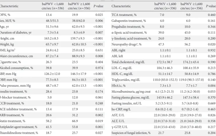
There were a total of 692 participants in this study. The partici- pants were divided into the baPWV ≥1,600 cm/sec group (n=
356) and the <1,600 cm/sec group (n=336). The characteris- tics of each group are summarized in Table 2.
There was a greater proportion of females in the ≥1,600 cm/
sec group compared to the <1,600 cm/sec group (62.0% vs.
51.5%, P<0.01). The mean age of patients was higher (62.0±
9.5 vs. 51.5±9.6, P<0.01), and the duration of diabetes was longer (8.5±6.9 vs. 7.3±5.4, P<0.01) in the ≥1,600 cm/sec group. Additionally, systolic blood pressure (146.5±17.9 mm Hg vs. 126.2±12.0 mm Hg, P<0.01), diastolic blood pressure (84.5±10.5 mm Hg vs. 77.5±8.5 mm Hg, P<0.01), pulse pres- sure (62.0±13.3 mm Hg vs. 48.7±8.7 mm Hg, P<0.01), and
glycated hemoglobin (7.7±1.7% vs. 7.3±1.5%, P<0.01) were higher in the ≥1,600 cm/sec group, and a greater proportion of this group used lipid lowering drugs (66.9% vs. 58.2%, P=
0.019) when compared to the <1,600 cm/sec group. There was no significant difference in the percent of individual neuropa- thy treatment drugs between the ≥1,600 cm/sec group and the
<1,600 cm/sec group, but there was a significant difference in the percent of the total neuropathy treatment drugs (56.2% vs.
47.3%, P<0.020) between the groups. Thus, lipid levels, fasting blood glucose, fasting insulin, and microalbuminuria were not significantly different between the ≥1,600 cm/sec group and the <1,600 cm/sec group.
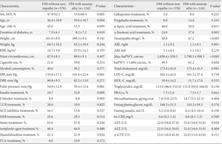
This study also evaluated the differences between the pa- tients with DPN (116 patients) and the patients without DPN (576 patients) (Table 3).
Characteristic DM without neu-
ropathy (n=576) DM with neurop- athy (n=116) P value
Sex, M/F, % 47.9/52.1 33.6/66.4 0.005
Age, yr 56.4±10.8 59.6±10.7 0.004
Age ≥60, % 45.0 53.5 0.095
Duration of diabetes, yr 7.9±6.1 8.2±7.2 0.610
Height, cm 161.6±8.9 160.3±9.4 0.141
Weight, kg 64.5±10.2 63.2±10.4 0.244
BMI, kg/m2 24.7±3.8 25.9±14.2 0.355
Waist circumference, cm 87.9±8.3 88.6±9.3 0.407
Cigarette use, % 21.0 19.0 0.214
Alcohol consumption, % 40.6 36.2 0.371
SBP, mm Hg 135.6±17.2 141.6±22.6 0.001
DBP, mm Hg 80.8±9.5 82.2±13.0 0.271
Pulse pressure, mm Hg 54.8±12.9 59.4±13.4 0.001
Insulin treatment, % 24.7 32.8 0.089
β-blocker treatment, % 8.0 8.0 0.903
CCB treatment, % 20.0 19.0 0.825
ACE inhibitor treatment, % 16.1 13.8 0.537
ARB treatment, % 25.6 28.5 0.514
Statin treatment, % 63.6 57.8 0.244
Antiplatelet agent treatment, % 48.4 44.9 0.480
Thiazolidinedione treatment, % 22.3 21.9 0.924
TCA treatment, % 8.0 10.0 0.372
Table 3. Comparison of clinical characteristics between type 2 diabetics with and without neuropathy
Values are presented as means±standard deviation for normally distributed data and as median (interquartile range) for non-normally distributed data (triglyc- erides, microalbuminuria, fasting insulin, hs-CRP, AST, ALT, and γ-GTP). P values <0.05 were considered significant.
DM, diabetes mellitus; BMI, body mass index; SBP, systolic blood pressure; DBP, diastolic blood pressure; CCB, calcium channel blocker; ACE inhibitor, angio- tensin converting enzyme inhibitor; ARB, angiotensin II receptor blocker; TCA, tricyclic antidepressant; ABI, ankle brachial index; baPWV, brachial-ankle pulse wave velocity; LDL-C, low density lipoprotein cholesterol; HDL-C, high density lipoprotein cholesterol; HbA1c, hemoglobin A1c; hs-CRP, high sensitive C-reac- tive protein; AST, aspartate transferase; ALT, alanine transferase; γ-GTP, gamma–glutamyltranspeptidase.
aNeuropathy drugs: TCA or gabapentin or pregabalin or α-lipoic acid or γ-linolenic acid.
Characteristic DM without neu-
ropathy (n=576) DM with neurop- athy (n=116) P value
Gabapentin treatment, % 5.0 8.0 0.225
Pregabalin treatment, % 8.0 14.0 0.103
α-lipoic acid treatment, % 40.0 50.0 0.057
γ-linolenic acid treatment, % 24.0 37.0 0.003
Neuropathy drugsa, % 48.8 67.2 <0.001
ABI, right 1.1±0.1 1.1±0.1 0.801
ABI, left 1.1±0.1 1.1±0.1 0.235
Max-baPWV, cm/sec 1,650.4±350.5 1,790.5±398.3 <0.001
baPWV ≥1,600 cm/sec, % 49.5 61.2 0.020
Total cholesterol, mg/dL 173.4±41.0 173.4±41.9 0.981
LDL-C, mg/dL 102.5±42.0 101.2±37.4 0.759
HDL-C, mg/dL 50.8±14.2 51.7±17.0 0.552
Triglycerides, mg/dL 113.0 (86.0-152.0) 131.0 (93.0-166.0) 0.130
HbA1c, % 7.5±1.6 7.5±1.7 0.662
Microalbuminuria, μg/mg creat 7.8 (3.0-23.5) 14.7 (5.5-42.3) 0.404 Fasting plasma glucose, mg/dL 146.3±51.5 143.3±59.3 0.576 Fasting insulin, mU/L 5.2 (3.6-8.6) 6.4 (4.5-10.4) 0.350
hs-CRP, mg/L 0.6 (0.2-1.4) 0.6 (0.1-1.5) 0.500
AST, U/L 22.0 (18.0-27.0) 25.0 (18.0-32.0) 0.028 ALT, U/L 22.0 (16.0-30.0) 21.0 (16.0-33.0) 0.408 γ-GTP, U/L 23.0 (16.0-41.0) 22.0 (15.0-43.0) 0.111
P<0.01) was significantly higher in the patients with DPN.
The max-baPWV (1,790±398.3 cm/sec vs. 1,650.4±350.5 cm/sec, P<0.01) and the percent of baPWV ≥1,600 cm/sec were significantly higher (61.2% vs. 49.5%, P=0.020) in the patients with DPN compare to the patients without DPN.
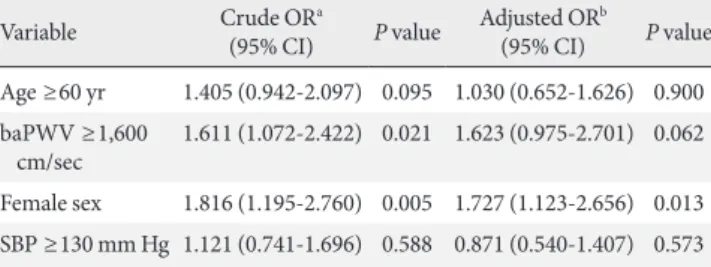
Univariate analysis and multivariate logistic regression analyses of risk factors for DPN such as age, blood pressure, gender, and baPWV were performed after conversion into bi- nary variables, and then odds ratios for each variable and CIs were calculated (Table 4).
The odds ratio of the baPWV ≥1,600 cm/sec was 1.611 (95%
CI, 1.072 to 2.422; P=0.021) and the odds ratio in females was 1.816 (95% CI, 1.195 to 2,760; P=0.005) in the univariate anal- ysis, which were both significantly higher than the control groups. However, the results of a multivariate logistic regres- sion analysis showed that female was independent risk factor of DPN.
When the associations among baPWV and symptoms or signs of DPN were evaluated, paresthesia and numbness in baPWV ≥1,600 cm/sec group was higher than <1,600 cm/sec group (10.1% vs. 5.7%, P=0.035; 13.5% vs. 6.9%, P=0.006, re- spectively) (Table 5). There was no significant difference in pain and burning. The monofilament test abnormality showed that the ≥1,600 cm/sec group (14.9%) was significantly higher Table 4. Univariate analysis and multiple logistic analysis for
the prediction of DPN in type 2 diabetics
Variable Crude ORa
(95% CI) P value Adjusted ORb
(95% CI) P value Age ≥60 yr 1.405 (0.942-2.097) 0.095 1.030 (0.652-1.626) 0.900 baPWV ≥1,600
cm/sec 1.611 (1.072-2.422) 0.021 1.623 (0.975-2.701) 0.062 Female sex 1.816 (1.195-2.760) 0.005 1.727 (1.123-2.656) 0.013 SBP ≥130 mm Hg 1.121 (0.741-1.696) 0.588 0.871 (0.540-1.407) 0.573 P values <0.05 were considered significant.
DPN, diabetic peripheral neuropathy; OR, odds ratio; CI, confidence interval;
baPWV, brachial-ankle pulse wave velocity; SBP, systolic blood pressure.
aSimple logistic regression, bMultiple logistic regression analysis (adjusted with other variables in the table).
There were more women in the patients with DPN compared to the patients without DPN (66.4% vs. 52.1%, P<0.01), and the patients with DPN were older (59.6±10.7 years vs. 56.4±
10.8 years, P<0.01), and had a higher systolic blood pressure (141.6±22.6 mm Hg vs. 135.6±17.2 mm Hg, P<0.01), and pulse pressure (59.4±13.4 mm Hg vs. 54.8±12.9 mm Hg, P<
0.01) than the patients without DPN. There was a significant difference in the percent of γ-linolenic acid (37.0% vs. 24.0%, P<0.01), one of the individual neuropathy treatment drugs, between the patients with/without DPN. The proportion of total neuropathy treatment drugs prescribed (67.2% vs. 48.8%,
Table 5. Prevalence of symptoms and signs of peripheral neuropathy in type 2 diabetics according to baPWV
Variable baPWV <1,600 cm/sec
(n=336) baPWV ≥1,600 cm/sec
(n=356) P value Age-adjusted
P valuea
TSS 0.00 (0.00-2.00) 1.00 (0.00-2.66) 0.067b -
TSS ≥2, % 28.3 36.8 0.017 0.088
Pain, % 38.7 43.5 0.216 0.481
Tingling or burning, % 11.3 12.1 0.813 0.851
Paresthesia, % 5.7 10.1 0.035 0.383
Numbness, % 6.9 13.5 0.006 0.026
Abnormal monofilament test, % 6.6 14.9 <0.001 0.009
Abnormal vibration sensation, % 22.0 20.5 0.643 0.798
Abnormal ankle jerk, % 1.2 1.7 0.753 0.464
Abnormal sign, % 27.1 33.1 0.097 0.329
TSS ≥2 and abnormal sign, % 13.4 19.9 0.025 0.081
TSS ≥2 or abnormal sign, % 47.0 53.1 0.128 0.268
TSS ≥2 or abnormal monofilament test, % 31.6 41.3 0.009 0.054
Values are presented as means±standard deviation for normally distributed data and as median (interquartile range) for non-normally distrib- uted data (TSS). P values <0.05 were considered significant.
baPWV, brachial ankle pulse wave velocity; TSS, total symptom score.
aCochran-Mantel-Hadnszel chi-square test, bMann-Whitney test.
than the <1,600 cm/sec group (P<0.001). There was no signif- icant difference in the vibration sensation and ankle reflex test between the two groups. Cases in which the total symptom score was 2 points or more, or the monofilament test abnor- mality, were significantly higher in the ≥1,600 cm/sec group (41.3%) compared to the <1,600 cm/sec group (31.6%) (P<
0.01). When Cochran and Mantel-Haenszel statistical meth- ods were used, after adjusting for age, numbness (P=0.026) and the monofilament test abnormality (P=0.009) were still significant.
DISCUSSION
The most common form of DPN is distal symmetry polyneu- ropathy (DSPN) [17]. Toronto Diabetic Neuropathy Expert Group proposed definitions of minimal criteria for DSPN [18].
1) Possible DSPN: The presence of symptoms or signs of DSPN
2) Probable DSPN: The presence of a combination of symp- toms and signs of neuropathy
3) Confirmed DSPN: The presence of an abnormality of nerve conduction and a symptom or symptoms or a sign or signs of neuropathy
The presence of an abnormality of nerve conduction was proposed to be the standard for confirmed diagnosis of DSPN [18]. However, the main pathological findings for early stage DPN are small nerve fiber damage [19,20], and this stage may not appear abnormality of nerve conduction reflected the function of large nerve fibers [21,22]. If nerve coduction is normal, a validated measure of small fiber neuropathy may be used for confirmed diagnosis of DSPN [18]. In this study, nerve conduction tests and small nerve dysfunction tests were not performed, so diagnosis could not be performed. Cases where the total symptom score is ≥2 and abnormal sensory functions are observed can be defined as probable DSPN.
Arterial pulse wave velocity reflects the stiffness of arteries, and is also an indicator of atherosclerosis [23]. By measuring baPWV, blood pressure and pulse wave velocity can be easily measured. The cutoff point of the baPWV for the prediction of cardiovascular disease based is varied, and a standard value has not been established. Kim and Kim [24] studied the rela- tionship between baPWV and the risk of cardiovascular dis- ease through a health screening of study subjects. baPWV
≥1,600 cm/sec was a independent risk factor for cardiovascu- lar disease defined with the systemic coronary risk evaluation
(SCORE) risk score [24]. Kim et al. [25] proposed that the cut- off point for the baPWV in type 2 diabetes patients with car- diovascular disease was 1,635 cm/sec (sensitivity 73%, speci- ficity 75%). In our study, a receiver operating characteristic curve was used for analysis, but significant cutoff values of baPWV could not be obtained. So the cutoff value was set at 1,600 cm/sec based on the previously mentioned study.
When the variables were compared between the patients with/without DPN, patients with DPN had higher baPWV and were more likely to have baPWV ≥1,600 cm/sec. This explains the association between baPWV and DPN. In a univariate analysis of the risk factors for DPN, the odds ratio was 1.611 (95% CI, 1.072 to 2.422; P=0.021) in cases where baPWV was
≥1,600 cm/sec, which is a meaningful risk factor for DPN. A multivariate logistic regression analysis was performed to ad- just for confounding variables and the adjusted odds ratio was 1.623 (95% CI, 0.975 to 2.701; P=0.062), which indicates that a baPWV ≥1,600 cm/sec was not an independent risk factor.
However, even though the P value was greater than 0.05, the 95% CI was 0.975 to 2.701; therefore, as a precaution, it was suspected that a baPWV ≥1,600 cm/sec is a risk factor.
The primary factors affecting baPWV are age, systolic blood pressure, and gender [12]. In a study on type 2 diabetes pa- tients, baPWV was significantly correlated with blood pres- sure, pulse pressure, age, waist circumference, and duration of diabetes [26]. In our study, when the ≥1,600 cm/sec group and the <1,600 cm/sec group were compared, gender, age, duration of diabetes, height, weight, waist circumference, systolic blood pressure, diastolic blood pressure, pulse pressure, and glycated hemoglobin were significantly different between the groups.
Lipid levels were similar in the groups. These were likely caused by the different effects of each atherosclerosis risk factor on PWV. In other words, age and blood pressure are independent factors that have a powerful effect on PWV, whereas total cho- lesterol and low density lipoprotein cholesterol have a minor effect on PWV [13,27]. Either PWV has a greater association with sclerosis of the blood vessels [28], or lipid metabolism has a greater association with atherosclerosis. Additionally, the increased prescription rate of lipid lowering drugs is thought to have a partial effect. Angiotensin II receptor blockers and antiplatelet agents were relatively widely prescribed for increas- es in arterial stiffness. Use of lipid lowering drugs, angiotensin II receptor blockers, and antiplatelet agents have been report- ed to lower hs-CRP [29,30], and it is estimated that there is no significant difference in hs-CRP between the two groups.
Antioxidant α-lipoic acid reduces oxidative stress in the pathogenesis of DPN and is effective in improving symptoms and in preventing neurovascular damage [31-33]. The total of neuropathy treatment drugs was higher in the DPN group and baPWV ≥1,600 cm/sec group, and there was no difference in the prescription of individual neuropathy treatment drugs in these groups. In cases where there are symptoms but there are no signs of early stage DPN, neuropathy treatment drugs may be used for prevention or to improve symptoms. This study is limited because it is a cross-sectional study, and patients may have experienced improved symptoms due to drugs taken pri- or to assessment, or as a result of being placed in the group without DPN. When comparing nerve fibers based on size, large nerve fibers transmit the sensation of light pressure and vibration, and small nerve fibers transmit the sensation of pain and temperature [34]. After DPN causes initial small nerve fi- ber damage, large nerve fibers become damaged, and damage to large nerve fibers is less common [19,20]. Typically, at the initial stages of the disease, pain and abnormal sensations oc- cur. Afterwards, sensation is lost, numbness occurs and pain and burning sensation can improve [35]. Monofilament tests are useful in measuring and detecting sensations of light pres- sure in large nerve fibers [36], and are inexpensive, easy to perform, and highly reproducible [37]. However, vibration sensory tests and ankle reflex tests are limited in their repro- ducibility. In this study, there was no increase in pain and burning sensations in baPWV ≥1,600 cm/sec group; however, there was a higher percent of numbness.
Among nerve function tests, monofilament tests showed a higher percent of abnormalities in the ≥1,600 cm/sec group compared to the <1,600 cm/sec group. Even after adjusting for age, the percent of the monofilament test abnormalities was significantly high. There were many patients in the ≥1,600 cm/sec group who showed relatively late stage symptoms of numbness and monofilament test abnormalities that reflected damage to large nerve fibers, which suggests that there is a correlation between baPWV and relatively advanced DPN.
There are several other limitations to this study. Nerve con- duction tests were not performed, so the diagnosis of DPN was used as clinical criteria. In order to confirm diagnosis, the To- ronto Diabetic Neuropathy Expert Group as well as Feldman et al. [38] claim that the Michigan Diabetes Neuropathy Score (MDNS) and a nerve conduction test (or validate measure of small fiber neuropathy) must be performed [18,37]. However, in this study, sufficient data of MDNS and nerve conduction
test could not be acquired due to the limitations of retrospec- tive studies. In addition, the baPWV need to do regression against neuropathy score. Thus, further research is required.
In conclusion, increased baPWV was correlated with DPN in patients with type 2 diabetes.
CONFLICTS OF INTEREST
No potential conflict of interest relevant to this article was re- ported.
ACKNOWLEDGMENTS
We thank Dr. Aaron I. Vinik for his review and advice.
REFERENCES
1. Boulton AJ, Gries FA, Jervell JA. Guidelines for the diagnosis and outpatient management of diabetic peripheral neuropathy.
Diabet Med 1998;15:508-14.
2. Carrington AL, Shaw JE, Van Schie CH, Abbott CA, Vileikyte L, Boulton AJ. Can motor nerve conduction velocity predict foot problems in diabetic subjects over a 6-year outcome peri- od? Diabetes Care 2002;25:2010-5.
3. Pascuzzi RM. Peripheral neuropathies in clinical practice. Med Clin North Am 2003;87:697-724.
4. Kumar S, Fernando DJ, Veves A, Knowles EA, Young MJ, Boulton AJ. Semmes-Weinstein monofilaments: a simple, ef- fective and inexpensive screening device for identifying dia- betic patients at risk of foot ulceration. Diabetes Res Clin Pract 1991;13:63-7.
5. Young MJ, Breddy JL, Veves A, Boulton AJ. The prediction of diabetic neuropathic foot ulceration using vibration percep- tion thresholds. A prospective study. Diabetes Care 1994;17:
557-60.
6. Vinik AI, Maser RE, Mitchell BD, Freeman R. Diabetic auto- nomic neuropathy. Diabetes Care 2003;26:1553-79.
7. Alexander CM, Landsman PB, Teutsch SM, Haffner SM; Third National Health and Nutrition Examination Survey (NHANES III); National Cholesterol Education Program (NCEP). NCEP- defined metabolic syndrome, diabetes, and prevalence of coro- nary heart disease among NHANES III participants age 50 years and older. Diabetes 2003;52:1210-4.
8. O’Neal DN, Dragicevic G, Rowley KG, Ansari MZ, Balazs N, Jenkins A, Best JD. A cross-sectional study of the effects of type
2 diabetes and other cardiovascular risk factors on structure and function of nonstenotic arteries of the lower limb. Diabe- tes Care 2003;26:199-205.
9. Cohn JN. Vascular wall function as a risk marker for cardio- vascular disease. J Hypertens Suppl 1999;17:S41-4.
10. van Popele NM, Grobbee DE, Bots ML, Asmar R, Topouchian J, Reneman RS, Hoeks AP, van der Kuip DA, Hofman A, Wit- teman JC. Association between arterial stiffness and athero- sclerosis: the Rotterdam Study. Stroke 2001;32:454-60.
11. Aso K, Miyata M, Kubo T, Hashiguchi H, Fukudome M, Fuku- shige E, Koriyama N, Nakazaki M, Minagoe S, Tei C. Brachial- ankle pulse wave velocity is useful for evaluation of complica- tions in type 2 diabetic patients. Hypertens Res 2003;26:807-13.
12. Kubo T, Miyata M, Minagoe S, Setoyama S, Maruyama I, Tei C.
A simple oscillometric technique for determining new indices of arterial distensibility. Hypertens Res 2002;25:351-8.
13. Benetos A, Waeber B, Izzo J, Mitchell G, Resnick L, Asmar R, Safar M. Influence of age, risk factors, and cardiovascular and renal disease on arterial stiffness: clinical applications. Am J Hypertens 2002;15:1101-8.
14. Yokoyama H, Shoji T, Kimoto E, Shinohara K, Tanaka S, Koya- ma H, Emoto M, Nishizawa Y. Pulse wave velocity in lower-limb arteries among diabetic patients with peripheral arterial dis- ease. J Atheroscler Thromb 2003;10:253-8.
15. Bastyr EJ 3rd, Price KL, Bril V; MBBQ Study Group. Develop- ment and validity testing of the neuropathy total symptom score-6: questionnaire for the study of sensory symptoms of diabetic peripheral neuropathy. Clin Ther 2005;27:1278-94.
16. Suzuki E, Kashiwagi A, Nishio Y, Egawa K, Shimizu S, Maega- wa H, Haneda M, Yasuda H, Morikawa S, Inubushi T, Kikkawa R. Increased arterial wall stiffness limits flow volume in the lower extremities in type 2 diabetic patients. Diabetes Care 2001;24:2107-14.
17. Vinik AI, Park TS, Stansberry KB, Pittenger GL. Diabetic neu- ropathies. Diabetologia 2000;43:957-73.
18. Tesfaye S, Boulton AJ, Dyck PJ, Freeman R, Horowitz M, Kem- pler P, Lauria G, Malik RA, Spallone V, Vinik A, Bernardi L, Valensi P; Toronto Diabetic Neuropathy Expert Group. Dia- betic neuropathies: update on definitions, diagnostic criteria, estimation of severity, and treatments. Diabetes Care 2010;33:
2285-93.
19. Donaghue VM, Giurini JM, Rosenblum BI, Weissman PN, Veves A. Variability in function measurements of three senso- ry foot nerves in neuropathic diabetic patients. Diabetes Res Clin Pract 1995;29:37-42.
20. Ziegler D, Mayer P, Wiefels K, Gries FA. Assessment of small and large fiber function in long-term type 1 (insulin-depen- dent) diabetic patients with and without painful neuropathy.
Pain 1988;34:1-10.
21. Holland NR, Crawford TO, Hauer P, Cornblath DR, Griffin JW, McArthur JC. Small-fiber sensory neuropathies: clinical course and neuropathology of idiopathic cases. Ann Neurol 1998;44:47-59.
22. Kennedy WR, Said G. Sensory nerves in skin: answers about painful feet? Neurology 1999;53:1614-5.
23. Laurent S, Boutouyrie P, Asmar R, Gautier I, Laloux B, Guize L, Ducimetiere P, Benetos A. Aortic stiffness is an independent predictor of all-cause and cardiovascular mortality in hyper- tensive patients. Hypertension 2001;37:1236-41.
24. Kim YK, Kim D. The relation of pulse wave velocity with fram- ingham risk score and SCORE risk score. Korean Circ J 2005;
35:22-9.
25. Kim HJ, Nam JS, Park JS, Cho M, Kim CS, Ahn CW, Kwon HM, Hong BK, Yoon YW, Cha BS, Kim KR, Lee HC. Usefulness of brachial-ankle pulse wave velocity as a predictive marker of multiple coronary artery occlusive disease in Korean type 2 di- abetes patients. Diabetes Res Clin Pract 2009;85:30-4.
26. Lee SW, Yun KW, Yu YS, Lim HK, Bae YP, Lee BD, Kim BH, Lee CW. Determinants of the brachial-ankle pulse wave veloc- ity (baPWV) in patients with type 2 diabetes mellitus. J Korean Endocr Soc 2008;23:253-9.
27. Kim YK, Lee MY, Rhee MY. A simple oscillometric measure- ment of pulse wave velocity: comparison with conventional tonometric measurement. Korean J Med 2004;67:597-606.
28. O’Rourke M. Mechanical principles in arterial disease. Hyper- tension 1995;26:2-9.
29. Ridker PM, Cushman M, Stampfer MJ, Tracy RP, Hennekens CH. Inflammation, aspirin, and the risk of cardiovascular dis- ease in apparently healthy men. N Engl J Med 1997;336:973-9.
30. Smith JK, Dykes R, Douglas JE, Krishnaswamy G, Berk S. Long- term exercise and atherogenic activity of blood mononuclear cells in persons at risk of developing ischemic heart disease.
JAMA 1999;281:1722-7.
31. Ziegler D, Hanefeld M, Ruhnau KJ, Meissner HP, Lobisch M, Schutte K, Gries FA. Treatment of symptomatic diabetic pe- ripheral neuropathy with the anti-oxidant alpha-lipoic acid. A 3-week multicentre randomized controlled trial (ALADIN Study). Diabetologia 1995;38:1425-33.
32. Reljanovic M, Reichel G, Rett K, Lobisch M, Schuette K, Moller W, Tritschler HJ, Mehnert H. Treatment of diabetic polyneu-
ropathy with the antioxidant thioctic acid (alpha-lipoic acid): a two year multicenter randomized double-blind placebo-con- trolled trial (ALADIN II). Alpha Lipoic Acid in Diabetic Neu- ropathy. Free Radic Res 1999;31:171-9.
33. Ziegler D, Schatz H, Conrad F, Gries FA, Ulrich H, Reichel G.
Effects of treatment with the antioxidant alpha-lipoic acid on cardiac autonomic neuropathy in NIDDM patients. A 4-month randomized controlled multicenter trial (DEKAN Study).
Deutsche Kardiale Autonome Neuropathie. Diabetes Care 1997;20:369-73.
34. Tavee J, Zhou L. Small fiber neuropathy: a burning problem.
Cleve Clin J Med 2009;76:297-305.
35. Boulton AJ, Malik RA, Arezzo JC, Sosenko JM. Diabetic so-
matic neuropathies. Diabetes Care 2004;27:1458-86.
36. Mason J, O’Keeffe C, McIntosh A, Hutchinson A, Booth A, Young RJ. A systematic review of foot ulcer in patients with type 2 diabetes mellitus. I: prevention. Diabet Med 1999;16:
801-12.
37. Bril V, Perkins BA. Validation of the Toronto Clinical Scoring System for diabetic polyneuropathy. Diabetes Care 2002;25:
2048-52.
38. Feldman EL, Stevens MJ, Thomas PK, Brown MB, Canal N, Greene DA. A practical two-step quantitative clinical and elec- trophysiological assessment for the diagnosis and staging of diabetic neuropathy. Diabetes Care 1994;17:1281-9.