Hypercalcemia and Extensive Osteolytic Lesion with Increased Plasma Prostaglandin E
2Level in a Child with
Acute Lymphoblastic Leukemia
Young-Ho Lee, M.D., Yeon-Jung Lim, M.D., Jung-Joon Bae, M.D., Jung-Yun Kim, M.D. and Jeh-Hoon Shin, M.D.
Department of Pediatrics, Hanyang University College of Medicine, Seoul, Korea
In this report, we present a rare case of childhood ALL with hypercalcemia and extensive osteolytic lesions. The case was a 7-year-old girl presenting with vomiting and aggravating bone pain. Radiologic examinations showed severe osteolytic lesions of the skull and extremities. Laboratory findings revealed low hemoglobin, normal WBC count with absent circulating blasts, and an increased serum calcium level.
Serum intact PTH and 1,25-(OH)2 vitamin D3 levels were below the normal ranges and parathyroid hor- mone-related peptide (PTHrP) was not detected, whereas serum levels of prostaglandin E2 were elevated.
The hypercalcemia resolved with specific antileukemic chemotherapy along with supportive care. The elevated plasma prostaglandin E2 levels decreased slightly after complete remission with induction chemotherapy. These findings suggest that increased plasma prostaglandin E2 levels may be one of the pathogenetic mechanisms responsible for the occurrence of hypercalcemia in this patient. (Korean J Hematol 2007;42:433-438.)
Key Words: Acute lymphoblastic leukemia, Prostaglandin E2, Hypercalcemia, Osteolytic lesion
433 접수:2007년 7월 27일, 수정:2007년 9월 27일
승인:2007년 10월 7일
교신저자:이영호, 서울시 성동구 행당동 산 17
133-792, 한양대학교 의과대학 소아청소년 과학교실
Tel: 02-2290-8383, Fax: 02-2297-2380 E-mail: [email protected]
Correspondence to:Young-Ho Lee, M.D.
Department of Pediatrics, Hanyang University College of Medicine 17, Haengdang-dong, Seongdong-gu, Seoul 133-792, Korea Tel: +82-2-2290-8383, Fax: +82-2-2297-2380
E-mail: [email protected] INTRODUCTION
Hypercalcemia is a rare complication in child- hood acute lymphoblastic leukemia (ALL), al- though it often presents with adult hematologic malignancies, such as multiple myeloma or adult T-cell leukemia/lymphoma.1,2) This presentation also commonly coincides with lytic bone lesions and absent circulating blasts.3) The underlying mechanism for hypercalcemia in malignancies is associated with paraneoplastic production of hu- moral factors, mainly parathyroid hormone-re-
lated peptide (PTHrP),4) or local osteolytic hyper- calcemia.5)
Here, we describe a case of childhood ALL pre- senting with hypercalcemia and extensive osteo- lytic lesions, which are probably mediated by in- creased plasma prostaglandin E2 levels.
CASE REPORT
A 7-year-old girl was admitted to the pediatric hematology-oncology service at our institution for evaluation of hypercalcemia and multiple osteo-
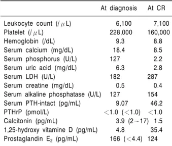
Table 1. Laboratory findings at diagnosis and upon com- plete remission (CR) of ALL
At diagnosis At CR Leukocyte count (/μL) 6,100 7,100
Platelet (/μL) 228,000 160,000
Hemoglobin (/dL) 9.3 8.8
Serum calcium (mg/dL) 18.4 8.5
Serum phosphorus (U/L) 127 2.2
Serum uric acid (mg/dL) 6.3 2.8
Serum LDH (U/L) 182 287
Serum creatine (mg/dL) 0.5 0.4 Serum alkaline phosphatase (U/L) 127 154 Serum PTH-intact (pg/mL) 9.07 46.2
PTHrP (pmol/L) <1.0 (<1.0) <1.0
Calcitonin (pg/mL) 3.9 (2∼17) 1.5 1,25-hydroxy vitamine D (pg/mL) 4.8 35.4 Prostaglandin E2 (pg/mL) 166 (<4.4) 124 Abbreviations: LDH, lactate dehydrogenase; PTH, parathy- roid hormone; PTHrP, parathyroid hormone-related peptide.
Fig. 1. Bone marrow aspiration (A: Wright stain, X1,000) and biopsy (B: H&E stain, X100). Nearly 30% of all hematopoietic cells were immature cells. The bone marrow section was hypocellular (40%) for her age and there were more immature cells than those in the aspirate. Immunophenotypic analysis was positive for CD45 and CD10, but negative for CD3, CD20, CD68, and CD79a. Cytogenetic analysis showed a normal female karyotype (46,XX).
limitation of motion with tenderness on the up- mg/dL, creatinine 0.5mg/dL, uric acid 6.3mg/dL, calcium 18.4mg/dL, and phosphorus 3.2mg/dL (Table 1). We performed bone marrow aspiration and biopsy (Fig. 1), which showed that nearly 30% of all hematopoietic cells were immature cells. These cells were medium to large in size with a high nuclear/cytoplasmic ratio, and a scanty bluish cytoplasm with fine nuclear chro- matin. The bone marrow section was hypocellular (40%) for her age and had more immature cells than those in the aspirate. The immunohisto- chemical staining was positive for CD45 and CD10, but negative for CD3, CD20, CD68, and CD79a. Cytogenetic study showed a normal fe- male karyotype (46,XX). The skeletal survey re- vealed multiple, extensive osteolytic lesions on the skull, upper and lower extremities, and pelvis (Fig. 2). MR imaging revealed a pathologic frac- ture on the metaphysis of the proximal humerus
Fig. 3. Simple x-ray and MR im- age of the left proximal hume- rus at diagnosis (A) and after CR (B). At diagnosis, a simple X-ray revealed an osteolytic le- sion of the left proximal hume- rus metaphysis. On MR imag- ing, an ill-defined low-signal in- tensity mass lesion was noted at the same level of the T1- weighted image coronal scan (A). However, simple x-ray and MR images of this lesion showed improvement after CR (B).
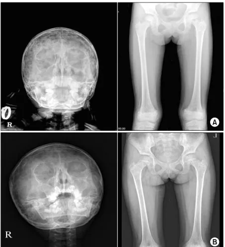
Fig. 2. Multiple osteolytic lesions at diag- nosis (A) and after CR (B). At diagnosis, multiple osteolytic lesions were seen in the skull, pelvic bone, both femurs, and both tibias (A), but after CR, the multiple osteo- lytic lesions were improved, but the osteo- porotic change remained (B).
(Fig. 3). A whole body bone scan showed an over- all decrease in activities of the growth plates, which were more severe at the left femoral head, right knee, and left humeral head (Fig. 4).
Further investigations to clarify the etiology of the hypercalcemia revealed the following (Table 1): 6.1pg/mL (9∼65) of intact parathyroid hor- mone (PTH), <1.0pmol/L (<1.0) of PTHrP, 3.9
Fig. 4. Whole body bone scan shows overall decreased ac- tivities of the growth plates, which were more severe at the left femoral head, right knee, and left humeral head.
Fig. 5. Clinical courses. The elevated calcium level was normalized after supportive care with the initiation of in- duction chemotherapy.
pg/mL (2∼17) of calcitonin, 4.8pg/mL (25∼45) of 1,25-(OH)2 vitamin D3, and 166pg/mL (<4.4) of prostaglandin E2.
Aggressive fluid therapy with furosemide and allopurinol slowly reduced the calcium and uric acid level. Immediately after the diagnosis of ALL, we started chemotherapy according to the standard risk protocol of the Children Cancer Group (CCG-1952), and the calcium and uric acid levels were rapidly normalized (Table 1, Fig.
5). The elevated plasma E2 level decreased slight- ly, yet not to normal after complete remission.
DISCUSSION
Hypercalcemia in childhood ALL is a rare
complication occurring at frequencies ranging from 0.6% to 4.8%.1,2,6) Hypercalcemia of malig- nancy is divided into two subgroups according to the underlying mechanisms for the elevated se- rum calcium levels. The first mechanism is local osteolytic hypercalcemia with osteolytic skeletal metastases and possible cytokine involvement.5) In multiple myeloma or adult T cell leukemia/
lymphoma, the primary mechanism for hyper- calcemia is increased osteoclastic bone resorption by tumor cells. The second subgroup of humoral hypercalcemia of malignancy is due to paraneo- plastic production of humoral factors, mainly parathyroid hormone-related peptide and some other factors such as vitamin-D-like sterols,7) pro- staglandin E2,8) tumor necrosis factor alpha
(TNF-α),9) and interleukin (IL)-6.10) TNF-α and IL-6 are known to promote osteoclastic bone re- sorption, but PTHrP-independent hypercalcemia with increased proinflammatory cytokines is rare- ly seen in childhood ALL.1,2) Osteoclasts are dif- ferentiated from hematopoietic progenitor cells in response to these bone-resorbing factors includ- ing prostaglandin E2, IL-6, and 1,25-(OH)2 vita- min D3, and increased osteoclasts results in in- creased bone resorption.
For our patient, serum intact PTH and 1,25- (OH)2 vitamin D3 levels were below the normal ranges and PTHrP was not detected, whereas se- rum levels of prostaglandin E2 were elevated be- fore treatment and then decreased after chemo- therapy. Based on these observations, we can spe- culate that the systemic overproduction of prosta- glandin E2, probably produced directly or in- directly by leukemic cells, may be the cause of the severe osteolytic lesions and hypercalcemia in our case. This suggests that hypercalcemia in our patient is associated with increased humoral fac- tor and prostaglandin E2, and is PTHrP-inde- pendent, which is rarely seen in childhood ALL.
In acute leukemia, some blasts can produce pros- taglandin E2,11) while in at least some in vitro sys- tems prostaglandin E2 appears to upregulate re- nal tubular 1-hydroxylation of 25-hydroxyvitamin D.12) Intravenous infusion of prostaglandin E2
was found to raise prlasma calcium concentra- tions in rats.13)
The clinical features of our patient, such as multiple osteolytic lesions with hypercalcemia and a normal white blood cell count without lym- phoblasts in the peripheral blood, are very sim- ilar to other reported cases.3,14) In our case, the hypercalcemia disappeared after chemotherapy.
In conclusion, we experienced a case of child- hood ALL with hypercalcemia and extensive os- teolytic lesions, which rapidly responded after the initiation of induction chemotherapy. The ele- vated plasma prostaglandin E2 levels, possibly re- lated to the hypercalcemia and bone lesions, de- creased after complete remission.
요 약
저자들은 구토와 골 통증을 주소로 내원한 7세 된 여자 환자에서 심한 골용해성병변과 고칼슘혈증을 동 반한 급성림프모구백혈병의 경우를 경험하였기에 보 고하는 바이다. 두개골과 사지의 방사선 소견상 심한 골용해성 병변이 있었다. 검사소견상 혈색소치가 감소 되어 있었고 백혈구수는 정상이면서 말초혈액에 모세 포는 발견되지 않았다. 혈청 칼슘은 증가되어 있었으 며, 부갑상성호르몬과 비타민 D3 농도는 약간 감소되 어 있었으나 부갑상선관련단백은 검출되지 않았고, 프 로스타글란딘 E2의 농도는 증가되어 있었다. 항암제치 료와 함께 보존적 치료로서 고칼슘혈증은 호전되었으 며, 프로스타글란딘 E2의 농도도 약간 감소되었다. 본 증례에서 고칼슘혈증의 원인은 증가된 프로스타글란 딘 E2의 농도가 관련이 있을 수 있다는 것을 시사한다.
REFERENCES
1) McKay C, Furman WL. Hypercalcemia complicating childhood malignancies. Cancer 1993;72:256-60.
2) Hibi S, Funaki H, Ochiai-Kanai R, et al. Hypercalce- mia in children presenting with acute lymphoblastic leukemia. Int J Hematol 1997;66:353-7.
3) Soni PN. Hypercalcemia and multiple osteolytic le- sions in childhood acute lymphoblastic leukaemia.
Postgrad Med J 1993;69:483-5.
4) Firkin F, Schneider H, Grill V. Parathyroid hor- mone-related protein in hypercalcemia associated with hematological malignancy. Leuk Lymphoma 1998;29:499-506.
5) Esbrit P. Hypercalcemia of malignancy - New in- sights into an old syndrome. Clin Lab 2001;47:67-71.
6) Wysolmerski JJ, Broadus AE. Hypercalcemia of ma- lignancy: the central role of parathyroid hormone- related protein. Ann Rev Med 1994;45:189-200.
7) Jacobson JO, Bringhurst FR, Harris NL, Weitzman SA, Aisenberg AC. Humoral hypercalcemia in Hodgkin's disease. Clinical and laboratory evalua- tion. Cancer 1989;63:917-23.
8) Todo S, Imashuku S, Inoda H, et al. Hypercalcemia in a case of childhood acute lymphoblastic leukemia.
Jpn J Clin Oncol 1987;17:357-62.
9) Antunovic P, Marisavljevic D, Kraguljac N, Jelusic V.
Severe hypercalcemia and extensive osteolytic lesions in an adult patient with T cell acute lymphoblastic
12) Wark JD, Taft JL, Michaelangeli VP, Veroni MC, Larkins RG. Biphasic action of prostaglandin E2 on conversion of 25-hydroxyvitamin D3 to 1, 25-dihy-
by lymphoblast-producing parathyroid hormone-re- lated peptide: a case report and review of the literature. Pediatr Blood Cancer 2005;45:333-9.