Prediction of survival by applying current prognostic models in diffuse large B-cell lymphoma treated with R-CHOP followed by autologous transplantation
Hong Ghi Lee
1, Sung-Yong Kim
1, Inho Kim
2, Yeo-Kyeoung Kim
3, Jeong-A Kim
4, Yang Soo Kim
5, Ho Sup Lee
5, Jinny Park
6, Seok Jin Kim
7, Hyeok Shim
8, Hyeon Seok Eom
9, Byeong-Bae Park
10, Junglim Lee
11, Sung Kyu Park
12, June-Won Cheong
13, Keon Woo Park
14Department of Internal Medicine, 1Konkuk University Medical Center, 2Seoul National University Hospital, Seoul, 3Chonnam National University Hwasun Hospital, Hwasun, 4The Catholic University of Korea College of Medicine, Suwon, 5Kosin University Gospel Hospital, Busan, 6Gachon University Gil Hospital, Incheon, 7Samsung Medical Center, Seoul, 8Wonkwang University School of Medicine, Iksan, 9National Cancer Center, Goyang, 10Hanyang University College of Medicine, Seoul, 11Daegu Fatima Hospital, Daegu, 12Soonchunhyang University Hospital, Bucheon, 13Yonsei University College of Medicine, Seoul, 14Dankook University Hospital, Cheonan, Korea
p-ISSN 2287-979X / e-ISSN 2288-0011 http://dx.doi.org/10.5045/br.2015.50.3.160 Blood Res 2015;50:160-6.
Received on May 21, 2015 Revised on July 16, 2015 Accepted on August 17, 2015
Background
Among the currently available prognostic models for diffuse large B-cell lymphoma (DLBCL), we investigated to determine which is most adoptable for DLBCL patients treat- ed with rituximab, cyclophosphamide, doxorubicin, vincristine, and prednisone (R-CHOP) followed by upfront autologous stem cell transplantation (auto-SCT).
Methods
We retrospectively evaluated survival differences among risk groups based on the International Prognostic Index (IPI), the age-adjusted IPI (aaIPI), the revised IPI (R-IPI), and the National Comprehensive Cancer Network IPI (NCCN-IPI) at diagnosis in 63 CD20-positive DLBCL patients treated with R-CHOP followed by upfront auto-SCT.
Results
At the time of auto-SCT, 74.6% and 25.4% of patients had achieved complete remission and partial remission after R-CHOP, respectively. As a whole, the 5-year overall (OS) and progression-free survival (PFS) rates were 78.8% and 74.2%, respectively. The 5-year OS and PFS rates according to the IPI, aaIPI, R-IPI, and NCCN-IPI did not significantly differ among the risk groups for each prognostic model (P-values for OS: 0.255, 0.337, 0.881, and 0.803, respectively; P-values for PFS: 0.177, 0.904, 0.295, and 0.609, respectively).
Conclusion
There was no ideal prognostic model among those currently available for CD20-positive DLBCL patients treated with R-CHOP followed by upfront auto-SCT.
Key Words Diffuse large B-cell lymphoma, Hematopoietic stem cell transplantation, Autologous transplantation, Rituximab, Prognostic groups
Correspondence to Hong Ghi Lee, M.D., Ph.D.
Division of Hematology-Oncology, Department of Internal Medicine, Konkuk University Medical Center, 120-1 Neungdong-ro, Gwangjin-gu, Seoul 05030, Korea
Tel: +82-2-2030-7538 Fax: +82-2-2030-7695 E-mail: [email protected]
Ⓒ 2015 Korean Society of Hematology
INTRODUCTION
Prior to the rituximab era, the International Prognostic Index (IPI) and the age-adjusted IPI (aaIPI) were developed by the International Non-Hodgkin’s Lymphoma Prognostic Factors Project to predict long-term survival for patients with aggressive non-Hodgkin’s lymphoma (NHL) [1]. Since
the advent of rituximab, Sehn et al. have proposed that the revised IPI (R-IPI) is a better predictor for 4-year pro- gression-free survival (PFS) and overall survival (OS) of dif- fuse large B-cell lymphoma (DLBCL) patients treated with rituximab, cyclophosphamide, doxorubicin, vincristine, and prednisone (R-CHOP) [2]. In addition, clinical data from the seven National Comprehensive Cancer Network (NCCN) member institutions demonstrated that the NCCN-IPI better
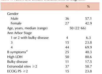
Table 1. Patient and disease characteristics at diagnosis.
N %
Gender
Male 36 57.1
Female 27 42.9
Age, years, median (range) 50 (22–66) Ann Arbor Stage
1 or 2 with bulky disease 4 6.3
3 15 23.8
4 44 69.9
B symptomsa) 25 40.3
High LDH 50 80.6
Bulky disease 11 17.5
Extranodal sites ≥2 37 58.7
ECOG PS ≥2 15 23.8
a)B symptoms indicate systemic symptoms such as fever, night sweats, and weight loss, which are associated with non-Hodgkin’s lymphoma.
Abbreviations: LDH, lactate dehydrogenase; ECOG, Eastern Coo- perative Oncology Group; PS, performance status.
discriminated low and high risk subgroups than the IPI for patients with DLBCL treated with rituximab-containing che- motherapy [3].
We confront uncertainty regarding the prognostic model that can differentiate the high risk group from the low risk group for patients with DLBCL treated with R-CHOP fol- lowed by upfront autologous stem cell transplantation (auto-SCT). Previously, we showed that the 5-year OS and PFS rates did not differ between the risk groups according to the aaIPI and R-IPI [4]. In this study, we aimed to de- termine which among the currently available prognostic models is most adoptable for DLBCL patients treated with R-CHOP followed by upfront auto-SCT.
MATERIALS AND METHODS
Data sources
Data were collected from the Korean Blood and Marrow Transplant Registry (KBMTR). The KBMTR is a voluntary organization comprised of 43 transplantation centers located in South Korea. The Transplant Registration Committee re- quires participating centers to submit detailed data from consecutive patients to the KBMTR. Informed consent is obtained on-site according to KBMTR regulations. The KBMTR database was used to identify adult patients with DLBCL who underwent upfront auto-SCT while in complete remission (CR) or partial remission (PR) after R-CHOP che- moimmunotherapy between January of 2005 and March of 2014. Additional data were obtained from each center to complete this study.
Patients
We analyzed data obtained from 63 CD20-positive DLBCL patients who underwent R-CHOP therapy followed by high-dose consolidation therapy with autologous stem cell rescue between January of 2005 and March of 2014 as re- ported to the KBMTR by 14 centers. Adult patients aged
≥20 years were included. In Korea, the majority of medical expenses are covered and tightly regulated by the National Health Insurance System. All types of hematopoietic stem cell transplantation, including auto-SCT, are reviewed in advance by the Health Care Review and Evaluation Committee. The regulations allow auto-SCT for patients ≤65 years old when their diseases are considered high risk.
Therefore, the majority of patients enrolled in this study were ≤65 years old and having Ann Arbor stage III or IV disease. Four stage I or II patients with bulky disease underwent upfront auto-SCT because their diseases were considered advanced. This study was approved by the institu- tional review board of Konkuk University Medical Center.
Assessment of responses
The 2007 revised guidelines of the International Harmoni- zation Project were adopted to describe the response criteria for DLBCL [5]. CR was defined as the complete disappearance of all detectable clinical evidence of disease and disease-
related symptoms if these were present before therapy. PR was defined as a ≥50% decrease in the sum of the product of the diameters (SPD) of up to 6 of the largest dominant nodes or nodal masses. Stable disease (SD) was defined as a case that failed to attain the criteria needed for CR or PR but did not fulfill those for progressive disease (PD).
OS was defined as the time interval from the date of diagnosis to the date of death as a result of any cause or to the last follow-up. PFS was defined as the time interval from the date of diagnosis to the date of lymphoma progression, relapse from CR, or death as a result of any cause.
Statistical analysis
The differences in the categorical variables among the study groups were analyzed with Pearson’s chi-square test and Fisher’s exact test. Survival curves were plotted using the Kaplan-Meier method and the confidence intervals were calculated using the standard error. The differences in surviv- al among the groups with respect to variables were analyzed with the log-rank test. The P-values reported were 2-sided;
a P< 0.05 was considered statistically significant. The stat- istical analyses were performed with the Statistical Package for the Social Sciences (SPSS) version 17.0 software (SPSS Inc., Chicago, IL).
RESULTS
Patient and disease characteristics
The patient and disease characteristics at the time of diag- nosis are summarized in Table 1. A total of 63 patients were evaluated in the study. The patients were classified as stage I/II with bulky disease (6.3%) or stage III/IV (93.7%). Bulky disease was defined by the presence of one of the following
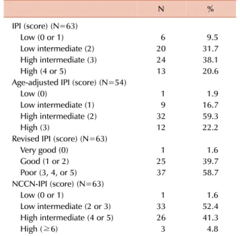
Table 2. The numbers and percentages of patients classified into the risk groups of each prognostic model.
N %
IPI (score) (N=63)
Low (0 or 1) 6 9.5
Low intermediate (2) 20 31.7
High intermediate (3) 24 38.1
High (4 or 5) 13 20.6
Age-adjusted IPI (score) (N=54)
Low (0) 1 1.9
Low intermediate (1) 9 16.7
High intermediate (2) 32 59.3
High (3) 12 22.2
Revised IPI (score) (N=63)
Very good (0) 1 1.6
Good (1 or 2) 25 39.7
Poor (3, 4, or 5) 37 58.7
NCCN-IPI (score) (N=63)
Low (0 or 1) 1 1.6
Low intermediate (2 or 3) 33 52.4
High intermediate (4 or 5) 26 41.3
High (≥6) 3 4.8
Abbreviations: IPI, International Prognostic Index; NCCN, National Comprehensive Cancer Network.
Table 3. Treatments and responses before auto-SCT.
N %
R-CHOP cycles
4 or 5 15 23.8
6 33 52.4
7, 8, or 9 15 23.8
Response to R-CHOP
CR 47 74.6
PR 16 25.4
RT before SCT Yes
Bulky disease 3 4.8
Remnant disease 3 4.8
No 57 90.5
Disease status at SCT
CR 47 74.6
PR 16 25.4
Abbreviations: auto-SCT, autologous stem cell transplantation;
R-CHOP, rituximab+cyclophosphamide+adriamycin+vincristine+
prednisolone; CR, complete remission; PR, partial remission; RT, radiation therapy; SCT, stem cell transplantation.
Table 4. Transplantation characteristics.
N %
Median time to SCT, months (range) 7.2 (3.4–40.3) Stem cell mobilization
Chemotherapy+G-CSF 60 95.2
G-CSF alone 3 4.8
Conditioning regimen
BU+CY+VP-16 20 31.7
BU+MEL+VP-16 22 34.9
MITO+VP-16+ARA-C+MEL 11 17.5
BCNU+VP-16+ARA-C+MEL 4 6.3
IFOS+CARB+VP-16 3 4.8
Others 3 4.8
CD34+ cell dose, ×106/kg (range) 5.3 (1.1–50.5) Median time to cell recovery after SCT
ANC, days (range) 12 (8–25)
PLT, days (range) 17 (2–391)
Abbreviations: SCT, stem cell transplantation; G-CSF, granulocyte- colony stimulating factor; BU, busulfan; CY, cyclophosphamide;
VP-16, etoposide; MEL, melphalan; MITO, mitoxantrone; ARA-C, cytosine arabinoside; BCNU, carmustine; IFOS, ifosfamide; CARB, carboplatin; CD, cluster of differentiation; ANC, absolute neutrophil count; PLT, platelet.
2 findings: (1) an abdominal node or nodal mass with a largest dimension of ≥10 cm as determined by an imaging study or (2) a mediastinal mass with a maximum width equal to or greater than one-third of the internal transverse diameter of the thorax at the T5/6 level as determined by a imaging study. At diagnosis, the IPI, R-IPI, and NCCN-IPI were available for 63 patients; however, the aaIPI was only available for 54 patients due to the age criteria. The numbers and percentages of patients classified into the risk groups of each prognostic model (IPI, aaIPI, R-IPI, and NCCN-IPI) are documented in Table 2.
Treatments before auto-SCT and transplantation characteristics
The treatments received by patients before auto-SCT and the transplantation characteristics are shown in Tables 3 and 4, respectively. The CR and PR rates following R-CHOP therapy were 74.6% and 25.4%, respectively. Six patients received involved field radiotherapy (IFRT) for bulky disease or remnant lymphoma prior to auto-SCT. One of the 6 patients receiving IFRT experienced an upgrade in response from PR to CR. However, the overall response rates were not improved at the time of auto-SCT. The median time from diagnosis to auto-SCT was 7.2 months (range, 3.4–40.3 months).
Response to treatment and outcomes
The median durations of follow-up after diagnosis and auto-SCT for all enrolled patients were 57.3 months (range, 6.3–110.7 months) and 46.3 months (range, 0.4–102.1 months), respectively. The median durations of follow-up after diagnosis and auto-SCT for the surviving patients were
60.2 months (range, 8.6–110.7 months) and 53.2 months (range, 3.6–102.1 months), respectively. During the fol- low-up period, 16 patients eventually relapsed after au- to-SCT. Four patients died of pneumonia or bacteremia, and 8 died of lymphoma relapse. The 5-year OS and PFS rates were 78.8±5.5% and 74.2±5.8%, respectively.
Survival rates according to the IPI at diagnosis
The 5-year OS rates of the patients in the low (N=6),
Fig. 1. Probability of overall survival (OS) (A) and progression-free survival (PFS) (B) after autologous stem cell transplantation according to the International Prognostic Index (IPI) score at diagnosis.
Fig. 2. Probability of overall survival (OS) (A) and progression-free survival (PFS) (B) after autologous stem cell transplantation according to the age-adjusted International Prognostic Index (aaIPI) score at diagnosis.
low intermediate (N=20), high intermediate (N=24), and high (N=13) risk groups were 100%, 71.2±11.0%, 86.7±7.2%, and 62.9±15.4%, respectively (P=0.255; Fig. 1A). The 5-year PFS rates of the patients in the same categories were 80±17.9%, 57.9±11.5%, 86.5±7.3%, and 67.1±13.5%, respectively (P=0.177;
Fig. 1B).
We reclassified the IPI risk groups into 2 groups: low and low intermediate vs. high intermediate and high. The 5-year OS rates of the patients in the low/low intermediate (N=26) and high intermediate/high (N=37) risk groups were 78.7±8.5% and 78.5±7.4%, respectively (P=0.997). The 5-year PFS rates of the patients in the same categories were 66.8±9.7% and 79.7±6.9%, respectively (P=0.186).
Survival rates according to the aaIPI at diagnosis
The 5-year OS rates of patients in the low (N=1), low intermediate (N=9), high intermediate (N=32), and high
(N=12) risk groups were 100%, 100%, 72.6±8.3%, and 62.5±
15.5%, respectively (P=0.337; Fig. 2A). The 5-year PFS rates of the patients in the same categories were 100%, 75.0±15.3%, 69.6±8.5%, and 66.7±1.36%, respectively (P=0.904; Fig. 2B).
We reclassified the aaIPI risk groups into 2 groups: low and low intermediate vs. high intermediate and high. The 5-year OS rates of patients in the low/low intermediate (N=10) and high intermediate/high (N=44) risk groups were 100% and 70.2±7.3%, respectively (P=0.078). The 5-year PFS rates of the patients in the same categories were 77.8±
13.9% and 68.8±7.2%, respectively (P=0.582).
Survival rates according to the R-IPI at diagnosis
The 5-year OS rates of the patients in the very good (N=1), good (N=25), and poor (N=37) risk groups were 100%, 77.7%, and 78.5±7.4%, respectively (P=0.881; Fig. 3A). The 5-year PFS rates of the patients in the same categories were 100%,
Fig. 3. Probability of overall survival (OS) (A) and progression-free survival (PFS) (B) after autologous stem cell transplantation according to the revised International Prognostic Index (R-IPI) score at diagnosis.
Fig. 4. Probability of overall survival (OS) (A) and progression-free survival (PFS) (B) after autologous stem cell transplantation according to the National Comprehensive Cancer Network International Prognostic Index (NCCN-IPI) score at diagnosis.
65.3±10.0%, and 79.7±6.9%, respectively (P=0.295; Fig. 3B).
We reclassified the R-IPI risk groups into 2 groups: very good and good vs. poor. The 5-year OS rates of the patients with very good/good (N=26) and poor risk (N=37) were 78.7±8.5% and 78.5±7.4%, respectively (P=0.977). The 5-year PFS rates of the patients in the same categories were 66.8±
10.3% and 79.7±6.9%, respectively (P=0.186).
Survival rates according to the NCCN-IPI at diagnosis The 5-year OS rates of the patients with low (N=1), low intermediate (N=33), high intermediate (N=26), and high risk (N=3) were 100%, 77.3±7.6%, 76.6±9.5%, and 100%, respectively (P=0.803; Fig. 4A). The 5-year PFS rates of the patients in the same categories were 100%, 66.6±8.7%, 79.3±8.3%, and 100%, respectively (P=0.609; Fig. 4B).
We reclassified the NCCN-IPI risk groups into 2 groups:
low and low intermediate vs. high intermediate and high.
The 5-year OS rates of the patients with low/low intermediate (N=34) and high intermediate/high risk (N=29) were 78.0±7.4% and 78.9±8.7%, respectively (P=0.742). The 5-year PFS rates of the patients in the same categories were 67.7±8.5% and 81.6±7.5%, respectively (P=0.401). The 5-year OS and PFS rates according to the IPI, aaIPI, R-IPI, and NCCN-IPI did not show statistically significant differences between the subgroups (P>0.05) when the subjects were stratified by each prognostic risk model.
DISCUSSION
High-dose chemotherapy with auto-SCT has been per- formed for curative purposes and is now the treatment of choice in patients with relapsed, chemotherapy-sensitive DLBCL. In addition, auto-SCT has been applied in patients
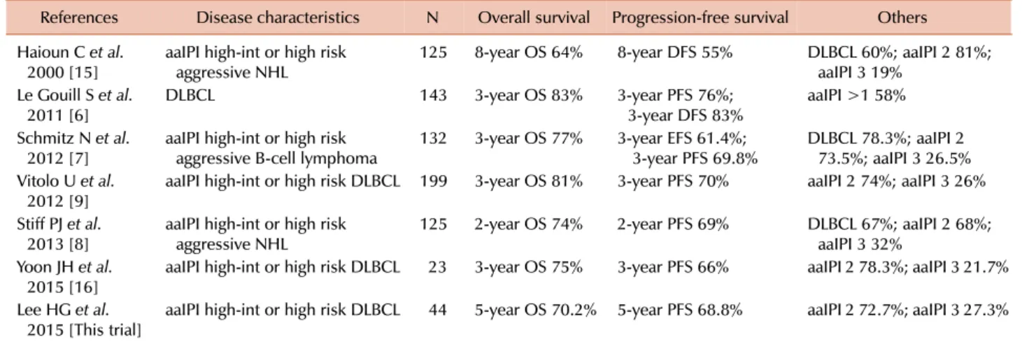
Table 5. Outcomes of high risk diffuse large B-cell lymphoma patients treated with upfront autologous stem cell transplantation.
References Disease characteristics N Overall survival Progression-free survival Others Haioun C et al.
2000 [15] aaIPI high-int or high risk
aggressive NHL 125 8-year OS 64% 8-year DFS 55% DLBCL 60%; aaIPI 2 81%;
aaIPI 3 19%
Le Gouill S et al.
2011 [6] DLBCL 143 3-year OS 83% 3-year PFS 76%;
3-year DFS 83% aaIPI >1 58%
Schmitz N et al.
2012 [7] aaIPI high-int or high risk
aggressive B-cell lymphoma 132 3-year OS 77% 3-year EFS 61.4%;
3-year PFS 69.8% DLBCL 78.3%; aaIPI 2 73.5%; aaIPI 3 26.5%
Vitolo U et al.
2012 [9] aaIPI high-int or high risk DLBCL 199 3-year OS 81% 3-year PFS 70% aaIPI 2 74%; aaIPI 3 26%
Stiff PJ et al.
2013 [8] aaIPI high-int or high risk
aggressive NHL 125 2-year OS 74% 2-year PFS 69% DLBCL 67%; aaIPI 2 68%;
aaIPI 3 32%
Yoon JH et al.
2015 [16] aaIPI high-int or high risk DLBCL 23 3-year OS 75% 3-year PFS 66% aaIPI 2 78.3%; aaIPI 3 21.7%
Lee HG et al.
2015 [This trial] aaIPI high-int or high risk DLBCL 44 5-year OS 70.2% 5-year PFS 68.8% aaIPI 2 72.7%; aaIPI 3 27.3%
Abbreviations: aaIPI, age-adjusted International Prognostic Index; high-int, high intermediate; NHL, non-Hodgkin’s lymphoma; OS, overall survival; DFS, disease-free survival; DLBCL, diffuse large B-cell lymphoma; PFS, progression-free survival; EFS, event-free survival.
with high risk aggressive NHL to improve the outcome of consolidative treatment. Recent randomized studies showed no significant survival benefit to upfront auto-SCT compared with rituximab-containing chemoimmunotherapy alone for aggressive B-cell lymphoma [6-8]. However, a prospective randomized trial performed by Vitolo et al. demonstrated that the 3-year PFS was significantly higher in the upfront auto-SCT group compared with the non-auto-SCT group in patients with high risk DLBCL [9]. The role of upfront auto-SCT remains to be established with long-term follow-up in patients with high risk DLBCL.
The IPI and aaIPI were developed to predict long-term survival for aggressive NHL, including DLBCL, before the introduction of rituximab to the medical field [1]. The IPI was also useful in predicting the outcome in patients with aggressive CD20-positive B-cell lymphoma treated with R-CHOP [10]. The IPI or aaIPI at diagnosis was found to be of value in predicting the OS and PFS in 25 patients with DLBCL who were treated with R-CHOP followed by upfront auto-SCT [11]. The 5-year OS and PFS of the high risk group were lower than those of the high intermediate risk group (P=0.04 and P=0.092, respectively). When the aaIPI was applied to 242 patients with relapsed CD20-positive DLBCL, an aaIPI score of 2 to 3 was a significant prognostic factor predicting 4-year OS and PFS after salvage auto-SCT (P<0.001) [12]. In contrast, a French prospective multi- center trial showed that 3-year PFS did not differ between the aaIPI high intermediate and high risk groups when 155 patients with DLBCL were treated with rituximab combined with ACVBP (doxorubicin, cyclophosphamide, vindesine, bleomycin, and prednisolone) and upfront auto-SCT [13].
Another retrospective study showed that the 5-year OS and PFS did not significantly differ between the high inter- mediate and high risk groups based on the aaIPI when 22 patients with DLBCL were treated with rituximab-contain- ing induction chemotherapy followed by upfront auto-SCT [14]. Here we summarize the representative survival data
of patients with aaIPI high intermediate or high risk ag- gressive NHL, including DLBCL, who were treated with upfront auto-SCT (Table 5) [7-9, 15, 16].
Sehn et al. reported that the R-IPI identified three distinct prognostic groups with a better predictive value of 4-year OS and PFS for patients with DLBCL treated with R-CHOP compared to the standard IPI [2]. The French trial, mentioned above, also analyzed the data regarding whether the out- comes were different when the subjects were stratified ac- cording to the R-IPI risk classification [13]. There were no differences in OS and PFS between the R-IPI good and poor risk groups.
Recently, the NCCN-IPI has been proposed as a model with better discrimination of 5-year OS and PFS for patients with DLBCL treated with rituximab-containing chemo- therapy as compared to the IPI for risk stratification [3].
So far, however, it has not been proved that the NCCN-IPI is a useful prognostic model for patients with DLBCL treated with rituximab-containing chemotherapy followed by up- front auto-SCT.
Because it was difficult to find an ideal prognostic model without controversy to predict outcomes for patients with DLBCL who were treated with chemoimmunotherapy fol- lowed by upfront auto-SCT, we applied 4 prognostic models to the prediction of survival in our study: the IPI, aaIPI, R-IPI, and NCCN-IPI. Unfortunately, none of the models demonstrated a statistically significant difference for OS and PFS among the risk groups when the patients were stratified by each risk classification.
In conclusion, the OS and PFS rates according to the IPI, aaIPI, R-IPI, and NCCN-IPI did not significantly differ among the subgroups. There was no ideal prognostic model among the established ones for CD20-positive DLBCL pa- tients who were treated with R-CHOP followed by upfront auto-SCT. A new prognostic model may be necessary to identify the patients who will gain the maximum benefit from upfront auto-SCT in the rituximab era.
ACKNOWLEDGMENTS
The clinical data for this study were collected from the Korean Blood and Marrow Transplantation Registry (KBMTR), the Korean Society of Blood and Marrow Transplantation.
AuthorsÊ Disclosures of Potential Conflicts of Interest
No potential conflicts of interest relevant to this article were reported.
REFERENCES
1. A predictive model for aggressive non-Hodgkin's lymphoma. The International Non-Hodgkin's Lymphoma Prognostic Factors Project. N Engl J Med 1993;329:987-94.
2. Sehn LH, Berry B, Chhanabhai M, et al. The revised International Prognostic Index (R-IPI) is a better predictor of outcome than the standard IPI for patients with diffuse large B-cell lymphoma treated with R-CHOP. Blood 2007;109:1857-61.
3. Zhou Z, Sehn LH, Rademaker AW, et al. An enhanced Interna- tional Prognostic Index (NCCN-IPI) for patients with diffuse large B-cell lymphoma treated in the rituximab era. Blood 2014;123:837-42.
4. Lee HG, Choi Y, Kim SY, et al. R-CHOP chemoimmunotherapy followed by autologous transplantation for the treatment of diffuse large B-cell lymphoma. Blood Res 2014;49:107-14.
5. Cheson BD, Pfistner B, Juweid ME, et al. Revised response criteria for malignant lymphoma. J Clin Oncol 2007;25:579-86.
6. Le Gouill S, Milpied NJ, Lamy T, et al. First-line rituximab (R) high-dose therapy (R-HDT) versus R-CHOP14 for young adults with diffuse large B-cell lymphoma: Preliminary results of the GOELAMS 075 prospective multicenter randomized trial. J Clin Oncol (ASCO Annual Meeting Abstracts) 2011;29(Suppl):abst 8003.
7. Schmitz N, Nickelsen M, Ziepert M, et al. Conventional chemo- therapy (CHOEP-14) with rituximab or high-dose chemotherapy (MegaCHOEP) with rituximab for young, high-risk patients with aggressive B-cell lymphoma: an open-label, randomised, phase 3 trial (DSHNHL 2002-1). Lancet Oncol 2012;13:1250-9.
8. Stiff PJ, Unger JM, Cook JR, et al. Autologous transplantation as consolidation for aggressive non-Hodgkin's lymphoma. N Engl J Med 2013;369:1681-90.
9. Vitolo U, Chiappella A, Brusamolino E, et al. Rituximab dose- dense chemotherapy followed by intensified high-dose chemo- therapy and autologous stem cell transplantation (HDC+ASCT) significantly reduces the risk of progression compared to standard rituximab dose-dense chemotherapy as first line treatment in young patients with high-risk (aa-IPI 2-3) diffuse large B-cell lymphoma (DLBCL): Final results of phase III randomized trial DLCL04 of the Fondazione Italiana Linfomi (FIL). Blood (ASH Annual Meeting Abstracts) 2012;120(Suppl):abst 688.
10. Ziepert M, Hasenclever D, Kuhnt E, et al. Standard International prognostic index remains a valid predictor of outcome for patients with aggressive CD20+ B-cell lymphoma in the rituximab era. J Clin Oncol 2010;28:2373-80.
11. Inano S, Iwasaki M, Iwamoto Y, et al. Impact of high-dose chemotherapy and autologous transplantation as first-line therapy on the survival of high-risk diffuse large B cell lymphoma patients: a single-center study in Japan. Int J Hematol 2014;99:
162-8.
12. Gisselbrecht C, Schmitz N, Mounier N, et al. Rituximab maintenance therapy after autologous stem-cell transplantation in patients with relapsed CD20(+) diffuse large B-cell lymphoma:
final analysis of the collaborative trial in relapsed aggressive lymphoma. J Clin Oncol 2012;30:4462-9.
13. Fitoussi O, Belhadj K, Mounier N, et al. Survival impact of rituximab combined with ACVBP and upfront consolidation autotransplantation in high-risk diffuse large B-cell lymphoma for GELA. Haematologica 2011;96:1136-43.
14. Takasaki H, Hashimoto C, Fujita A, et al. Upfront autologous stem cell transplantation for untreated high-risk diffuse large B-cell lymphoma in patients up to 60 years of age. Clin Lymphoma Myeloma Leuk 2013;13:404-9.
15. Haioun C, Lepage E, Gisselbrecht C, et al. Survival benefit of high-dose therapy in poor-risk aggressive non-Hodgkin's lymphoma: final analysis of the prospective LNH87-2 protocol--a groupe d'Etude des lymphomes de l'Adulte study. J Clin Oncol 2000;18:3025-30.
16. Yoon JH, Kim JW, Jeon YW, et al. Role of frontline autologous stem cell transplantation in young, high-risk diffuse large B-cell lymphoma patients. Korean J Intern Med 2015;30:362-71.