Vol.12, No.2, April, 1999
불안정성 콜레스 골절에서 외고정장치 시술과 경피성 핀고정술 치료결과 비교
윤석웅・황태성・이종행
서울 적십자 병원 정형외과
= Abstract =
External fixation versus percutaneous pinning for unstable Colles’f r a c t u r e
Suk-Woong Yoon, M.D., Tae-Sung Hwang, M.D., Jong-Haeng Lee, M.D.
Department of Orthopaedic Surgery, Seoul Red Cross Hospital, Seoul, Korea.
The twenty-three cases of unstable Colles fracture were treated from Jan. 1994 to May 1998 at the department of orthopaedic surgery of Seoul Red Cross Hospital. Among them, the sixteen cases were treated with closed reduction with percutaneous pinning, others were treated with external fixator(Ace colles quadrilateral devices, USA). A retrospective study was made and evaluated using the Modification of Gartland and Werley’s scoring system.
The result of this study were as follow:
1. The ratio of male to female was 9 cases(39%) to 14 cases(61%).
2. The causes were falling down from a height 12 cases(50%), slip down 8 cases(35%) and traffic accident 3 cases(13%).
3. The reduction loss did not occur with the patients treated external fixation using Ace colles qredrilateral device, however three cases of the reduction loss have occurred with the patients using percuteneous pinning.
4. According to the Modification of Gartland and Werley’s scoring system, the results of
※통신저자 : 윤석웅
서울특별시종로구평동164 (110-102) 서울적십자병원정형외과
Tel : (02) 398 - 9441 Fax : (02) 398 - 9439
서 론
콜레스 골절8 )은 요골 원위부 1 . 5인치내의 후측방 전위골절로신전외력에의해생기며, 성인골절의약 1 0 %와요골 원위부 골절의 9 0 %를 차지하여 가장흔 한 골절중의 하나이다3 5 ). 원위요골골절이 응급실에 서모든골절의약 2 0 %를차지하는가장흔한골절이
나2 6 )분류, 치료 및 방사선과 기능적 결과의 연관성
에는 논란이 많다2 2 ). 그럼에도 불구하고, 최근에 골 절형을 고려하여 불안정 분쇄골절에 대한 부가고정 과함께다른치료계획안이발전되어왔다. 콜레스골 절은 대부분5 0대여성에많다고보고되고있으며이 는 골다공증과 활동성이 많은 나이로 수상의 기회가 많아발생되며골절의 양상또한불완전하고심한분 쇄골절로 흔히나타난다. 이에 대한치료법은다양하 며, 가장많이권고되는방법으로 K -강선고정법및석 고붕대 또는 외고정기구에 의한 외고정법이다1 , 1 1 , 2 1 ). 본서울적십자병원정형외과에서는 5 0세이상환자 의 불안정 분쇄상 골절 2 3례에 대해 도수정복 후 경 피성 K -강선 고정술과 외고정 장치인 Ace Colles quadrilateral frame devices 고정법에 의한 방사선 그리 고임상적 치료결과를비교검토하여문헌고찰과함 께보고하는바이다.
대상 및 방법
연령대상은폐경기이후 5 0세이상 환자를대상으 로 1 9 9 4년 1월부터 1 9 9 8년 5월까지서울적십자병원 정형외과에서 원격조사가 가능한 5 0세 이상의 콜레 스골절 환자 총 2 3례 중 도수정복과 경피성 K -강선
고정술을이용하여치료한1 6례와외고정장치인A c e colles quadrilateral device로 치료한 7례를 대상으로 하 였다.
1. 연령및성별분포
Colles 골절 환자의 5 0세 이상에서 연령분포는 5 0 - 60 대가 1 2례( 5 2 % )로 가장 많았으며, 남녀 발생비율 은 남자가 9례(39%), 여자가 1 4례( 6 1 % )로 여자가 많 았다. 좌우빈도는우측이1 3례로좌측보다많았다.
2. 골절의분류
완관절면으로부터2.5cm 이내에발생한모든신전 성 골절을 콜레스 골절로 정의하였고 Gartland &
Werley 방법1 4 )으로 분류하였다(Table 1). 본 논문에서 대상으로 한불안전성골절은 심한분쇄골절로서 관 절을 침범하였으며 도수정복후 석고고정에 의하여 해부학적 위치를 계속 유지하기 어려운 Gartland and Werley 분류 3군(intra-articular , displaced)을 예로 정하 였다.
3. 수상원인
수상원인은Gartland & Werley 분류 3군에해당되는 총 2 3례중추락사고가1 2례( 5 2 % )로가장많았고실족 사고 8례(35%), 교통사고가3례( 1 3 % )였다.
exteral fixation were excellent 4 cases(58%), good 1 case(14%), fair 1 case(14%) and poor 1 case(14%). and for percutoneous pinning, excellent 6 cases(38%), good 4 cases(25%), fair 2 cases(12%) and poor 4 cases(25%).
5. The complications of cases using external fixation were pin site infection 1 case and wrist stiffness 1 case, but for percutaneous pinning, reduction loss 3 cases. pin site infection 2 cases, wrist stiffness 2 cases, and decreased external rotation of forearm 3 cases.
Key Words: Colles’fracture, external fixation, percutaneous pinning.
Table 1.Classification of Gartland & Werley
Stage
1 extra-articular, displaced
2 intra-articular, undisplaced
3 intra-articular, displaced
4. 골절의치료방법
Ace Colles quadrilateral frame device에의한외고정법 은 부분마취나 전신마취후 수지견인장치에 의한 8 - 12 pound로 5 - 1 0분간부드럽게원위부골절편을견인 하여 수상부 피질면의 해부학적 정렬과 요골길이를 회복하고 원위요척관절의 정복을 확인한후 만족할 만한정복을얻었을때첫번째원위부핀은신전근을 피하여 제 3중수골 기저부에 약간의 절개후 삽입하 였다. Colles frame을 부착후 두 번째 핀은 첫 번째 핀 에대해 6 0 - 9 0도각도로요골골절부위로부터근위부 7-8cm 위치에 삽입하였고 세 번째는 제 2 중수골 기 저부에 네 번째 핀은 두 번째와 같은 위치에서 척골 에삽입하여고정하였다. 해부학적정복과핀고정상 태는 C - a r m으로전후방과측면방사선상에서확인하 였으며또한요측길이, 요골관절면경사와전방경사 의 회복을 확인하였다. 6-8주 후에 외고정 장치를제 거한후 2 - 3주간단상지부목으로고정하면서간헐적 완관절운동을 시작하였으며 단상지 부목제거후 적 극적인 완관절운동을 시작하였다. 도수정복후 경피 적 핀 삽입방법은 부분마취나 전신마취하에서 무균
조작으로 C - a r m하에서도수정복을실시하고 정복을 확인한후 K -강선으로 골절부를 고정하였다. K-강선 의 크기는 1 . 3 m m와 1 . 6 m m를 이용하였으며 대개 2 - 3 개의 K -강선으로유지가가능하였다. 첫번째 K -강선 은요골경상돌기부위에서요골장축에대해 4 0 - 6 0도 및 전방 3 0도 방향으로 삽입하였고 두 번째와 세 번 째 K -강선은 불안정성과 분쇄골절의 정도에 따라 ulnar pinning등다양한방법으로삽입하였다. K -강선 삽입시 표재성 요골 동맥과 신경 및 건의 손상은 피 하였으며 K -강선은 골절부를 지나 반대편 골피질을 통과하도록 하였다. 삽입후 골절의 안정성은 C - a r m 하에서 관절의 굴곡 및 신전에 의한 운동성 여부로 판정하였고 U자 석고부목으로 고정하였다. 완관절 은 1 0 - 2 0도굴곡위치로고정하였으며가능한 수지운 동은 조기에 실시하였다. 술후 3 - 4주 경에 단상지 석 고부목으로바꾸었으며 술후 6 - 7주경에석고부목을 제거하고 K -강선을 제거한후 완관절 운동을 시작하 였다.
Table 2. Degree of final range of motion
Op. method Ace colles qudrilateral Percutaneous device (degree) pinning(degree) Dorsiflexion 60.2(35-70) 57.8(35-70) Plantar flexion 53.4(35-70) 50.3(30-60)
Pronation 66.5(40-80) 65.6(40-80)
Supination 65.2(40-80) 63.5(40-80) Ulnar deviation 18.8(15-20) 18.2(15-20) Radial deviation 21.7(15-30) 18.3(15-25)
Table 3.Average of final range of motion (percentage of unaffected side)
Op. method Ace colles qudrilateral Percutaneous device (degree) pinning (degree) Dorsiflexion-
palmar flexion 77 75
Pronation-supination 86 81
Ulnar-radial deviation 89 86
Table 4.3 cases treated with percutaneous pinning of alignment loss between Postop.
Table 4.and final follow up is shown in reontgenogram.
Cases dorsal angle (degree) radial angle (degree) radial length (mm) Postop. final change Postop. final change Postop. final change case 1 4 3 1 19 15 4 10 6 4 case 2 3 2 1 18 13 5 9 6 3 case 3 5 3 2 22 15 8 11 8 3
결 과
추시과정에서 관절의 동통유무와 치료된 관절과 건측관절의 운동범위를 신전, 굴곡, 회내전, 회외전, 척사위, 요사위로분류하여각도를측정하였으며치 료된 관절운동 범위를 비교하여 백분율로 표시하였 다(Table 2,3). 마지막추시결과완관절평균운동범위 는 Ace Colles quadrilateral devices 고정술에서 신전 Table 5Modified Gartland and Werley scoring system to evaluate Colles’fracture
Result Points Residual deformity 1 Residual dorsal tilt 2 Radial deviation of hand 2-3 Maximum 6 Subjective evaluation
Excellent - no pain, disability, or motion limitation 0 Good - occasional pain, slight limitation, no disability 2 Fair - occasional pain, some limitation, some disability 4 Poor - pain, limitation of movement, marked disability 6 Maximum 6 Objective evaluation 5
Loss of dorsiflexion < 45° 3
Loss of ulnar deviation < 30° 2
Loss of supination < 50° 1
Loss of palmar flexion < 30° 1
Loss of radial deviation < 15° 1
Pain in the distal radioulnar joint 1 Maximum 14 Complications
Arthritic change
Minimal 1 Moderate 2 Severe 3 Nerve complications 1-3 Poor finger function 1-2 Grip strength < 50% normal side 1 Poor finger function 1-2 Maximum 14 Point range on a deduction scale
Excellent 0-2 Good 3-8 Fair 9-20 Poor 21+
Table 6.Overall results of treatment with external fixation & percutaneous pinning for colles’fracture
Result\Op. method Ace colles Percutaneous quadrilateral device pinning
Excellent 4 6
Good 1 4
Fair 1 2
Poor 1 4
6 0 . 2°(35-70), 굴곡 5 3 . 4°(35-70), 회내전 6 6 . 5°(40-80) , 회외전 6 5 . 2°( 40-80) , 척사위 1 8 . 8°(15-20), 요사위 2 1 . 7°( 1 5 - 3 0 )이며 경피적 K -강선 고정술에서는 신전 5 7 . 8°(35-70), 굴곡 5 0 . 6°(30-65), 회내전6 5 . 6°(40-80), 회 외전 6 3 . 5°(40-80), 척사위 1 8 . 2°(15-20), 요사위 2 0 . 6°
( 1 5 - 2 5 )로 나타났다 . 방사선 관찰은 G a r t l a n d와 W e r l e y1 4 ), Van Der Linden과 Ericson 방법3 4 )에 의해서 측면방사선상에서 요골관절면 경사각과 요골길이 를 측정한 후, 방사선 촬영의 결과를 술후와 마지막 추시로 나누어서 표시하였다. 마지막 추시결과는
Ace Colles quadrilateral devices 고정술에서요골관절면 경사각이 평균 2 0 . 6°( 1 6 - 2 5°), 요골길이는 평균 8.3(5.2-14.1), 수장측 경사각은 평균 9 . 3°( - 1 2 - 1 6 )였고 경피적 K -강선 고정술에서는 요골관절면 경사각이 평균 1 9 . 2°(14-27), 요골길이는 평균 8.5mm( 4.8-14.5), 수장측경사각이평균 8 . 8°(-14 - 17)로나타났다. 방사 선상정복소실은방사선 촬영의결과를술후와마지 막 추시로 나누어서 표시하였는데 Ace Colles quadrilateral devices 고정술에서는없었으나 도수정복 및경피성 K -강선고정술에서는3례에서정복소실을 Table 7.합병증
ACQD* Percutaneous pinning Total
Reduction loss 3 3
Pin site infection 1 1 2
Wrist stiffness 1 2 3
Decreased exteranl rotaition of forearm 2 3
Total 2 9 11
* Ace colles quadrilateral device
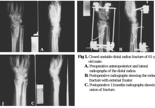
A
C
B
Fig 1.Closed unstable distal radius fracture of 61-year- old male.
Fig A.Preoperative anteroposterior and lateral radiographs of the distal radius.
Fig B.Postoperative radiographs showing the reduced fracture with external fixator.
Fig C.Postoperative 11months radiographs showing union of fracture.
보였다(Table 4).
치료 성적
치료성적은 주관적 객관적 판정을 겸한 s a r m i e n t o 에 의한 Modification of Gartland and Werley’s scoring s y s t e m으로평가하였다(Table 5). 최종평가는점수의 합계에의해 0 - 2점은우수, 3-8점은양호, 9-20점은보 통, 그리고 2 1점 이상은 불량으로 하였으며, Ace colles quadrilateral device 고정술은우수 4례(58%), 양호 1례(14%), 보통 1례(14%), 불량 1례(14%), 경피성K -강 선고정술에서는우수 6례(38%), 양호 4례(25%), 보통 2례(12%), 불량은 4례( 2 5 % )로 나타났다. Ace colles quadrilateral device 고정술 에서 예후가 불량했던 1례 는분쇄가심하고 수근골탈구가동반되었던경우로 요수근관절고정술로치료하였으며, 경피성 핀고정 술에서 우수의 예들은 ulnar pinning등 안정된 정복을 유지할수있는부가고정술을시행했던환자들이였 으며, 불량의예들은골조송증과분쇄가심했던환자
들이었다(Table 6).
합병증
술후합병증은 Ace colles quadrilateral devices 고정술 을 시행한 예에서 핀삽입부 감염 1례, 완관절강직 1 례였으며 경피성 K -강선 고정술에서는 강선의 전이 와 정복소실 3례, 핀삽입부 감염 1례, 완관절 강직 2 례, 전박의 외회전 운동의 감소가 3례 였다. 감염은 K -강선과외고정장치제거후완쾌되었다(Table 7).
고 찰
콜레스 골절8 )은 1 8 1 4년에 처음 기술되었으며, 신 전외력에 의하여 발생하여 전위가 일어났을 경우로 정의하였으나 현재는 비전위성 골절과 분쇄 관절내 골절도포함하게되었다.
Gantland 와 W e r l e y1 4 )는 골절선의 관절침범여부와
A
C
B
Fig 2. Closed unstable distal radius fracture of 61-year- old female.
Fig A.Preoperative anteroposterior and lateral radiographs of the distal radius.
Fig B.Postoperative radiographs showing the reduced fracture with external fixator.
Fig C.Postoperative 8weeks radiographs showing union of fracture.
A
C
B
Fig 3.Closed unstable distal radius fracture of 71-year- old female.
Fig A.Preoperative anteroposterior and lateral radiographs of the distal radius.
Fig B.Postoperative radiographs showing the reduced fracture with percutaneous pinning.
Fig C.Postoperative 7weeks radiographs showing reduction loss and union of fracture.
A
C
B
Fig 4.Closed unstable distal radius fracutre of 51-year- old female.
Fig A.Preoperative anteroposterior and lateral radiographs of the distal radius.
Fig B.Postoperative radiographs showing the reduced fracture with percutaneous pinning.
Fig C.Postoperative 2months radiographs showing reduction loss and union of fracture.
골절편의 전위정도에 따라 3군으로 나누었으며 , F r y k m a n n1 3 )은골절의 양상에 따른 치료방법및 예후 를제시하기위하여 관절의침범여부와척골의경상 돌기의 골절 동반여부에 따라 분류하였고 M e l o n e2 4 ) 은 관절내 골절을 4형으로 분류하여 각각의 치료법 을제시하고요골내측면의골절을보다적극적인방 법으로고정하여우수한기능적 결과를얻으려고하 였다.
골절의기전은 대부분과신전및압박력으로 일어 난다고하였으며이때압박력은관절내골절을유발 시킨다고 하였다. 즉 월상골에 의해서 압박력이 골 절에 전달되어 대부분의 관절면을 침범한다고 하였
다23 ,29).
콜레스골절중 관절외 원위요골 골절은 도수정복 및석고고정으로대부분만족할만한 결과를얻을수
있으나3 , 4 , 1 8 , 1 9 )불안정하고 심한 분쇄 관절내 골절은
치료가어렵다2 , 3 , 5 , 1 5 , 2 0 ).
S o l g a a r d ,3 3 )V i l l a r3 5 )그리고 A b b a s z a d e g e n1 )은요골단 축을 콜레스골절의 주된 골절변형이라고 하였으며, 불안정 분쇄원위요골골절에서 변형을 막기위한 치 료법으로 많은 저자들은 외고정장치를 권고하였다
1 , 1 1 , 2 1 ). 그러나 관절강직, 핀삽입부감염, 핀이완 그리
고 반사성 교감신경 이영양증 (reflex sympathetic d y s t r o p h y )등합병증이보고되었다7 , 3 0 ). 경피성K -강선 고정술은처음 G r e e n1 6 )에의해간단하고값싼시술로 권고하였으며, Habernek , Pritcheff and Lenoble등은 다 양한 핀 고정술로 좋은 결과를 보여 주었다. 그러나 다른저자들은 특히, 불안정한 분쇄관절내골절에서 경피성 K -강선 고정술로는 골절골편들의 재전이를 막을 수 없다고하였다. Cooney1 0 )등은 금속외고정이 좋다고보고하였으며, 예후에있어서수상당시전위 정도, 분쇄정도, 관절침범이심할수록결과가저조할 것이라고 하였으며 약 3 1 %에서 불량한 결과를 보고 하였다. 본저자들은 Ace colles quadrilateral device고정 술에서 불량이 14%, 경피성K -강선 고정술에서 1 9 % 로 두 방법 모두에서 다른 저자들에 의해 보고된 것 보다 좋은 결과를 얻었으며, Saffar and Cooney,2 6 ) Fernadez and Jupiter1 2 )의보고에서와같이 외고정장치 에서는골편재전이에의한정복소실이없었으나, 경 피성 K -강선 고정술에서는 골편 재전이에의한 정복 소실이 3례에서 발생하였다. 저자들은 골조송증과
분쇄가심한관절내골절환자의치료에서정복의유 지는 외고정장치에 의한 치료법이 더 좋은 것으로 사료된다. 최근에는 과다신연과 정복소실의 문제들 은 경피성 K -강선 사용과 함께 외고정술,6 , 3 1 )연골하 골이식술(Subchondral bone grafting)2 3 )의 보강술식이 권고되고있다.
합병증은 콜레스 골절중에서도 골조송증과 분쇄 가심한관절내골절환자에서이의유지에각별한관 심이요구되며여러합병증이발생하게된다. 저자들 은 Ace colles quadrilateral device 외고정에서핀삽입부 감염1례, 완관절강직1례가발생하였으며, 경피성K - 강선 고정술에서는 정복소실 2례, 핀 삽입감염 1례, S u d e k’s atrophy 1례, 완관절강직 2례, 전박의 외회전 감소 2례를경험하였다.
결 론
1 9 9 4년 1월부터 1 9 9 8년 5월까지 4년 5개월간 서울 적십자병원 정형외과에서경험한 5 0세이상불안정 분쇄상 관절내 콜레스 골절 2 3례에 대해 Ace Colles quadrilateral devices 외고정술과경피성 K -강선고정술 로 치료한후 S a r m i e n t o에의한 Modification of Gartland and Werley’s scoring system으로 평가하여 다음과 같 은결과를얻었다.
1. G a r t l a n d와 W e r l e y의주관적및 객관적평가에 의 한결과는 Ace Colles quadrilateral 고정술은우수 3 례(57%), 양호가2례(29%), 보통 1례(14%), 불량 1 례( 1 4 % )였으며 도수정복 및 K -강선 고정술에서 는우수 7례(44%), 양호 4례(25%), 보통 2례( 1 2 % ) , 불량 3례( 1 9 % )로 Ace Colles quadrilateral devices 고 정술로치료한예에서더좋게평가되었다 2. Ace Colles quadrilateral devices 외고정술에의한합
병증은핀삽입부감염 1례, 완관절강직 1례였으 나도수정복및경피적 K -강선고정술에서는 정 복소실 3례, 핀 삽입부 감염 1례, 완관절 강직 2 례, 전박의외회전운동의감소가 3례었다.
이상으로5 0세이상고령의콜레스골절에서골조 송증및관절내분쇄정도가심한경우에는 Ace Colles quadrilateral device 고정술이도수정복및경피적 K -강 선고정술에비해 Modification of Gartland and Werley’s
scoring system평가에서 더 좋은 결과로 나타났으며 합병증또한적어좋은치료방법이라사려된다.
R E F E R E N C E S
1) Abbaszadegan H, Jonsson U, Von Sivers K..
Prediction of instability of Colles’fractures. Acta Orthop Scand., 60:646-50, 1989.
2) Agee JM, Distal radius fractures: multiplanar ligamentotaxis. Hand Clin., 9:577-585, 1993.
3) Agee JM, Szabo RM, Chidgey LK, King FC, Kerfoot C. Treatment of comminuted distal radius fractures: an approach based on pathomechanics.
Orthopedics., 17:1115-1122, 1994.
4) Axelrod TS, McMurty RY. Open reduction and internal fixation of comminuted, intraarticular fractures of the distal radius. J Hand Surg, 15A:1 - 11, 1990.
5) Bassett RL. Displaced intraarticular fractures of the distal radius. Clin Orthop., 214:148-152, 1987.
6) Braun RM, Gellman H.. Dorsal pin placement and external fixation for correction of dorsal tilt in fractures of the distal radius. J Hand Sur., 19A:6 5 3 - 655, 1994.
7) Clyburn TA. Dynamic external fixation for comminuted intra-articular fractures of the distal end of the radius. J Bone Joint Surg(Am), 69:2 4 8 - 5 4 , 1 9 8 7 .
8) Colles, A. On the fracture of the carpal extremity of the radius. Edinb Med Surg J., 10:182-186, 1814.
9) Cooney WP.: Extrenal fixation of distal radial fractures. Clin Orthop., 180:44-49, 1983.
10) Cooney, WP Ⅲ , Dobyns JH, Linscheid, R.L.
Complication of Colles’fracture. J. Bone Joint Surg., 62A:613, 1980.
11) E d i t o r i a l . Differentiated treatment of Colles’ fracture. Acta Orthop Scand 60:511-2, 1989.
12) Femandez D L. Jupiter J B.: Fractures of the distal radius. Springer Verlag., New York 1996.
13) Frykman, G. Fracture of the distal radius including
sequelae--shoulder-hand-finger syndrome, disturbance in the distal radio-ulnar joint and impairment of nerve function: a clinical and experimental study. Acta Orthop. Scand, 1 0 8 ( s u p p l ):1-153, 1967.
14) Gartland, J.J. and Werley, C.W.: Evaluation of healed Colles’fractures. J. Bone Joint Surg., 33 A:
895, 1951.
15) Geissler WB, Fernandez DL. Percutaneous and limited open reduction of the articular surface of the distal radius. J Occup Trauma., 5:255-264, 1991.
16) Green, D P. Pins and plaster treatment of comminuted fractures of the distal end of the radius.
J Bone Joint Surg(AM), 57:304-10, 1975.
17) Habernek H, Weinstabl R. Fialka C, Schmid L.
Unstable distal radius fractures treated by modified Kirschner wire pinning: Anatomic considerations.
technique. and results. J Trauma., 36:83-88, 1994.
18) Jupiter JB. Current concepts review: fractures of the distal end of the radius. J Bone Joint Surg., 7 3 A:461-469, 1991.
19) Jupiter JB, Lipton H.. The operative treatment of intraarticular fractures of the distal radius. Clin Orthop, 292:48-61, 1993.
20) Kaempffe FA, Wheeler DR, Peimer CA, Hvisdak KS, Ceravolo J, Senall J. Severe fractures of the distal radius: effect of amount and duration of external fixator distraction on outcome. J Hand Surg, 1 8 A:33-41, 1993.
21) Kaukonen JP, Karaharju E, L thje P, Porras M.
Extemal fixation of colles’fracture. Acta Orthop Scand., 60:54-6, 1989.
22) Lenoble E, Dumontier C, Goutallier D. Apoil A.
Fracture of the distal radius. J. Bone Joint Surgery (Br) 77(4):562-7, 1995.
23) Leung KS, Shen WY, Tsang HK, Chiu KH, Leung PC, Hung LK. An effective treatment of comminuted fractures of the distal radius. J Hand Surg., 15:11-17, 1990.
24) Melone, C.P., Jr.: Articular fractures of the distal radius. Orthopedic Clinics of North America.,
Vol.15, No. 2, April 1984.
25) Pritchett J W. Extemal fixation or closed medullary inning for unstable Colles fractures? J BoneJoint Surg(Br), 77:267-9, 1995.
26) Saffar P, Cooney WP. Fractures of the distal radius.
Martin Dunitz, London 1995.
27) Sahlin Y. Occurrence of fractures in a defined population: a 1-year study. Injury 21: 1 5 8 - 1 6 0 , 1 9 9 0 .
28) Samriento. A.: Pratt, GW., Berry, NC, et al.:
C o l l e s’fractures. Functional bracing in supination. j.
Bone Joint Surg., 57A: 311, 1975.
29) Scheck, M. Long-term follow-up of treatment of comminuted fractures of the distal end of the radius by transfixation with Kirschner wires and cast. J Bone Joint Surg, 44A:337-351, 1962.
30) Schuind F, Donkerwolcke M, Rasquin C. Burny F. External fixation of fractures of the distal radius: a study of 225 cases. J Hand Surg(Am)., 14:4 0 4 - 7 , 1 9 8 9 .
31) Seitz WH Jr, Froimson Al, Leb R, Shapiro JD.
Augmented external fixation of unstable distal radius fractures. J Hand Surg., 16A:1010-1016, 1991.
32) Sommerkamp TG, Seeman M, Silliman J, Jones A. Patterson S, Walker J, Semmler M, Browne R, Ezaki M. Dynamic external fixation of unstable fractures of the distal part of the radius. J Bone Joint Surg(Am)., 76(8):1149-61, 1994..
33) Solgaard S. Classification of distal radius fractures.
Acta Orthop Scand., 56:249-52, 1985.
34) Van Der Linden, W. and Ericson, R.: Colles fracture. How should its displacement be measured and How should it be immobilized? J. Bone Joint Surg., 63-A: 1285-1288, Oct. 1981.
35) Villar RN, Marsh D, Rushdon N, Greatorex R A.
Three years after colles fracture. A prospective review. J Bone Joint Surg(Br), 69(4):635-8, 1987.
36) Wilson, J.N.: Watson-Jones, Fracture and Joint injuries. 6th Ed. pp. 690, Beccles and London, Churchill Livingstone. 1982.