Received:April 15, 2019, Revised:July 4, 2019, Accepted:July 6, 2019 Corresponding to:Hei-Cheul Jeung http://orcid.org/0000-0003-0952-3679
Division of Medical Oncology, Department of Internal Medicine, Gangnam Severance Hospital, Yonsei University College of Medicine, 211 Eonju-ro, Gangnam-gu, Seoul 06237, Korea. E-mail:[email protected]
Copyright ⓒ 2019 by The Korean College of Rheumatology. All rights reserved.
This is an Open Access article, which permits unrestricted non-commerical use, distribution, and reproduction in any medium, provided the original work is properly cited.
Immune-related Adverse Events: Overview and Management Strategies for the Use of Immune Checkpoint Inhibitors
Hei-Cheul Jeung, Se Eung Oh, Jee Hung Kim
Division of Medical Oncology, Department of Internal Medicine, Gangnam Severance Hospital, Yonsei University College of Medicine, Seoul, Korea
Recent studies on T cell immunology have been instrumental in developing therapies to overcome cancer immune escape, and immune checkpoint inhibitors have emerged as one of the most promising therapeutic tools in advanced cancer patients.
Immune checkpoint inhibitors (ICPIs) are monoclonal antibodies that modulate the effects of immune checkpoints. These in- clude cytotoxic T lymphocyte antigen 4 and programmed cell death protein 1, which are co-inhibitory signals responsible for immune suppression. Despite their clinical benefits, ICPIs behave as general immune activators, exerting to several toxic effects called immune-related adverse events attributed to organ-specific inflammation. Here, we review ICPI toxicities, highlighting the importance of their early identification and proper management. (J Rheum Dis 2019;26:221-234)
Key Words. Immune checkpoint inhibitors, Immune-related adverse events, Adrenal cortex hormones, Programmed cell death protein 1, CTLA-4 antigen
INTRODUCTION
Concept of cancer immunotherapy
Cancer immunotherapy is a cancer therapeutic para- digm that has been rapidly changing in recent years, espe- cially for metastatic disease treatment. Immunotherapy encompasses the development and use of new com- pounds to harness the patient’s own immune system to fight cancer. The immune system’s main function is not to fight cancer because cancer is a “recent” event. However, the concept of cancer immunotherapy starts from the fol- lowing premise. Cancer arises from our own cells; the cells are not “foreign” like an infection. However, cancers arise because of gene mutations that may result in the ex- pression of abnormal proteins on cancer cells. The abnor- mal cancer cell proteins could be recognized by the im- mune system. In such a case, does the immune system
“recognize” cancer and destroy cancer cells?
The immune system exerts a strong selective pressure
during cancer progression, leading to immune tumor editing. Cancer cells often co-opt for immune suppressive and tolerance mechanisms to avoid immune destruction.
The emergence of cancer immunotherapy is supported by accumulating knowledge about immunology in the carci- nogenic process, which is to be summarized as follows.
During the early phase of carcinogenesis, genetic and ge- nomic alterations or aberrations drive the advent of tu- mor-associated antigens (TAAs) or determinants, which are normally recognized and removed by the host im- mune system. However, the accumulation of TAAs and immune-escaped cancer cells leads to immune evasion or disruption. Thus, cancer cells can evade immunosurveillance and progress through mechanisms like (1) loss of major histocompatibility complex or co-stimulatory molecules (including transforming growth factor and interleukin- 10), (2) ineffective presentation of tumor antigens, (3) release of immunosuppressive factors, (4) T cell ex- haustions (anergy), and (5) recruitment of immune sup-
pressive cells (myeloid derived suppressor cells, tumor associated macrophage, neutrophil, regulatory T cell). In this review, we particularly focus on T cell checkpoint dys- regulation and the suppression of antitumor immune re- sponse [1,2].
Definition of immune checkpoint inhibitors (ICPIs) We already know that cancer cells can hijack immune checkpoints that act as “brakes” to escape immune detection. We hypothesize that we can make drugs, ICPIs, to “release the brake” and remove inhibitory T-cell activa- tion signals that empower tumor-reactive T cells to over- come regulatory mechanisms and mount an effective an- titumor response [3,4]. The immune checkpoint block- ade suppresses T cell-negative co-stimulation to unleash antitumor T cell responses that recognize TAAs [5].
Importantly, the development of immune checkpoint blockade therapies was predicated by basic research that identified key regulatory mechanisms in T-cell activation.
However, much remains to be elucidated, and further in- sight would be essential to rationally develop im- munotherapeutic approaches. To summarize, immune checkpoints represent immunological brakes that block activating effects. They are crucial for self-tolerance, which prevents the immune system from attacking cells indiscriminately.
For the present, two molecules–cytotoxic T-lymphocyte antigen 4 (CTLA-4) and programmed cell death protein 1 (PD-1) or its ligand (PD-L1)–are active targets for ther- apeutic interventions to augment immunologic reaction against malignant cells. CTLA-4 (CD152) is expressed on the surface of CD4+ and CD8+ lymphocytes. It trans- mits an inhibitory signal to T cells by competing with co-stimulatory receptor CD28 for its B7 ligands, CD80 (B7-1) and CD86 (B7-2), which are present on anti- gen-presenting cells [6]. CTLA-4 binds CD80 and CD86 with greater affinity and avidity than CD28, enabling it to outcompete CD28 for its ligands. Blocking CTLA-4 using therapeutic CTLA-4 monoclonal antibodies like ipilimu- mab and tremelimumab releases CD28, which binds to B7 ligands to produce a stimulatory signal.
Another target, PD-1 (CD279), is a cell surface protein on T cells, B cells, and natural killer cells. Interacting with its ligands, PD-1 promotes programmed cell death of an- tigen-specific T-cells in lymph nodes. It also inhibits apoptosis in Treg cells. PD-L1 and PD-L2 promotes T cell exhaustion, which prevents cytotoxicity in cancer cells [5]. Its ligand, PD-L1 is expressed in a variety of cells in-
cluding cancer cells. High expression of PD-L1 is corre- lated with poor prognosis in several type of cancers. The PD-1:PD-L1 interaction results in T cell suppression (anergy, exhaustion, and death). Overexpression of PD-L1 by cancer cells activates the PD-1 pathway, which subsequently blocks the immune response by down-reg- ulating T cell effector functions [7]. Therapeutic anti- bodies against PD-1, pembrolizumab and nivolumab, and those against PD-L1, durvalumab, avelumab, and atezoli- zumab, all block immune checkpoints from binding to their ligands. This releases the brakes and unleashes T cells. These antibodies–called ICPIs–have recently re- shaped the therapeutic landscape in a variety of cancers including malignant melanoma, non-small cell lung can- cer, colorectal cancer, and urothelial cancers [2]. Moreover, different conventional cancer treatment modalities–che- motherapy, radiotherapy, and anti-angiogenic therapy–
are being tested in combination with ICPIs to achieve syn- ergistic effects and overcome ICPI-related resistance.
Definition of immune-related adverse events (irAEs) In contrast to conventional chemotherapy and targeted therapies, ICPIs can cause inflammatory side effects by promoting the activity of the immune system and causing a unique constellation of inflammatory toxicities that are known as irAEs. irAEs may warrant the cessation of ther- apy or the administration of immunosuppressive agents.
irAEs most commonly involve the gastrointestinal (GI) tract, endocrine glands, skin, and liver. Less often, the central nervous system and cardiovascular, pulmonary, musculoskeletal, and hematologic systems are also involved. The incidence of irAEs of any grade is reported to range from 15% to 90% in single agent trials. The rate of severe irAEs requiring medical treatment and with- drawal of immunotherapy is approximate 0.5%∼13%.
They are sometimes fatal unless appropriately managed [8].
Here, we review the current literature on appropriate steps for patient evaluation with an eye toward the prompt diagnosis of irAEs. We also describe strategies for optimizing patient outcome with suitable treatments.
MAIN SUBJECTS
Assessment and general management principles for irAEs
1) Timing of irAE occurrence
The major pathophysiology of irAE is the non-specific
enhancement of the immune response driven by T-cell ac- tivation and subsequent inflammation in normal tissues.
Knowing when suspected irAEs began manifestation is sometimes helpful in therapy. Most ipilimumab-related irAEs occur during the induction period, (1) cutaneous manifestation is the earliest to appear at 2∼3 weeks after the first dose of ipilimumab, (2) GI toxicities like enter- ocolitis and hepatitis present approximately 5∼10 and 12∼
16 weeks after the second and the third dose, respectively, (3) endocrinopathy appears from week 9 onwards after the fourth dose, (4) immune-mediated pneumonitis is documented 8∼14 weeks after treatment initiation, and (5) nephritis appears much later after 14∼42 weeks on immunotherapy [9].
However, similar temporal associations with the appear- ance of irAEs has not been described for PD-1/PDL-1 antagonists. Pembrolizumab has a median onset of mod- erate to severe toxicity around nine weeks compared to six weeks with ipilimumab. In a pooled analysis of pa- tients treated with nivolumab, 54% of whom had been treated with ipilimumab, the median onset of skin irAEs was 5 weeks, gastrointestinal at 7.3 weeks, hepatic at 7.7 weeks, pulmonary at 8.9 weeks, endocrine at 10.4 weeks, and renal irAEs at 15.1 weeks [9]. Some patients exposed to ICPIs develop late-onset irAEs that may occur after treatment has been completed.
2) Patient selection and baseline assessment
Patients should be assessed for susceptibility to irAE de- velopment before treatment. Initial assessment consists of the patient/family history, general physical condition, underlying autoimmune diseases, baseline laboratory tests and radiological exams (mostly computed tomog- raphy [CT] scans of the chest, abdomen/pelvis and often brain magnetic resonance imaging [MRI]). Patients with a history of autoimmune disease or who are being actively treated for it are at risk for worsening the underlying au- toimmune disease while on immunotherapy.
The standard system to describe toxicity in clinical trials is to assess and grade according to the Common Terminology Criteria for Adverse Events (CTCAE), and these may also be applied at the bedside. The severity of adverse events is graded on a scale from grade 1 to 5 (1=mild, 2=moderate, 3=severe, 4=life threatening, and 5=mortality related to toxicity). While the CTCAE provides a useful framework, they may underestimate some irAEs like pituitary dysfunction.
3) Clinical landscape of irAEs
Grade 3/4 toxicities occur in 5%∼22% of patients un- dergoing ipilimumab therapy [10-12]. The most common irAE are dermatological (rash, pruritus) or GI (diarrhea, enterocolitis) toxicity. It is noteworthy that ipilimumab toxicity is dose-dependent, with toxicity frequency in- creasing at a 10 mg/kg dose (e.g., adjuvant setting in mel- anoma).
In contrast, anti-PD-1 therapies are generally less toxic compared to anti-CTLA4 therapy, with grade 3/4 events reported in approximately 5%∼11% of patients treated with nivolumab and 8%∼14% of patients treated with pembrolizumab [13-16]. Nivolumab-related irAEs are not dose-dependent; grade 3/4 pembrolizumab toxicities are somewhat higher at a dose of 10 mg/kg every 2 weeks than 2 mg/kg (U.S. Food and Drug Administration [FDA]-approved dose) or 10 mg/kg every 3 weeks [14].
The most common irAEs seen in anti-PD-1 therapy in- volve rash, pruritus, diarrhea, nausea, loss of appetite and arthralgia [16,17].
Certain irAEs are more common according to cancer types. Pneumonitis is more prevalent in lung cancer pa- tients in whom pre-existing pulmonary inflammation (possibly from tobacco, previous lung damage or carci- nogen exposure) can be implicated. The exclusive devel- opment of vitiligo is noteworthy in melanoma patients.
Melanoma patients also have higher rates of colitis due in part to greater experience with ipilimumab. Hematologic toxicity is more commonly attributed to ICPIs in malig- nant lymphoma studies, which may represent a reporting bias though. Discovering and validating predictive bio- markers for other irAEs is an area of ongoing research. We might unearth novel irAEs as ICPIs become more widely used in other malignancies. Briefly, anti-PD-L1 anti- bodies do not seem to cause as many high-grade irAEs compared to anti-CTLA4 antibodies, but this is not con- clusive due to the small number of studies.
4) Management strategy for irAEs
It is imperative for oncology personnel to be well-versed in the heterogeneous presentations of irAEs and to do comprehensive assessment for patients. Patient and care- giver education about the significance of early detection and prompt reporting is important for timely recognition and management. Education should include the dis- cussion of key points about irAEs, criteria for early inter- vention, and confirmation of the patient’s ability to ver- balize symptoms. It should emphasize that symptoms
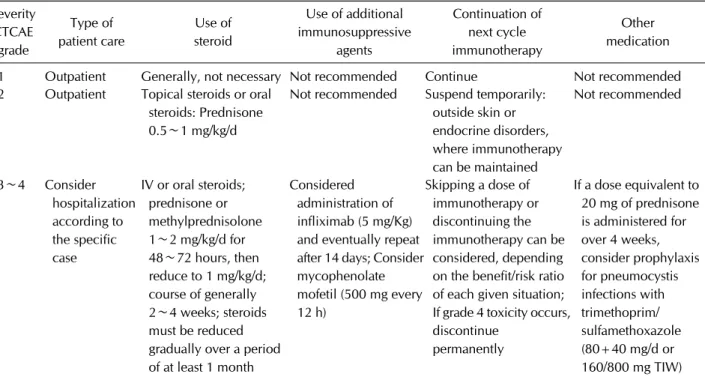
Table 1. General management guideline of immune-related adverse events Severity
CTCAE grade
Type of patient care
Use of steroid
Use of additional immunosuppressive
agents
Continuation of next cycle immunotherapy
Other medication
1 Outpatient Generally, not necessary Not recommended Continue Not recommended 2 Outpatient Topical steroids or oral
steroids: Prednisone 0.5∼1 mg/kg/d
Not recommended Suspend temporarily:
outside skin or endocrine disorders, where immunotherapy can be maintained
Not recommended
3∼4 Consider hospitalization according to the specific case
IV or oral steroids;
prednisone or methylprednisolone 1∼2 mg/kg/d for 48∼72 hours, then reduce to 1 mg/kg/d;
course of generally 2∼4 weeks; steroids must be reduced gradually over a period of at least 1 month
Considered administration of infliximab (5 mg/Kg) and eventually repeat after 14 days; Consider mycophenolate mofetil (500 mg every 12 h)
Skipping a dose of immunotherapy or discontinuing the immunotherapy can be considered, depending on the benefit/risk ratio of each given situation;
If grade 4 toxicity occurs, discontinue
permanently
If a dose equivalent to 20 mg of prednisone is administered for over 4 weeks, consider prophylaxis for pneumocystis infections with trimethoprim/
sulfamethoxazole (80+40 mg/d or 160/800 mg TIW) CTCAE: common terminology criteria for adverse events, TIW: 3 times a week.
may be intermittent and can even occur weeks to months after completion of the treatment. Consultation with oth- er specialties is also advisable. General guidelines for the management of irAEs using ipilimumab are described in the FDA Risk Evaluation and Management Strategies [18]. A similar approach is expected to apply to PD-1 inhibitors. Recently, Clinical Practice Guidelines were de- veloped in accordance with the European Society for Medical Oncology (ESMO) standard operating proce- dures for Clinical Practice Guidelines development [19,20].
As a rule, the management of severe irAEs requires im- mediate interruption of the drug and initiation of corticosteroids. For patients with grade 2 toxicities, ICPIs should be withheld and not resumed until toxicity is grade 1 or lower. If symptoms do not resolve within a week, corticosteroids (prednisone 0.5 mg/kg/day or equivalent) must be initiated. On the other hand, for pa- tients experiencing grade 3/4 irAEs, the drug should be permanently discontinued and high doses of cortico- steroids (prednisone 1 to 2 mg/kg/day or equivalent) should be started. When the symptoms subside to grade 1 or lower, steroids should be gradually tapered off over a month. However, in intractable cases, administration of infliximab (for GI toxicity) or mycophenolate (for hep- atotoxicity) should be considered [21]. Following is sum-
mary of several general rules that are widely accepted irre- spective of the affected organs, and a general irAE man- agement guideline is summarized in Table 1.
• For grade 2 irAEs: ICPI should be interrupted and re- sumed when symptoms or laboratory values decrease to grade 1 or lower. Glucocorticoids (prednisone 0.5∼1 mg/kg/day or equivalent) should be considered if symp- toms persist for more than 1 week.
• For grade 3/4 irAEs: High doses of glucocorticoids (prednisone 1∼2 mg/kg/day or equivalent) should be started. The glucocorticoids should be tapered off gradu- ally when symptoms subside to grade 1 or lower. Patients on 20 mg or equivalent prednisone doses for at least 4 weeks should also consider prophylactic management against Pneumocystis jirovecii.
• If symptoms continue for more than 3 days on intra- venous (IV) glucocorticoids, alternative immunosuppressive agents should be considered (infliximab 5 mg/kg; myco- phenolate mofetil for hepatitis). Infliximab 5 mg/kg should be repeated after 2 weeks for persistent symptoms.
• For grade 4 irAEs: ICPIs should be discontinued per- manently (except in endocrinopathies when controlled by proper hormone replacement). Therapy can be re- sumed in selected patients with grade 3 toxicities (see be- low organ-specific toxicities section).
ICPIs should also be stopped permanently in the follow- ing circumstances:
• Grade 2 reactions lasting for 6 weeks or more.
However, anti-PD-1/PD-L1 therapies can be continued in endocrinopathies when controlled by proper hormone replacement.
• Inability to reduce glucocorticoid dose to 7.5 mg pre- dnisone or daily equivalent for patients treated with an- ti-CTLA-4 antibodies and less than 10 mg/day within 12 weeks for anti-PD-1 therapy.
• Grade 2∼4 ocular toxicities not improving to grade 1 within 2 weeks after treatment with topical immuno- suppression or requiring systemic treatment.
The mechanisms of action of commonly used im- munomodulatory medications are summarized as fol- lows, and communication with other specialists is highly recommended to combine expertise on non-steroid im- munosuppression strategies.
(1) Steroids have various effects on T cells, B cells and macrophages, which include inhibition of cytokine or in- terleukin production, inhibition of neutrophil apoptosis, and reduced macrophage function.
(2) Infleximab is a monoclonal antibody that prevents tumor necrosis factor alpha (TNF-α) from binding to its receptors.
(3) Mycophenylate mofetil (MMF) suppresses inosine monophosphate dehydrogenase, an enzyme involved in nu- cleotide production, particularly in activated lymphocytes.
(4) Tacrolimus and cyclosporine are calcineurin in- hibitors that limit transcription of interleukin-2, which is involved in T cell proliferation.
The association between irAEs and the efficacy of ICPIs is still controversial, and a recent retrospective analysis reported similar overall survival in patients who received immunosuppression. Combination immunotherapy has only been approved for metastatic melanoma. The onset of G3/4 toxicities for either monotherapy with nivolumab or combination immunotherapy differs, since irAEs may develop earlier in combination therapy and may start over a prolonged period of time.
Organ-specific adverse events and management 1) Cutaneous toxicity
Cutaneous toxicity is the most common and the earliest developing irAE, and it shows myriad clinical manifes- tations, including maculopapular, follicular, pustular, ve- sicular and acneiform eruptions. These were earlier cate- gorized by Curry et al. [22] into 4 groups, according to
their histomorphology: (1) inflammatory, (2) im- munobullous, (3) alterations of epidermal keratinocytes and (4) alterations of epidermal melanocytes. Approximately half of patients treated with ipilimumab will experience rash or pruritus, and rashes are often mild in severity and present after the first or second dose [23]. Skin biopsy usually reveals perivascular lymphocytic infiltrates ex- tending deep into the dermis [24]. Rare cases of Steven- Johnson syndrome have also been reported [10,25].
Combination immunotherapy was associated with a higher rate of severe skin toxicity (2.9%) [26].
Regarding the maculopapular rash, the most frequent cutaneous toxicity, CTCAE classification proposes:
• Grade 1: macules/papules covering less than 10% the body surface area (BSA) with or without symptoms (e.g., pruritus, burning, tightness);
• Grade 2: macules/papules covering 10%∼30% BSA with or without symptoms (like pruritus, burning, tight- ness); limiting instrumental activities of daily living (ADL);
• Grade 3: macules/papules covering more than 30%
BSA with or without associated symptoms; limiting self-care ADL;
• Grade 4: papulopustular rash associated with life- threatening superinfection; Stevens–Johnson syndrome, toxic epidermal necrolysis, and bullous dermatitis cover- ing more than 30% of BSA and requiring intensive care unit (ICU) admission.
Dermatological toxicity is also observed with anti-PD1 therapy (30%∼40%) [27]. Interestingly, skin toxicity mainly develops in patients with melanoma. For example, vitiligo was reported in approximately 10% of patients [13,26], but it was not described in lung or renal cell car- cinoma studies [28,29]. Notably, vitiligo was also shown to be predictive of a durable response [30]. Treatment management for skin toxicity can be summarized as follows.
• Grade 1/2 toxicity: ICPIs should be continued for at least 1 week. Treatment with topical emollients, antihist- amines in the case of pruritus or topical mild strength cor- ticosteroid creams should be started. ICPI should be re- initiated when at or below G1.
• Grade 3 toxicity: ICPI should be interrupted and treat- ment with topical emollients, antihistamines, and high strength corticosteroid creams should be start immediately.
• Grade 4 toxicity: ICPI should be discontinued perma- nently, the patient should be hospitalized and dermato- logical consultation for skin biopsy should be scheduled.
IV corticosteroids (1∼2 mg/kg [methyl]prednisone) should be started, and steroids should be tapered off over 1 month after resolution of AE to grade 1 [11,25].
GI toxicity: diarrhea and enterocolitis
Diarrhea is a common clinical manifestation in ICPI treatment. Diarrhea is clinically different from enter- ocolitis, which involves abdominal pain and radiological and laboratory signs of colonic inflammation. Diarrhea most commonly arise within 6 weeks on ipilimumab or nivolumab and as late as 6 months on pembrolizumab [25,31]. Both conditions should be excluded from enter- ocolitis caused by Clostiridium difficile and other pathogens.
In a phase III ipilimumab trial for melanoma, diarrhea of any grade was reported in 30% of patients. Grade 3/4 tox- icity, defined as 7 or more stools above baseline, fever, ileus or peritoneal signs, was observed in 10% and was dose-dependent (10% at a 10 mg/kg dose vs. 1% at a 3 mg/kg dose) [10]. Meanwhile, enterocolitis appears to be less common, with an incidence of approximately 5%
[10]. Endoscopic findings revealed mucosal edema with biopsies demonstrating neutrophilic, lymphocytic or mixed neutrophilic-lymphocytic infiltrates [32]. CT scan findings associated with ICPI-induced colitis included mesenteric engorgement and bowel wall thickening [33].
Diarrhea or colitis was less frequent with PD-1 blockers compared to CTLA-4 blockers, and G3/4 toxicity was seen in about 1% to 2% of cases [13,16,34]. Interestingly, patients who developed significant diarrhea and enter- ocolitis during CTLA-4 blockade were subsequently been treated with PD-1 antibodies without experiencing com- plications [35].
Management starts with maintaining oral hydration. In patients with mild diarrhea, administration of ICPIs should be continued without starting corticosteroids.
Low residual diet and anti-motility agents like loper- amide can help. Colonoscopy is reserved for patients with G2 or higher diarrhea or in cases where diagnosis is unclear. If G3/4 enterocolitis develops, ipilimumab should be permanently discontinued and high dose IV corticosteroids should be started [23,36]. If symptoms do not resolve in 3 days, treatment with 5 mg/kg anti-TNF antibody infliximab once every two weeks is recom- mended [37]. In infliximab-refractory cases, mycopheno- late mofetil can be used. In rare cases, enterocolitis can re- sult in perforation and require colostomy. Treatment management can be summarized as follows.
• Grade 1 toxicity, defined as fewer than 4 liquid stools per day over baseline; ICPI can be continued. High fi- ber/lactose diet should be avoided. Treatment with anti- diarrheal medication, like loperamide, should be prescribed.
• Grade 2 toxicity, defined as 4 to 6 liquid stools per day over baseline or abdominal pain or blood in stool or nau- sea or nocturnal episodes, ICPI should be interrupted and the patient should start with corticosteroids depending on the severity and other symptoms (either budesonide or oral corticosteroids 1 mg/kg). If the patient shows no improvement within 3 to 5 days, colonoscopy should be done and, in the case of colitis, infliximab 5 mg/kg should be administered.
• Grade 3/4 toxicity, defined as 7 or more liquid stools per day, is life-threatening. It requires hospitalization and isolation until the infection is treated. ICPI should be per- manently discontinued and (methyl)prednisone 2 mg/kg IV should be initiated. Patients who respond to IV corti- costeroids within 3 to 5 days should be switched to the or- al formula, which should be tapered off over 8 to 12 weeks. Patients who do not respond to corticosteroids within 3 to 5 days should be transitioned to infliximab, unless it is contraindicated.
GI toxicity: hepatotoxicity
Hepatotoxicity has been observed with both an- ti-CTLA-4 and anti-PD-1 inhibitors. It occurs within 8∼
12 weeks after treatment initiation and generally mani- fests as elevation of levels of the liver enzymes aspartate aminotransferase (AST) and alanine aminotransferase (ALT). Elevation of total bilirubin level is a rare con- sequence of prolonged periods of elevated liver enzyme levels [11]. The phase III melanoma trial of ipilimumab reported grade 2 hepatotoxicity in 2.5% in patients and the incidence of grade 3 to 5 events reached 2% [10]. In the large phase I trial of anti-PD-1 blockers, grade 1/2 liv- er dysfunction was noted in approximately 5% of pa- tients, and grade 3∼5 events were rare (1%) [16]. The in- cidence of hepatotoxicity is higher with the combination of ipilimumab and nivolumab, with grade 3/4 events up to 20% [26]. Liver biopsy may be considered to assist dif- ferential diagnosis. Lobular hepatitis indistinguishable from autoimmune hepatitis is commonly reported, and most cases are panlobular but inflammation may be con- fined to zone 3. Additional sinusoidal histiocytosis and central vein endothelitis may help identify ipilimu- mab-associated inflammation [26].
In the clinic, liver enzymes should be monitored regu-
larly prior to each dose of ICPIs. Management of hep- atotoxicity starts by ruling out other liver injury etiol- ogies, including concomitant medications, alcohol con- sumption, and combined viral hepatitis. Radiological studies should be performed to exclude metastatic disease. In the case of immune-related hepatitis, CT scan can show surface nodularity, intrahepatic biliary dilata- tion, varices, and ascites.
Treatment with systemic corticosteroids should not be delayed [25]. As immune-related hepatitis may persist, a minimum of a three-week treatment with subsequent ta- pering of the dose is required. For grade 2 hepatotoxicity, defined as AST or ALT values 2.5 to 5 times the upper lim- it of normal (ULN) and total bilirubin values between 1.5 to 3 ULN, treatment with ICPIs should be withheld.
Steroid therapy should continue until toxicity resolves to grade 0 or 1. For grade 3/4 immune-related hepatitis (AST/ALT values above 5 times the ULN or total bilirubin level above 3 times ULN) the drug should be permanently discontinued and liver biopsy could be considered.
Hospitalization is required if liver enzyme levels are above 8 times ULN. In rare cases of corticosteroid re- fractory hepatitis, mycophenolate mofetil (500 mg every 12 hours) should be given [11,25]. Infliximab should not be administered, since it carries a risk of hepatotoxicity.
• Grade 2 hepatitis: ICPI should be withheld and AST/ALT levels should be closely monitored (1∼2 times/week). If there is no improvement in a week, treat- ment with (methyl)prednisone (0.5∼1 mg/kg) should be started. This should be tapered off over several weeks under close monitoring of AST/ALT and bilirubin levels.
• Grade 3 hepatitis: ICPI should be discontinued and treatment with (methyl)prednisone 1∼2 mg/kg should be immediately started. If there is no improvement in 2 to 3 days, MMF (1,000 mg 3x daily) should be added.
Immunosuppression should be tapered off over 4 to 6 weeks under close monitoring of AST/ALT and bilirubin levels.
• Grade 4 hepatitis: ICPI should be permanently dis- continued, hospitalization and initiation of (methyl)pre- dnisone 2 mg/kg IV is needed. MMF should be added if no improvement is observed within 2 to 3 days. A hepatol- ogy specialist should be consulted if no improvement is seen under double immunosuppression. Other im- munosuppressive drugs to consider are anti-thymocyte globulin and tacrolimus. Sometimes the patient should be referred to experienced centers. Treatment should be tapered off over 6 weeks under close liver function
monitoring.
Endocrinopathies
Immunotherapy has been causatively associated with several endocrinopathies, including hypophysitis, hypo- pituitarism, adrenal insufficiency, and hypothyroidism.
The median onset of endocrinopathy is about 7 weeks for ipilimumab and 10 weeks for nivolumab [11,38], and clinically significant endocrinopathies typically occur in less than 5% of patients on ipilimumab. For anti-PD-1 therapies, grade 3/4 events are rarer (approximately 1%) [39,40].
1) Hypophysitis
Hypophysitis, an inflammation of the anterior lobe of the pituitary gland, has been associated with ICPIs in pa- tients with various malignancies. Large differences (0%∼
25%) in the incidence of ipilimumab-induced hypo- physitis have been reported in different trials [41]. In the phase III trial of ipilimumab, severe hypophysitis was re- ported in 1.8% of patients [10]. On the other hand, trem- elimumab has been much less frequently shown to be re- lated to hypophysitis (0.5%∼4%) [42]. Similarly, hypo- physitis is a rare event with anti-PD-1 antibodies [34,43].
The median time to onset is approximately 2 to 4 months after treatment initiation, but delayed presentations can occur [44,45].
The pathophysiology of anti-CTLA4-induced hypo- physitis remains unclear. In a mouse model, researchers noted low-level ectopic CTLA4 RNA and protein ex- pression on thyrotropin and prolactin secreting cells of the pituitary gland [32]. Anti-CTLA4 treatment could lead to mononuclear cell infiltration in the pituitary gland, anti-pituitary antibodies and activation of the com- plement cascade in these animals, causing inflammation of the gland like what was observed in full-blown hypo- physitis patients [46].
Hypophysitis presents with non-specific symptoms like headache, fatigue, and visual disturbances. A swollen or enlarged pituitary gland may be visible in brain MRIs [47]. Diagnosis is based on low pituitary hormone levels (adrenocorticotropic hormone [ACTH], follicle stimulat- ing hormone, thyroid-stimulating hormone [TSH], lutei- nizing hormone and growth hormone), and differential diagnosis includes primary hypothyroidism (low thyro- xine, high TSH) and primary adrenal insufficiency (low cortisol).
In grade 1 hypophysitis, defined as asymptomatic or
mild symptoms, only clinical or diagnostic observation is recommended. In any grade 2 or higher hypophysitis which limits self-care ADL (headache, fatigue, sepsis, se- vere ataxia), ICPI should be interrupted and high-dose corticosteroids should be started (prednisone 1 mg/kg daily). Permanent discontinuation of the drug is recom- mended for grade 3/4 toxicities. Endocrinology con- sultation and long-term supplementation of affected hor- mones due to secondary hypothyroidism and hypoa- drenalism is necessary [25]. Long-term supplementation of affected hormones is often required, including secon- dary hypothyroidism requiring levothyroxine replace- ment or secondary hypoadrenalism requiring replace- ment hydrocortisone. In most cases, the immune check- point inhibition can be continued. Long-term HRT is re- quired in most patients.
2) Thyroid gland dysfunction
Both hyper- and hypothyroidism have been reported, but hypothyroid disorders are more common than hyper- thyroidism [25]. Still, little is known about the patho- genesis of thyroid disorders following ICPIs. It is thought to be mediated by T cells rather than B cell autoimmunity.
Hypothyroidism is more common with anti-PD-1 thera- pies than with ipilimumab 3 mg/kg, but a higher in- cidence can be seen at high doses (10 mg/kg) [26, 34,43,48].
Hypothyroidism is usually subclinical; hormone re- placement therapy with levothyroxine can be initiated and ICPI treatment can be continued without interruption.
If concurrent hypophysitis is suspected (low TSH, low T4), treatment should not be solely based on TSH mea- surement. Instead, free thyroxine and T3 levels should al- so be considered [39]. On the other hand, hyper- thyroidism is less common and might be a result of tran- sient thyroiditis preceding hypothyroidism or TSH-re- ceptor antibodies and Grave’s disease [38]. Persistent hy- perthyroidism should be treated as primary hyper- thyroidism.
According to the ESMO guidelines, substitution with thyroid hormone should be considered in the case of fa- tigue or other complaints that could be attributed to hy- pothyroidism even with subclinical hypothyroidism. In symptomatic patients, especially in the case of hyper- thyroidism, treatment with beta-blockers should be start- ed (propranolol or atenolol). Rarely, carbimazole or ste- roids are used. In those cases, treatment with ICPIs should be interrupted until symptom recovery. Hormone
replacement therapy is usually long-lasting [19].
3) Adrenal insufficiency
Primary adrenal insufficiency was reported in 1.5%∼
2% of patients [39]. It should be distinguished from hy- pophysitis based on measurement of ACTH levels. Acute adrenal insufficiency (adrenal crisis) constitutes an emer- gency manifesting as dehydration, hypotension, hyper- kalemia, and hyponatremia. Patients suspected to have adrenal crisis should be immediately hospitalized with prompt initiation of IV corticosteroids [11]. Endocrinology consultation is also warranted.
4) Type I diabetes mellitus
Type I diabetes mellitus (DM) can occur with both an- ti-CTLA-4 and anti-PD-1 blockade at a rate of less than approximately 1% [39]. Incidence is doubled with the use of combined ipilimumab and nivolumab [26]. Insulin treatment is recommended. Finally, cases of low testos- terone have been reported in patients treated with ipili- mumab [45].
5) General guideline of endocrinopathies
Below is the summary of management principles for en- docrinopathy according to ESMO guidelines [19];
• In symptomatic hyperthyroidism of grade 1/2, ICPI should be interrupted and beta-blocker therapy (propranolol or atenolol/metoprolol) should be started. ICPI should be restarted when asymptomatic.
• In the case of hypothyroidism (rarely higher than grade 2), HRT should be started depending on the se- verity (50∼100 μg/day). The dose should be increased until TSH level is normal. In the case of thyroid gland in- flammation, treatment with 1 mg/kg prednisone should be started orally. This should be tapered based on recov- ery from clinical symptoms. Interruption of ICPI treat- ment should be considered when symptomatic.
• In the case of hypophysitis (rarely higher than grade 2), when headache, diplopia or other neurological symp- toms are present, treatment with (methyl)prednisone 1 mg/kg should be started orally and taper over 2 to 4 weeks. Start HRT depending on the affected hormonal ax- is (levothyroxine, hydrocortisol, testosterone).
• In patients with type I diabetes mellitus grade 3/4 (ketoacidosis [sub]coma), hospitalization is needed to start treatment of newly onset type I DM. The role of cor- ticosteroids in preventing complete loss of insulin pro- ducing cells is unknown and not recommended.
Immune-mediated pneumonitis
Pneumonitis is an uncommon toxicity of ICPIs, but the implicated mechanisms are still undetermined. The hy- pothesis is that dysregulated effector T cells are accumu- lated in the pulmonary interstitial, leading to an increased inflammatory response [49]. It can occur with both an- ti-CTLA-4 and anti-PD-1 treatment. Ipilimumab- in- duced pneumonitis has been reported in up to 5% of pa- tients in monotherapy [13,34]. A combination of nivolu- mab and ipilimumab results in pneumonitis at a higher incidence (5% to 10%, with 2% G3/4) [26]. Pneumonitis can occur in 2% to 5% of patients with nivolumab, and more commonly in patients with renal cell and lung can- cer than in patients with melanoma [34,50,51].
The most acute form is acute interstitial pneumo- nitis/diffuse alveolar damage syndrome, which is some- times life-threatening [52]. Organizing inflammation and sarcoidosis-like granulomatosis has been described [53-55]. Rarely, pneumonitis worsens despite im- munosuppression, and may be fatal due to superimposed infection or progressive disease. Patients presenting with symptoms of upper respiratory infection like coughing or shortness of breath should be evaluated using appro- priate imaging. However, radiological findings of pneu- monitis are not pathognomonic, and may demonstrate ground glass opacities, a cryptogenic organizing pneumo- nia-like appearance and interstitial pneumonia pattern, and characteristics of hypersensitivity pneumonitis.
Although lung biopsy or bronchoscopy is not usually re- quired for decision, it may assist in discriminating acute infection, or lepidic or lymphangitic spread of lung cancer, from a variety of inflammatory changes described above.
The treatment strategy is summarized according to the ESMO guidelines [19];
• Grade 1 pneumonitis: defined as radiographic change only and non-symptomatic, delay ICPI treatment, consid- er steroids (e.g., prednisone 1 mg/kg/day orally [PO] or methylprednisolone 1 mg/kg/day IV). To follow-up, re- assessment should be done after 3 weeks. If it worsens, patients should be treat as grade 2 or 3/4.
• Grade 2 pneumonitis: defined as mild-to-moderate symptoms (dyspnea, cough, shortness of breath) limiting instrumental ADL, ICPI treatment should be delayed and hospitalization considered. Symptoms should be moni- tored daily. Steroids are recommended (prednisone 1∼2 mg/kg/day PO or methylprednisolone 1∼2 mg/kg/day IV). Empiric antibiotics (if suspicious for concurrent in- fection) should be considered. To follow-up, manage-
ment should be reassessed every 1 to 3 days. If improve- ment is observed, steroids is tapered and treatment con- tinued if symptoms resolve completely. If it worsens, the condition should be treated as grade 3/4.
• Grade 3/4 pneumonitis: defined as severe symptoms limiting self-care ADL, ICPI is discontinued permanently and hospitalization is needed, even in ICU if necessary, and high-dose (methyl)prednisone 2∼4 mg/kg IV should be immediately started. Infliximab should be added, and MMF or cyclophosphamide should be added in the case of deterioration under steroids. The drugs should be ta- pered over a period of 4 to 6 weeks. Prophylactic anti- biotics should be added for opportunistic infections.
Bronchoscopy with biopsy should be considered.
Management should be reassessed daily.
Renal toxicity
The prevalence of renal insufficiency was reported to be low (0%∼4%) but it has been reported from both an- ti-CTLA-4 and anti-PD-1 blockers [38]. The highest in- cidence reported was in the phase II trial with nivolumab in non-small-cell lung cancer [56]. The histopathological findings of CTLA-4-induced nephrotoxicity include acute granulomatous interstitial nephritis and lupus mem- branous nephropathy [57,58]. In the case of nephritis, first rule out other causes of renal failure. Interrupt or permanently discontinue ICPI depending on the severity of the renal insufficiency. Stop other nephrotoxic drugs.
Start (methyl)prednisone 1∼2 mg/kg. Consider renal bi- opsy to confirm diagnosis.
• Grade 1 toxicity: defined as creatinine above 1∼1.5x baseline; proteinuria 1+, less than 1.0 g/24 h: monitor re- nal function, promote hydration and cessation of neph- rotoxic drugs.
• Grade 2 toxicity: defined as creatinine above 1.5∼
3.0x baseline; proteinuria 2+, 1.0∼3.4 g/24 h: exclude non-immune causes, commence prednisolone 0.5∼1 mg/kg. If it worsens, manage as per grade 3 and dis- continue ICPI.
• Grade 3/4: defined as creatinine above 3.0x baseline;
proteinuria at or above 3.5 g/24 h: initiate prednisolone 1∼
2 mg/kg or IV equivalent. Consider renal biopsy.
Discontinue ICPI.
Neurological toxicity
Neurological AEs were reported to be as rare as 1%
[43,56]. However, recent analysis involving 9,208 pa- tients demonstrated a somewhat higher incidence: 3.8%
in patients receiving anti-CTLA4, 6.1% in patients receiv- ing anti-PD-1 agents and 12% in patients with an- ti-CTLA4 in combination with anti-PD-1 drugs [48].
Symptoms are varied; polyneuropathy, facial nerve palsy, demyelination, myasthenia gravis, Guillain–Barre syn- drome, posterior reversible leukoencephalopathy, trans- verse myelitis, enteric neuropathy, encephalitis and asep- tic meningitis [59,60]. Depending on the clinical pre- sentations, a work-up including nerve conduction studies and lumbar puncture may assist in diagnosis. Early con- sultation with a neurologist is warranted.
In the case of mild neurological AEs, ICPI should be withheld and work-up should be performed to define the nature of the neurotoxicity. In the case of deterioration or severe neurological symptoms, the patient should be ad- mitted and started on (methyl)prednisone 1∼2 mg/kg orally or IV. In the case of Guillain–Barre or myasthe- nia-like symptoms, adding plasmapheresis or IV im- munoglobulin should be considered.
Rheumatological or musculoskeletal toxicity Rheumatologic or musculoskeletal irAEs observed in patients treated with ICPIs have been mainly focused on arthralgia and myalgia, with a prevalence ranging from 1% to 43% and from 2% to 20%, respectively [61]. It is more common with an anti-PD-1 agent. However, the prevalence may have been underestimated because only high-grade irAEs were reported in some trials [32]. A pat- tern associated with inflammatory rheumatological con- ditions (morning stiffness, synovitis, proximal weak- ness) may be elicited. In addition, arthritis, seropositive rheumatoid arthritis (RA), (poly)myositis, sicca symp- toms, Sjögren's syndrome, and less commonly (temporal) vasculitis, cryoglobulinemic vasculitis and lupus neph- ritis are also reported [25,56,61,62]. Time to onset of rheumatic manifestations varies from weeks to months after ICPI initiation. Belkhir et al. [63] suggested there be a contrast between irAEs occurring after blockade with CTLA-4 (colitis, endocrine disorders, skin rashes) and PD-1/PD-L1 (non-specific arthritis, sicca syndrome, RA, polymyalgia rheumatica, and other connective tissue dis- eases).
ICPI treatment may aggravate pre-existing autoimmune rheumatic disease (ARD) in 40%∼50% of cases. The most frequent is psoriasis exacerbation, but exacerbation or aggravation of other disease-RA, scleroderma, chronic cutaneous lupus or systemic lupus erythematosus, vascu- litis, Sjögren’s syndrome, ankylosing spondylitis, my-
ositis, sarcoidosis-have all been reported [64,65].
Moreover, rheumatologic patients seem to have a higher proportion of flares compared with patients affected by non-rheumatologic autoimmune diseases.
For treatment, non-steroidal anti-inflammatory drugs should be started under ICPI continuation for mild symp- tom of arthralgia. Sometimes, rheumatic irAEs sponta- neously disappear. In the case of no improvement, low-dose steroids (10∼20 mg prednisone) can be considered. For severe or fatal cases, multidisciplinary ex- pertise of oncologists and rheumatologists is crucial for successful management. Treatment with 1 mg/kg pre- dnisone should be started. Infliximab or another an- ti-TNF-α inhibitor drug is sometimes required for arthri- tis improvement. There seem no differences in adverse events between patients with active disease and those with inactive disease, whereas patients receiving im- munosuppressive therapy at initiation of ICPI seem to have fewer irAEs than those not receiving treatment [65].
Cardiac toxicity
The incidence of cardiac AEs is less than 1%, but a wide range of toxicities including myocarditis, pericarditis, ar- rhythmias, cardiomyopathy, and impaired ventricular function have been reported after treatment with ipilimu- mab, pembrolizumab and nivolumab [25,31,48,59].
However, the incidence of cardiac toxicity is higher with the combination of ipilimumab and nivolumab (0.27%) compared with nivolumab alone (0.06%). Early con- sultation with a cardiologist is recommended. According to the recent ESMO guidelines, when myocarditis is sus- pected, admit the patient and immediately start high- dose (methyl)prednisone (1∼2 mg/kg). In the case of de- terioration, consider adding another immunosuppressive drug (MMF or tacrolimus).
Ocular toxicity
Ocular irAEs are rare (less than 1%) treated with ICPIs.
The clinical presentation can be one of two types: ocular inflammation, like peripheral ulcerative keratitis, uveitis and Vogt– Koyanagi–Harada syndrome, orbital in- flammation, including thyroid-associated orbitopathy and idiopathic orbital inflammation (scleritis, myositis, neuritis, dacryoadenitis); or retinal and choroidal disease (choroidal neovascularization and melanoma-associated retinopathy) [25,31,48,59].
The treatment of these rare toxicities depends on their severity, with topical corticosteroids in the case of epis-
cleritis and anterior uveitis, and systemic corticosteroids in the case of severe ocular inflammation and orbital inflammation. Intra-vitreal anti-vascular endothelial growth factor is indicated for choroidal neova- scularization.
Hematological toxicity
Fatigue is the most commonly reported AE in many of the ICPI trials, although rarely at a high grade. It is crucial to rule out secondary causes like hypothyroidism and hypoadrenalism. If other causes are excluded, short-term application of prednisolone (10∼20 mg) may be helpful in managing intolerable cases.
Red cell disorders–hemolytic anemia, red cell aplasia and acquired hemophilia A–are all reportedly related to ipilimumab [66,67]. Mortality from probable drug-in- duced neutropenia was reported in one patient on nivolu- mab [10]. Grade 3 myelodysplasia was also reported in one case with Hodgkin’s lymphoma on nivolumab.
Pembrolizumab has only been associated with low grade anemia and neutropenia [5]. ICPI-associated sarcoidosis has been reported with ipilimumab at both 3 mg/kg and 10 mg/kg. Visceral and even neurosarcoid findings have been noted [68,69]. If new lymphadenopathy is docu- mented in an otherwise well and responding patient, a tissue diagnosis should be obtained to avoid unnecessary antineoplastic treatment. Treatment with steroids (prednisolone 1∼2 mg/kg) may be required, as in the case of symptomatic neuro-sarcoidosis. In an asymptomatic patient, observation may be appropriate [70].
CONCLUSION
Although immune-checkpoint inhibitors have im- proved survival for a range of cancer types, they are com- monly associated with various types of irAEs. Some irAEs are severe and sometimes fatal. However, they are usually reversible if recognized early and promptly and properly managed. Management algorithms for irAEs have been developed and provide a framework within which in- dividual clinicians may exercise their discretion. Thoughtful management of short and medium-term irAEs is im- portant in optimizing quality of life and long-term outcomes.
Prospective management strategy trials are lacking in this field and are required to advance knowledge.
Therefore, where possible, histological or serological confirmation would be helpful to enlighten the nature of
the autoimmune process, since this may differ. For exam- ple, hyperthyroidism may be mediated by thyroiditis or Grave’s disease, and this can impact the management.
One challenge lies in deciding when to instigate next-line immunosuppression after steroids. Given the potential for fatal perforation in those with colitis, we advocate the use of infliximab sooner rather than later. These need to incorporate prolonged durations of follow-up to identify possible late complications.
Lastly, the field must establish true contraindications to use second-line ICPI when the first has been complicated by severe irAEs, or whether patients experiencing grade 3 or 4 toxicity were included in the re-treated cohorts.
While most clinical trials of ICPIs after first-line treat- ment excluded patients who had a prior grade 3/4 toxicity and mandated a minimum 4-week rest between different agents, the practice outside of trials differs under the dis- cretion of the physicians. Moreover, the best way is an in- formed discussion with the patient regarding the risk ver- sus benefit of rechallenge with an ICPI in the context of prior severe irAEs, especially when balanced with the chance of death from metastatic disease. Patients may al- so be prepared to accept greater toxicity than that pre- dicted by their physicians for a relatively small perceived benefit.
ACKNOWLEDGMENTS
This research was supported by Basic Science Research Program through the National Research Foundation of Korea (NRF) funded by the Ministry of Education (grant no. 2017R1D1A1B04035691).
CONFLICT OF INTEREST
No potential conflict of interest relevant to this article was reported.
AUTHOR CONTRIBUTIONS
H.C.J. conception and design of study, acquisition of da- ta, analysis and/or interpretation of data, drafting the manuscript, revising the manuscript critically for im- portant intellectual content. S.E.O. acquisition of data, drafting the manuscript, revising the manuscript crit- ically for important intellectual content. J.H.K. drafting the manuscript, revising the manuscript critically for im- portant intellectual content.
REFERENCES
1. Lesterhuis WJ, Haanen JB, Punt CJ. Cancer immunotherapy-- revisited. Nat Rev Drug Discov 2011;10:591-600.
2. Dolan DE, Gupta S. PD-1 pathway inhibitors: changing the landscape of cancer immunotherapy. Cancer Control 2014;
21:231-7.
3. Sharma P, Allison JP. The future of immune checkpoint therapy. Science 2015;348:56-61.
4. Topalian SL, Drake CG, Pardoll DM. Immune checkpoint blockade: a common denominator approach to cancer therapy.
Cancer Cell 2015;27:450-61.
5. Dunn GP, Old LJ, Schreiber RD. The three Es of cancer immunoediting. Annu Rev Immunol 2004;22:329-60.
6. Linsley PS, Brady W, Urnes M, Grosmaire LS, Damle NK, Ledbetter JA. CTLA-4 is a second receptor for the B cell acti- vation antigen B7. J Exp Med 1991;174:561-9.
7. Sznol M, Chen L. Antagonist antibodies to PD-1 and B7-H1 (PD-L1) in the treatment of advanced human cancer-- response. Clin Cancer Res 2013;19:5542.
8. Xu C, Chen YP, Du XJ, Liu JQ, Huang CL, Chen L, et al.
Comparative safety of immune checkpoint inhibitors in cancer: systematic review and network meta-analysis. BMJ 2018;363:k4226.
9. Weber JS, Antonia SJ, Topalian SL, Schadendorf D, Larkin JMG, Sznol M, et al. Safety profile of nivolumab (NIVO) in patients (pts) with advanced melanoma (MEL): a pooled analysis. J Clin Oncol 2015;33(15 Suppl):9018.
10. Hodi FS, O'Day SJ, McDermott DF, Weber RW, Sosman JA, Haanen JB, et al. Improved survival with ipilimumab in pa- tients with metastatic melanoma. N Engl J Med 2010;363:
711-23.
11. Weber JS, Kähler KC, Hauschild A. Management of im- mune-related adverse events and kinetics of response with ipilimumab. J Clin Oncol 2012;30:2691-7.
12. O'Day SJ, Maio M, Chiarion-Sileni V, Gajewski TF, Pehamberger H, Bondarenko IN, et al. Efficacy and safety of ipilimumab monotherapy in patients with pretreated ad- vanced melanoma: a multicenter single-arm phase II study.
Ann Oncol 2010;21:1712-7.
13. Robert C, Long GV, Brady B, Dutriaux C, Maio M, Mortier L, et al. Nivolumab in previously untreated melanoma with- out BRAF mutation. N Engl J Med 2015;372:320-30.
14. Topalian SL, Hodi FS, Brahmer JR, Gettinger SN, Smith DC, McDermott DF, et al. Safety, activity, and immune corre- lates of anti-PD-1 antibody in cancer. N Engl J Med 2012;366:2443-54.
15. Ribas A, Puzanov I, Dummer R, Schadendorf D, Hamid O, Robert C, et al. Pembrolizumab versus investigator-choice chemotherapy for ipilimumab-refractory melanoma (KEYN OTE-002): a randomised, controlled, phase 2 trial. Lancet Oncol 2015;16:908-18.
16. Robert C, Ribas A, Wolchok JD, Hodi FS, Hamid O, Kefford R, et al. Anti-programmed-death-receptor-1 treatment with pembrolizumab in ipilimumab-refractory advanced mela- noma: a randomised dose-comparison cohort of a phase 1 trial. Lancet 2014;384:1109-17.
17. Topalian SL, Sznol M, McDermott DF, Kluger HM, Carvajal RD, Sharfman WH, et al. Survival, durable tumor remission, and long-term safety in patients with advanced melanoma
receiving nivolumab. J Clin Oncol 2014;32:1020-30.
18. Immune-mediated adverse reactions management guide [Internet]. Princeton (NJ): Bristol-Myers Squibb, 2013 [cited 2019 Mar 20]. Available from: http://www.hcp.yer voy.com/servlet/servlet.FileDownload?file=00Pi000 000PI1ZVEA1.
19. Haanen JBAG, Carbonnel F, Robert C, Kerr KM, Peters S, Larkin J, et al.; ESMO Guidelines Committee. Management of toxicities from immunotherapy: ESMO Clinical Practice Guidelines for diagnosis, treatment and follow-up. Ann Oncol 2017;28(suppl_4):iv119-42.
20. ESMO Guidelines Methodology [Internet]. Lugano: ESMO, 2019 [cited 2019 Mar 20]. Available from: https://www.es- mo.org/Guidelines/ESMO-Guidelines-Methodology.
21. Michot JM, Bigenwald C, Champiat S, Collins M, Carbonnel F, Postel-Vinay S, et al. Immune-related adverse events with immune checkpoint blockade: a comprehensive review. Eur J Cancer 2016;54:139-48.
22. Curry JL, Tetzlaff MT, Nagarajan P, Drucker C, Diab A, Hymes SR, et al. Diverse types of dermatologic toxicities from immune checkpoint blockade therapy. J Cutan Pathol 2017;44:158-76.
23. Villadolid J, Amin A. Immune checkpoint inhibitors in clin- ical practice: update on management of immune-related toxicities. Transl Lung Cancer Res 2015;4:560-75.
24. Hodi FS, Mihm MC, Soiffer RJ, Haluska FG, Butler M, Seiden MV, et al. Biologic activity of cytotoxic T lympho- cyte-associated antigen 4 antibody blockade in previously vaccinated metastatic melanoma and ovarian carcinoma patients. Proc Natl Acad Sci U S A 2003;100:4712-7.
25. Bristol-Myers Squibb. Yervoy (Ipilimumab) injection [Internet]. Princeton (NJ): Bristol-Myers Squibb, 2015 [cited 2019 Feb 20]. Available from: http://packageinserts.
bms.com/pi/pi_yervoy.pdf.
26. Larkin J, Chiarion-Sileni V, Gonzalez R, Grob JJ, Cowey CL, Lao CD, et al. Combined nivolumab and ipilimumab or monotherapy in untreated melanoma. N Engl J Med 2015;
373:23-34.
27. Naidoo J, Page DB, Li BT, Connell LC, Schindler K, Lacouture ME, et al. Toxicities of the anti-PD-1 and an- ti-PD-L1 immune checkpoint antibodies. Ann Oncol 2015;
26:2375-91.
28. Brahmer J, Reckamp KL, Baas P, Crinò L, Eberhardt WE, Poddubskaya E, et al. Nivolumab versus docetaxel in ad- vanced squamous-cell non-small-cell lung cancer. N Engl J Med 2015;373:123-35.
29. Motzer RJ, Escudier B, McDermott DF, George S, Hammers HJ, Srinivas S, et al. Nivolumab versus everolimus in ad- vanced renal-cell carcinoma. N Engl J Med 2015;373:
1803-13.
30. Hua C, Boussemart L, Mateus C, Routier E, Boutros C, Cazenave H, et al. Association of vitiligo with tumor re- sponse in patients with metastatic melanoma treated with pembrolizumab. JAMA Dermatol 2016;152:45-51.
31. Merck Sharp & Dohme Corp. Highlights of prescribing in- formation [Internet]. County Cork: Merck Sharp & Dohme Corp, 2015 [cited 2019 Feb 20]. Available from: http://www.
merck.com/product/usa/pi_circulars/k/keytruda/keytru da_pi.pdf.
32. Berman D, Parker SM, Siegel J, Chasalow SD, Weber J, Galbraith S, et al. Blockade of cytotoxic T-lymphocyte anti-
gen-4 by ipilimumab results in dysregulation of gastro- intestinal immunity in patients with advanced melanoma.
Cancer Immun 2010;10:11.
33. Kim KW, Ramaiya NH, Krajewski KM, Shinagare AB, Howard SA, Jagannathan JP, et al. Ipilimumab-associated colitis: CT findings. AJR Am J Roentgenol 2013;200:
W468-74.
34. Weber JS, D'Angelo SP, Minor D, Hodi FS, Gutzmer R, Neyns B, et al. Nivolumab versus chemotherapy in patients with advanced melanoma who progressed after an- ti-CTLA-4 treatment (CheckMate 037): a randomised, con- trolled, open-label, phase 3 trial. Lancet Oncol 2015;16:
375-84.
35. Weber JS, Kudchadkar RR, Yu B, Gallenstein D, Horak CE, Inzunza HD, et al. Safety, efficacy, and biomarkers of nivolu- mab with vaccine in ipilimumab-refractory or -naive melanoma. J Clin Oncol 2013;31:4311-8.
36. Fecher LA, Agarwala SS, Hodi FS, Weber JS. Ipilimumab and its toxicities: a multidisciplinary approach. Oncologist 2013;18:733-43.
37. Minor DR, Chin K, Kashani-Sabet M. Infliximab in the treat- ment of anti-CTLA4 antibody (ipilimumab) induced im- mune-related colitis. Cancer Biother Radiopharm 2009;24:
321-5.
38. Spain L, Diem S, Larkin J. Management of toxicities of im- mune checkpoint inhibitors. Cancer Treat Rev 2016;44:
51-60.
39. Corsello SM, Barnabei A, Marchetti P, De Vecchis L, Salvatori R, Torino F. Endocrine side effects induced by im- mune checkpoint inhibitors. J Clin Endocrinol Metab 2013;
98:1361-75.
40. Corsello SM, Salvatori R, Barnabei A, De Vecchis L, Marchetti P, Torino F. Ipilimumab-induced endocrinopathies:
when to start corticosteroids (or not). Cancer Chemother Pharmacol 2013;72:489-90.
41. Torino F, Corsello SM, Salvatori R. Endocrinological side-effects of immune checkpoint inhibitors. Curr Opin Oncol 2016;28:278-87.
42. Ribas A, Kefford R, Marshall MA, Punt CJ, Haanen JB, Marmol M, et al. Phase III randomized clinical trial compar- ing tremelimumab with standard-of-care chemotherapy in patients with advanced melanoma. J Clin Oncol 2013;31:
616-22.
43. Robert C, Schachter J, Long GV, Arance A, Grob JJ, Mortier L, et al. Pembrolizumab versus Ipilimumab in Advanced Melanoma. N Engl J Med 2015;372:2521-32.
44. Weber JS, Yang JC, Atkins MB, Disis ML. Toxicities of im- munotherapy for the practitioner. J Clin Oncol 2015;33:
2092-9.
45. Ryder M, Callahan M, Postow MA, Wolchok J, Fagin JA.
Endocrine-related adverse events following ipilimumab in patients with advanced melanoma: a comprehensive retro- spective review from a single institution. Endocr Relat Cancer 2014;21:371-81.
46. Iwama S, De Remigis A, Callahan MK, Slovin SF, Wolchok JD, Caturegli P. Pituitary expression of CTLA-4 mediates hypophysitis secondary to administration of CTLA-4 block- ing antibody. Sci Transl Med 2014;6:230ra45.
47. Dillard T, Yedinak CG, Alumkal J, Fleseriu M. Anti-CTLA-4 antibody therapy associated autoimmune hypophysitis: se- rious immune related adverse events across a spectrum of
cancer subtypes. Pituitary 2010;13:29-38.
48. Eggermont AM, Chiarion-Sileni V, Grob JJ, Dummer R, Wolchok JD, Schmidt H, et al. Adjuvant ipilimumab versus placebo after complete resection of high-risk stage III mela- noma (EORTC 18071): a randomised, double-blind, phase 3 trial. Lancet Oncol 2015;16:522-30.
49. Tabchi S, Messier C, Blais N. Immune-mediated respiratory adverse events of checkpoint inhibitors. Curr Opin Oncol 2016;28:269-77.
50. Borghaei H, Paz-Ares L, Horn L, Spigel DR, Steins M, Ready NE, et al. Nivolumab versus docetaxel in advanced non- squamous non-small-cell lung cancer. N Engl J Med 2015;373:1627-39.
51. Herbst RS, Baas P, Kim DW, Felip E, Pérez-Gracia JL, Han JY, et al. Pembrolizumab versus docetaxel for previously treated, PD-L1-positive, advanced non-small-cell lung can- cer (KEYNOTE-010): a randomised controlled trial. Lancet 2016;387:1540-50.
52. Nishino M, Sholl LM, Hodi FS, Hatabu H, Ramaiya NH.
Anti-PD-1-related pneumonitis during cancer immunotherapy.
N Engl J Med 2015;373:288-90.
53. Vogel WV, Guislain A, Kvistborg P, Schumacher TN, Haanen JB, Blank CU. Ipilimumab-induced sarcoidosis in a patient with metastatic melanoma undergoing complete remission. J Clin Oncol 2012;30:e7-10.
54. Eckert A, Schoeffler A, Dalle S, Phan A, Kiakouama L, Thomas L. Anti-CTLA4 monoclonal antibody induced sar- coidosis in a metastatic melanoma patient. Dermatology 2009;218:69-70.
55. Wilgenhof S, Morlion V, Seghers AC, Du Four S, Vanderlinden E, Hanon S, et al. Sarcoidosis in a patient with metastatic melanoma sequentially treated with anti- CTLA-4 monoclonal antibody and selective BRAF inhibitor.
Anticancer Res 2012;32:1355-9.
56. Rizvi NA, Mazières J, Planchard D, Stinchcombe TE, Dy GK, Antonia SJ, et al. Activity and safety of nivolumab, an an- ti-PD-1 immune checkpoint inhibitor, for patients with ad- vanced, refractory squamous non-small-cell lung cancer (CheckMate 063): a phase 2, single-arm trial. Lancet Oncol 2015;16:257-65.
57. Izzedine H, Gueutin V, Gharbi C, Mateus C, Robert C, Routier E, et al. Kidney injuries related to ipilimumab.
Invest New Drugs 2014;32:769-73.
58. Fadel F, El Karoui K, Knebelmann B. Anti-CTLA4 anti- body-induced lupus nephritis. N Engl J Med 2009;361:
211-2.
59. Bristol-Myers Squibb. Highlights of prescribing in- formation [Internet]. Princeton (NJ): Bristol-Myers Squibb, 2015 [cited 2019 Feb 20]. Available from: http://package inserts.bms.com/pi/pi_opdivo.pdf.
60. Wilgenhof S, Neyns B. Anti-CTLA-4 antibody-induced Guillain-Barré syndrome in a melanoma patient. Ann Oncol 2011;22:991-3.
61. Cappelli LC, Gutierrez AK, Bingham CO 3rd, Shah AA.
Rheumatic and musculoskeletal immune-related adverse events due to immune checkpoint inhibitors: a systematic review of the literature. Arthritis Care Res (Hoboken) 2017;69:1751-63.
62. Goldstein BL, Gedmintas L, Todd DJ. Drug-associated poly- myalgia rheumatica/giant cell arteritis occurring in two pa- tients after treatment with ipilimumab, an antagonist of
ctla-4. Arthritis Rheumatol 2014;66:768-9.
63. Belkhir R, Burel SL, Dunogeant L, Marabelle A, Hollebecque A, Besse B, et al. Rheumatoid arthritis and polymyalgia rheumatica occurring after immune checkpoint inhibitor treatment. Ann Rheum Dis 2017;76:1747-50.
64. Benfaremo D, Manfredi L, Luchetti MM, Gabrielli A.
Musculoskeletal and rheumatic diseases induced by im- mune checkpoint inhibitors: a review of the literature. Curr Drug Saf 2018;13:150-64.
65. Calabrese LH, Calabrese C, Cappelli LC. Rheumatic im- mune-related adverse events from cancer immunotherapy.
Nat Rev Rheumatol 2018;14:569-79.
66. Simeone E, Grimaldi AM, Esposito A, Curvietto M, Palla M, Paone M, et al. Serious haematological toxicity during and after ipilimumab treatment: a case series. J Med Case Rep
2014;8:240.
67. Delyon J, Mateus C, Lambert T. Hemophilia A induced by ipilimumab. N Engl J Med 2011;365:1747-8.
68. Vogel WV, Guislain A, Kvistborg P, Schumacher TN, Haanen JB, Blank CU. Ipilimumab-induced sarcoidosis in a patient with metastatic melanoma undergoing complete remission. J Clin Oncol 2012;30:e7-10.
69. Tissot C, Carsin A, Freymond N, Pacheco Y, Devouassoux G. Sarcoidosis complicating anti-cytotoxic T-lymphocyte- associated antigen-4 monoclonal antibody biotherapy. Eur Respir J 2013;41:246-7.
70. Andersen R, Nørgaard P, Al-Jailawi MK, Svane IM. Late de- velopment of splenic sarcoidosis-like lesions in a patient with metastatic melanoma and long-lasting clinical re- sponse to ipilimumab. Oncoimmunology 2014;3:e954506.