Received:October 11, 2017, Revised:January 1, 2018, Accepted:January 23, 2018 Corresponding to:Ji Seon Oh http://orcid.org/0000-0002-0205-6492
Clinical Research Center, Asan Medical Center, University of Ulsan College of Medicine, 88 Olympic-ro 43-gil, Songpa-gu, Seoul 05505, Korea. E-mail:[email protected]
Copyright ⓒ 2018 by The Korean College of Rheumatology. All rights reserved.
This is a Open Access article, which permits unrestricted non-commerical use, distribution, and reproduction in any medium, provided the original work is properly cited.
Is the Serum Uric Acid Level Independently Associated with Incidental Urolithiasis?
Doo-Ho Lim1, Min-ho Kim2, Seokchan Hong3, Yong-Gil Kim3, Chang-Keun Lee3, Seung Won Choi1, Bin Yoo3, Ji Seon Oh4
1Division of Rheumatology, Department of Internal Medicine, Ulsan University Hospital, University of Ulsan College of Medicine, Ulsan, 2Ewha Institute of Convergence Medicine, Ewha Womans University Mokdong Hospital, 3Division of Rheumatology, Department of Internal Medicine and 4Clinical Research Center, Asan Medical Center, University of Ulsan College of Medicine, Seoul, Korea
Objective. Urolithiasis is one of the manifestations of gout and the risk is higher in gouty patients. On the other hand, an in- dependent association between the urinary stone and serum uric acid (UA) level has not been established. This study examined whether the risk of urolithiasis increases with increasing serum UA level. Methods. Among the people who visited a tertiary hos- pital from 2010 to 2013, 13,964 individuals who underwent both ultrasonography and a laboratory test were recruited in the study. The risk of urolithiasis on ultrasonography was analyzed in association with the serum UA level by multiple logistic re- gression analysis with an adjustment for age, sex, body mass index, estimated glomerular filtration rate, and known underlying diseases, including diabetes mellitus and hypertension. Results. Among the 6,743 men (48.3%) and 7,221 women (51.7%), the age was 51.3±13.5 and the serum UA level was 4.5±2.1 mg/dL. Hyperuricemia (>7 mg/dL) was observed in 1,381 cases (9.9%). Urolithiasis was detected by ultrasonography in 608 cases (4.4%). The detection rates of urolithiasis in individuals with hyperuricemia and normouricemia were 5.9% and 4.1%, respectively (p=0.001). Multiple logistic regression analysis showed that individuals with hyperuricemia had a significantly higher risk of urolithiasis (adjusted odds ratio [OR]=1.54; 95% con- fidence interval [CI], 1.20∼1.96; p=0.001). A comparison of the highest with the lowest quartile of serum UA revealed a multi- variable-adjusted OR of 3.17 (95% CI, 1.98∼5.11) for men and 1.79 (1.08∼2.93) for women. Conclusion. These results sug- gest that individuals with a higher serum UA level have a higher risk of subclinical and clinical urolithiasis. (J Rheum Dis 2018;25:116-121)
Key Words. Uric acid, Hyperuricemia, Urolithiasis, Urinary calculi, Gout
INTRODUCTION
Urolithiasis is a relatively common problem observed in medical practice with a lifetime risk of approximately 8.8% to 10%, and its prevalence has been increasing in many industrialized countries [1,2]. Although calcium-con- taining renal stones are the most common type of renal calculi, uric acid (UA) stones are the second most fre- quent component of a renal stone (about 10%∼15% of all renal calculi) and can contribute to calcium-containing stone formation [3,4].
Hyperuricemia, which occurs when the concentration of
urate in the serum is above the solubility limit, is the main predisposing factor for gout. Several studies have re- ported that the prevalence of urolithiasis in gout patients was higher than that found in the general population [5-7]. However, gout and urolithiasis shared common risk factors such as hypertension, diabetes and obesity [8,9]. Additionally, it is not yet known with certainty whether hyperuricemia itself is independently associated with the risk for incidental urolithiasis. Until now, only gout patients have been included in most studies asso- ciated with urolithiasis except for those with asympto- matic hyperuricemia [5-7].
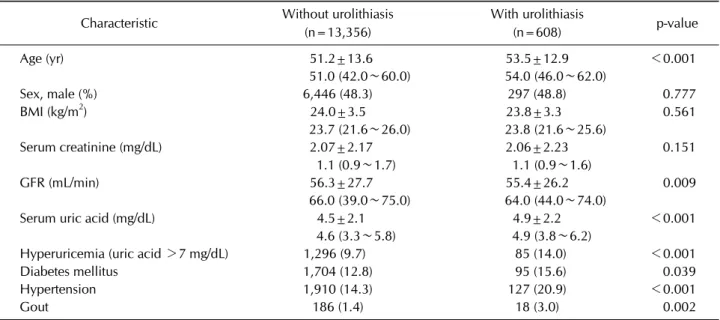
Table 1. Clinical characteristics of the study population
Characteristic Without urolithiasis (n=13,356)
With urolithiasis
(n=608) p-value
Age (yr) 51.2±13.6 53.5±12.9 <0.001
51.0 (42.0∼60.0) 54.0 (46.0∼62.0)
Sex, male (%) 6,446 (48.3) 297 (48.8) 0.777
BMI (kg/m2) 24.0±3.5 23.8±3.3 0.561
23.7 (21.6∼26.0) 23.8 (21.6∼25.6)
Serum creatinine (mg/dL) 2.07±2.17 2.06±2.23 0.151
1.1 (0.9∼1.7) 1.1 (0.9∼1.6)
GFR (mL/min) 56.3±27.7 55.4±26.2 0.009
66.0 (39.0∼75.0) 64.0 (44.0∼74.0)
Serum uric acid (mg/dL) 4.5±2.1 4.9±2.2 <0.001
4.6 (3.3∼5.8) 4.9 (3.8∼6.2)
Hyperuricemia (uric acid >7 mg/dL) 1,296 (9.7) 85 (14.0) <0.001
Diabetes mellitus 1,704 (12.8) 95 (15.6) 0.039
Hypertension 1,910 (14.3) 127 (20.9) <0.001
Gout 186 (1.4) 18 (3.0) 0.002
Values are presented as mean±standard deviation, median (interquartile range) or number (%). BMI: body mass index, GFR:
glomerular filtration rate.
Therefore, the primary objective of this study is to inves- tigate the independent association between serum UA level and the prevalence of subclinical and clinical ur- olithiasis detected by ultrasonography.
MATERIALS AND METHODS
Among people visited a tertiary hospital from January 2010 to December 2013, we selected 13,964 eligible in- dividuals (≥20-year old) who performed both laboratory test and abdominal or kidney ultrasonography. Retro- spectively, we reviewed all clinical data from the elec- tronic medical records according to the permission of re- gional Institutional Review Board. Clinical information was collected in the following areas: demographic charac- teristics (sex, age, weight and height), underlying dis- eases (diabetes mellitus, hypertension and gout), labo- ratory data (serum UA and serum creatinine) and the presence of urolithiasis, as ascertained by ultrasonography.
Comorbidities were identified using the Korean Classification of Disease, 6th edition (KCD-6), a mod- ification of the International Classification of Disease and Related Health Problems, 10th revision (ICD-10). Mean values of laboratory data were used when serum UA or se- rum creatinine levels were repeatedly measured within the study period. If ultrasonography was performed more than once, a single positive result was regarded as a pos- itive urinary stone diagnosis.
Hyperuricemia was defined as having serum UA level above 7.0 mg/dL. Body mass index (BMI) was calculated by the formula of weight in kilograms divided by the square of height in meters. The estimated glomerular fil- tration rate (eGFR) was calculated using the re-expressed 4-variable modification of diet in renal disease (MDRD) study equation: eGFR (mL/min/1.73 m2)=175×stand- ardized serum creatinine concentration (mg/dL)−1.154×
age−0.203×0.742 if patient is female.
This study was performed in accordance with the ethical guidelines of the Declaration of Helsinki (as revised in Brazil 2013) and received approval from the Institutional Review Board (IRB) of Ulsan University Hospital (IRB no. 2014-12-018).
Statistical analysis
All continuous variables were expressed as mean±
standard deviation and median (interquartile range), as appropriate, and compared using Student’s t-test or Mann-Whitney U-test for parametric and non-parametric data, respectively. Categorical variables were reported as numbers (percentages) and compared by the Chi-square test. The Cochran-Armitage trend test was used to assess the trend for the proportion of urolithiasis according to the UA categories. The risk of urolithiasis was analyzed in association with serum UA level by multiple logistic re- gression analysis with adjustment for age, sex, BMI, GFR and known underlying diseases including diabetes melli-
Table 2. Clinical characteristics of the study population according to gender Characteristic
Female
p-value
Male
p-value Without urolithiasis
(n=6,910)
With urolithiasis (n=311)
Without urolithiasis (n=6,446)
With urolithiasis (n=297)
Age (yr) 51.5±13.5 53.0±12.7 0.015 50.9±13.6 54.0±13.2 <0.001
51.0 (43.0∼60.0) 53.0 (46.0∼61.0) 51.0 (41.0∼60.0) 55.0 (45.0∼63.0)
BMI (kg/m2) 23.9±3.8 23.6±3.3 0.449 24.0±3.3 24.0±3.3 0.985
23.5 (21.4∼26.0) 23.7 (21.2∼25.5) 23.9 (22.0∼26.0) 24.1 (22.0∼25.8)
Serum creatinine (mg/dL) 1.8±1.9 1.8±2.0 0.699 2.4±2.4 2.4±2.4 0.015
0.9 (0.8∼1.2) 0.9 (0.8∼1.1) 1.2 (1.0∼2.4) 1.2 (1.1∼1.8)
GFR (mL/min) 56.1±26.7 56.5±26.2 0.512 56.5±28.7 54.2±26.2 0.003
65.0 (45.0∼74.0) 65.0 (49.0∼74.0) 67.0 (28.0∼77.0) 63.0 (42.0∼73.0)
Serum uric acid (mg/dL) 3.9±1.8 4.2±1.7 0.011 5.0±2.3 5.6±2.3 <0.001
4.2 (3.2∼4.9) 4.3 (3.4∼5.1) 5.5 (3.8∼6.6) 5.8 (4.6∼6.8) Hyperuricemia
(uric acid >7 mg/dL)
222 (3.2) 19 (6.1) 0.009 1,074 (16.7) 66 (22.2) 0.015
Diabetes mellitus 790 (11.4) 44 (14.1) 0.169 914 (14.2) 51 (17.2) 0.175
Hypertension 959 (13.9) 66 (21.2) <0.001 951 (14.8) 61 (20.5) 0.008
Gout 32 (0.5) 1 (0.3) 1.000 154 (2.4) 17 (5.7) 0.001
Values are presented as mean±standard deviation, median (interquartile range) or number (%). BMI: body mass index, GFR:
glomerular filtration rate.
Figure 1. Proportion of urolithiasis according to serum uric acid levels.
tus and hypertension.
Statistical significance was defined as p<0.05. Data were analyzed using PASW statistical software, version 18.0 (IBM Co., Armonk, NY, USA) and R 3.2.4 statistical software (The R Foundation for Statistical Computing, Vienna, Austria). Predicted probabilities for urolithiasis according to serum UA level and other risk factors were generated based on the multiple logistic regression model by using R software and effects package [10].
RESULTS
Among a total of 13,964 cases (6,743 men [48.3%] and 7,221 women [51.7%]), the mean age was 51.3±13.5 (range, 20∼95), the mean serum UA level was 4.5±2.1 mg/dL (range, 0.4∼21.8), and the mean serum crea- tinine level was 2.1±2.2 mg/dL (range, 0.4∼18.9). The characteristics of individuals with or without urolithiasis are shown in Tables 1 and 2. Among the total of 13,964 cases, renal stone was detected by ultrasonography 608 cases (4.4%). Age and the mean serum UA levels were found to be higher in cases with urolithiasis. Additionally, the proportion of each underlying disease (diabetes melli- tus, hypertension and gout) was also higher in the ur- olithiasis group. There was no significant difference in the gender, BMI, and serum creatinine level between the two groups.
Hyperuricemia was found in 1,381 cases (9.9%). The rates of urolithiasis in individuals with hyperuricemia and normal serum UA level were 5.9% and 4.1%, re- spectively (p=0.001). Moreover, the overall detection rate of urolithiasis increased proportionally to serum UA level (p<0.001) (Figure 1).
In multiple logistic regression analysis adjusted by age, sex, BMI, GFR, diabetes mellitus and hypertension, in- dividuals with hyperuricemia had significantly higher
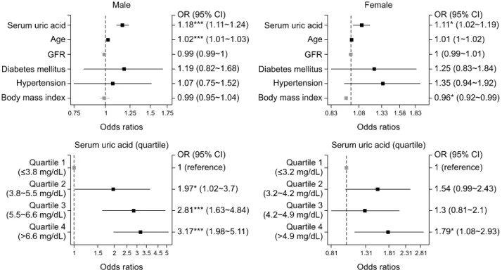
Figure 2. Plots of odds ratios (ORs) for urolithiasis according to serum uric acid level, age, glomerular filtration rate (GFR), presence of diabetes mellitus, presence of hypertension, body mass index, and serum uric acid quartile. Squares and horizontal bars repre- sent ORs and 95% confidence intervals (CIs), respectively. *p<0.05, **p<0.01, ***p<0.001.
Table 4. Age-adjusted and Multivariable-adjusted odds ratio for the presence of urolithiasis by serum uric acid level
Variable Age-adjusted
p-value Multivariable-adjusted*
p-value
Odds ratio (95% CI)† Odds ratio (95% CI)†
Total 1.11 (1.07∼1.15) <0.001 1.15 (1.09∼1.20) <0.001
Male 1.13 (1.08∼1.19) <0.001 1.18 (1.11∼1.24) <0.001
Female 1.10 (1.03∼1.17) 0.003 1.11 (1.02∼1.19) 0.012
CI: confidence intervals. *Adjusted by age, body mass index, glomerular filtration rate, diabetes mellitus and hypertension. †Odds ratio and 95% CI was impressed by 1 mg/dL increase of serum uric acid.
Table 3. Age-adjusted and multivariable-adjusted odds ratio for the presence of urolithiasis by hyperuricemia
Variable Age-adjusted
p-value Multivariable-adjusted*
p-value
Odds ratio (95% CI) Odds ratio (95% CI)
No hyperuricemia 1.00 (Reference) 1.00 (Reference)
Hyperuricemia
(serum uric acid >7 mg/dL)
Total 1.54 (1.21∼1.94) <0.001 1.54 (1.20∼1.96) 0.001
Male 1.50 (1.12∼1.98) 0.005 1.47 (1.09∼1.94) 0.009
Female 1.89 (1.13∼2.99) 0.010 1.89 (1.11∼3.02) 0.012
CI: confidence intervals. *Adjusted by age, body mass index, glomerular filtration rate, diabetes mellitus and hypertension.
risk of urolithiasis than those without hyperuricemia (adjusted odds ratio [OR]=1.54; 95% confidence interval [CI], 1.20∼1.96; p=0.001) (Table 3). The OR for ur-
olithiasis was 1.15 (95% CI, 1.09∼1.20; p<0.001) whenever the serum UA level was increased by 1 mg/dL (Table 4). The odds ratios for urolithiasis also signifi-
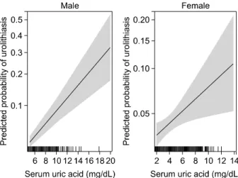
Figure 3. Effect plots showing the predicted probabilities and 95% confidence intervals (represented by shaded areas or er- ror bars) for urolithiasis based on the multivariable logistic re- gression model.
cantly increased in both male and female subgroup (Figure 2). Comparing the highest with the lowest quar- tile of serum UA, the multivariable-adjusted OR were 3.17 (95% CI, 1.98∼5.11) for men and 1.79 (1.08∼2.93) for women (Figure 2). Predicted probability for ur- olithiasis based on the multivariable logistic regression model was shown in Figure 3.
DISCUSSION
Whereas a relatively large number of studies showed the association between gout and urolithiasis [5-7,11], few reports have investigated the independent association be- tween serum UA level and urolithiasis. Hyperuricemia is known to be an underlying metabolic disorder causing monosodium urate crystal formation and it is reasonable to hypothesize that the prevalence of urolithiasis is re- lated to the serum UA levels. Our investigation of 13,964 cases revealed that the overall detection rate of ur- olithiasis by ultrasonography increased proportionally to serum UA level. After adjusting for potential confounders such as age, sex, BMI, GFR, diabetes mellitus and hyper- tension, we found that serum UA level was independently associated with the presence of urolithiasis.
Most of the daily uric acid excretion (65%∼75%) occurs through kidneys. Previous study showed that hyper- uricemia, in about 90% of cases, was due to an impaired renal excretion [12]. Conversely, it was known that hy- peruricemia affects kidney function deterioration. There were several mechanisms by which uric acid may be caus- ing these effects. Firstly, uric acid was associated with ac-
tivation of the renin–angiotensin system, with the devel- opment of arteriolosclerosis and glomerular hypertrophy [13]. Moreover, uric acid might cause mitochondrial dys- function by acting a pro-oxidant inside the cell to induce stimulation of nicotinamide adenine dinucleotide phos- phate (NADPH) oxidases and uric acid can also induce endothelial dysfunction via stimulating the release of alarmins from endothelial cells that activate Toll-like re- ceptor pathways [14].
Several imaging modalities are used for investigating the prevalence of urolithiasis. Many factors have an effect on choosing the imaging tool including the clinical setting, patient’s preference, cost and tolerance of radiation.
Although by no means as sensitive as computed tomog- raphy (CT), ultrasonography is a frequently used tool be- cause of its lower expense compared to CT and lack of ra- diation exposure, in spite of several disadvantages such as user dependency or poor visualization of stones in the ureter. Furthermore, detection of stones by ultrasono- graphy is relatively easy, regardless of their radiolucency [15].
However, while ultrasonography could detect the radio- lucent stone, it is difficult to use it distinguish the type of urolithiasis. About 80 percent of patients with urolithiasis formed calcium containing stones, in which most stones were composed primarily of calcium oxalate, followed by calcium phosphate [16]. Pure UA stones account for about 10%∼15% of urolithiasis [3]. Some patients may have more than one type of stone concurrently, for exam- ple mixed calcium oxalate and UA stones. Moreover, in- creased UA could well contribute to the formation of cal- cium oxalate stones [17]. Therefore, the higher detection rate of urolithiasis in individuals with the higher serum UA level in our study was thought to be related to the in- crease in mixed urolithiasis as well as pure UA stone.
Uric acid is a frequent component of urinary stones and can also contribute to calcium containing stone formation as well as pure uric acid stone. The main mechanism of uric acid stone was the supersaturation of urinary uric acid, which was more likely to occur when urinary pH was less than 5.5 and when urinary volume was decreased.
The solubility of urate salts is also affected by the relative concentrations of cations in the urine. Increased urinary sodium concentrations promote formation of the mono- sodium urate complex [4]. In addition to uric acid stone, hyperuricemia may contribute to calcium oxalate stone formation via heterogeneous nucleation, the primary mechanism of uric acid containing calcium nephroli-
thiasis [18].
Our present study had some limitations. First, our anal- ysis was a retrospective investigation from a single ter- tiary hospital. Therefore, even though a relatively large number of individuals participated in our study, there might be some selection bias: for instance, more in- dividuals who have one or more co-morbidities might be included in this study than those in the general population.
Thus, this study population could not fully represent a general population. Second, as with previous epidemio- logical studies, we could not distinguish the type of urolithiasis. So, the nephrolithiasis, which was less re- lated with hyperuricemia (for example, magnesium am- monium phosphate stone, cystine stone and cholesterol stone), may also be included in our results. Third, we could not adjust for medication use or dietary habits which could also influence the serum UA level and stone formation: medications as such as diuretics, salicylates, warfarin and xanthine oxidase inhibitor or dietary factors such as alcohol, meat, sodium and calcium. Forth, be- cause we only checked the presence of urolithiasis at the time of ultrasonographic examination, regardless of ur- olithiasis symptoms, it is possible that passed stones were not detected, resulting in an underestimation for the detection rate of urolithiasis.
However, this study was based on objective data ob- tained from laboratory and radiographic evaluation, not on survey data which could have recall bias. Also, this study is from a large number of individuals with asympto- matic hyperuricemia and those with subclinical urolithiasis.
Therefore, in this study we demonstrated an independent relationship between serum UA level and urolithiasis, which has not yet been well established.
CONCLUSION
This study suggests that individuals with a higher serum UA level had a higher risk of having incidental urolithiasis proportionally. This finding may provide a basis for estab- lishing appropriate screening and preventive strategies for hyperuricemia and its complication.
ACKNOWLEDGMENTS
This work was funded by Ulsan University Hospital, Biomedical Research Center Promotion Fund (number.
1503027).
CONFLICT OF INTEREST
No potential conflict of interest relevant to this article was reported.
REFERENCES
1. Scales CD Jr, Smith AC, Hanley JM, Saigal CS. Prevalence of kidney stones in the United States. Eur Urol 2012;62:160-5.
2. Parmar MS. Kidney stones. BMJ 2004;328:1420-4.
3. Mehta TH, Goldfarb DS. Uric acid stones and hyper- uricosuria. Adv Chronic Kidney Dis 2012;19:413-8.
4. Shekarriz B, Stoller ML. Uric acid nephrolithiasis: current concepts and controversies. J Urol 2002;168:1307-14.
5. Yü T, Gutman AB. Uric acid nephrolithiasis in gout.
Predisposing factors. Ann Intern Med 1967;67:1133-48.
6. Kramer HM, Curhan G. The association between gout and nephrolithiasis: the National Health and Nutrition Examination Survey III, 1988-1994. Am J Kidney Dis 2002;
40:37-42.
7. Kramer HJ, Choi HK, Atkinson K, Stampfer M, Curhan GC.
The association between gout and nephrolithiasis in men:
The Health Professionals' Follow-Up Study. Kidney Int 2003;64:1022-6.
8. Madore F, Stampfer MJ, Willett WC, Speizer FE, Curhan GC. Nephrolithiasis and risk of hypertension in women. Am J Kidney Dis 1998;32:802-7.
9. Roubenoff R, Klag MJ, Mead LA, Liang KY, Seidler AJ, Hochberg MC. Incidence and risk factors for gout in white men. JAMA 1991;266:3004-7.
10. Fox J. Effect displays in R for generalised linear models. J Stat Softw 2003;8:1-27.
11. Roughley MJ, Belcher J, Mallen CD, Roddy E. Gout and risk of chronic kidney disease and nephrolithiasis: meta-analysis of observational studies. Arthritis Res Ther 2015;17:90.
12. Richette P, Bardin T. Gout. Lancet 2010;375:318-28.
13. Mazzali M, Hughes J, Kim YG, Jefferson JA, Kang DH, Gordon KL, et al. Elevated uric acid increases blood pressure in the rat by a novel crystal-independent mechanism.
Hypertension 2001;38:1101-6.
14. Johnson RJ, Nakagawa T, Jalal D, Sánchez-Lozada LG, Kang DH, Ritz E. Uric acid and chronic kidney disease: which is chasing which? Nephrol Dial Transplant 2013;28:2221-8.
15. Brisbane W, Bailey MR, Sorensen MD. An overview of kid- ney stone imaging techniques. Nat Rev Urol 2016;13:654- 62.
16. Teichman JM. Clinical practice. Acute renal colic from ure- teral calculus. N Engl J Med 2004;350:684-93.
17. Coe FL. Uric acid and calcium oxalate nephrolithiasis.
Kidney Int 1983;24:392-403.
18. Coe FL, Lawton RL, Goldstein RB, Tembe V. Sodium urate accelerates precipitation of calcium oxalate in vitro. Proc Soc Exp Biol Med 1975;149:926-9.