Breast Cancer
O R I G I N A L A R T I C L E
INTRODUCTION
Nonpalpable breast cancers have a better prognosis than palpable breast cancers, and a higher incidence of ductal carcinoma in situ (DCIS), a lower tumor stage, and a lower incidence of lymph node metastasis.(1-7) Currently, mammography has been used with clinical breast examination and breast self-examination for screening of breast cancer.
Mammography has been successfully used as a screen- ing test for breast cancer over the past 20-30 yr, but has substantial limitations. Approximately 10-20% of
palpable breast cancers are not visible on mammograph- ic images, mainly due to insufficient contrast between normal and abnormal breast tissue.(8-10) Moreover, the sensitivity of mammography for the diagnosis of breast cancer is variable and known to be influenced by age, breast density, family history, and other factors.(11) False negative rates for mammographic breast cancer detec- tion are higher in women with dense breast parenchy- ma, and the risks of subsequent breast cancer are also higher, particularly in women with a first-degree family history of breast cancer.(12-14)
Breast ultrasound (US) is widely used as a supplemen- tary modality for evaluating mammographically detected abnormalities,(15-17) and as an effective screening modal- ity for detecting occult breast cancers in mammographi- cally determined dense breasts.(15, 17-21) A study by Stavros et al.(22) reported that US has a high sensitivity and negative predictive value for diagnosing breast cancer, i.e., 98.4% and 99.5%, respectively, and recent advances Purpose: The aim of this study was to investigate any dif-
ference of ultrasound findings for palpable and nonpalpable breast cancers.
Methods: Two hundred breast cancer patients that had undergone preoperative ultrasound and surgery were en- rolled in the study. A total of 126 cancers were palpable, and the remaining 74 cancers were nonpalpable. We com- pared lesion characteristics using ultrasound images accord- ing to the BI-RADS��-Ultrasound guidelines of the American College of Radiology. A crude odds ratio (OR) and 95% con- fidence interval (CI) were calculated for a comparison of the palpable and nonpalpable breast cancers.
Results: Nonpalpable cancers displayed more often an oval shape (OR=0.35, 95% CI=0.17-0.70), no posterior acoustic features (OR=0.50, 95% CI=0.28-0.89), and a parallel ori- entation (OR=0.50, 95% CI=0.28-0.89). An irregular shape (OR=2.98, 95% CI=1.60-5.54), a spiculated margin (OR=
2.66, 95% CI=1.23-5.74), and a combined pattern of poste- rior acoustic features (OR=7.20, 95% CI=1.64-31.66) were more commonly observed in the palpable cancers.
Conclusion: Palpable and nonpalpable breast cancers were found to have different ultrasound characteristics.
Key Words : Breast, Breast neoplasms, Palpation, Ultrasonography, Mammary
A Comparative Study of Palpable and Nonpalpable Breast Cancers determined by Ultrasonography
Kyu Ran Cho, Bo Kyoung Seo
1, Juneyoung Lee
2, Ki Yeol Lee
1, Bo Kyung Je
1, Baek Hyun Kim
1, Sang Hoon Cha
1, Yu Whan Oh, Seokjin Kim
3, Jeoung Won Bae
4Department of Radiology, Korea University Anam Hospital, Korea University College of Medicine, Seoul; 1Department of Radiology, Korea University Ansan Hospital, Korea University College of Medicine, Ansan; 2Department of Biostatistics, Korea University College of Medicine, Seoul; 3Division of Oncology/Hematology, Departments of Internal Medicine and 4Surgery, Korea University Anam Hospital, Korea University College of Medicine, Seoul, Korea
Correspondence : Bo Kyoung Seo
425-707 Department of Radiology, Korea University College of Medicine, Korea University Ansan Hospital, 516 Kozan 1-dong, Danwon-gu, Ansan, Korea
Tel: 031-412-5228, Fax: 031-412-5224 E-mail : [email protected]
Received : March 6th 2008 Accepted : March 27th 2008
*This study was supported by a Korean Institute of Medicine.
64
in US technology and transducer design permit greater spatial and contrast resolution. Therefore, we decided to investigate the correlation between breast US and clinical findings in patients with breast cancer. Specifically, this study was undertaken to evaluate US findings in palpable and nonpalpable breast cancers and to identify US features that differentiate these two cancer groups.
METHODS
1. Patients
This study was approved by the institutional review board for human investigation. We searched patients who underwent surgery for primary breast cancers at our cancer center from January 2002 to August 2004 using a computer data base system. The total number of patients that had undergone breast cancer surgery during this period was 370. Two hundred nineteen pati- ents underwent modified radical mastectomy, 151 breast conservation therapy, and 309 axillary lymph node dis- section. Of these 370 patients, 201 patients underwent breast US before surgery and 191 patients had an abnor- mal US finding, and these 191 patients were included in this study. All patients were female and ages ranged from 27 to 82 yr (mean, 48 yr).
Breast cancers were considered palpable if they were detected by physical examination by at least one physi- cian before treatment.(6, 18) Patients at our institution are routinely examined by multiple experienced clinicians before treatment, i.e., radiologists, surgeons, medical oncologists, and plastic surgeons. If a lesion was detect- ed by any physician, then it was scored as a palpable breast cancer. Nonpalpable cancers were identified using abnormal imaging study findings. One hundred twenty one of the 191 patients had palpable breast cancer and the remaining 70 patients, nonpalpable breast cancer. Of the 70 patients with nonpalpable breast cancer, 14 patients had nipple discharge and 3 patients had skin eczema. In total the 191 patients had 200 breast cancers, 167 of which were invasive carcinomas and 33 were DCIS cases. Nine patients had ipsilateral multiple breast cancers.
One hundred four of 191 patients obtained mammo-
graphy and 99 patients were available for evaluation of mammography. The mammographic findings of these 99 patients were: mass in 38, mass with calcifications in 22, focal asymmetry in 10, architectural distortion in 1, calcifications only in 8, and no lesion in 20.
2. Evaluation points
In our institute, US is routinely performed as an initial examination for palpable masses in women younger than 35 yr, and for nonpalpable masses detected by mammo- graphy, to allow mass characterization. US is also per- formed to screen women with mammographically dense breasts. One hundred ninety one patients in this study underwent breast US to further evaluate; abnormal mammographic findings (n=73), to screen mammograph- ically dense breasts (n=47), and those with symptoms but without specific mammographic abnormalities (n=10), and as an initial examination in young women (n=61).
All patients were examined using a Logiq9 unit (General Electronic Medical System, Milwaukee, USA) or a HDI 5000 unit (Advanced Technology Laboratories, Bothell, USA) using a broad-bandwidth (14-5 MHz) and a linear scanhead. Entire breasts were scanned by one of three radiologists, their experiences of breast imaging ranged from 3 to 10 yr. When a lesion was detected, it was saved in two different projections (transverse and longitudinal projections). A breast radiologist, 7 yr experience in breast imaging, reviewed the soft copies of US. Lesion sizes and characteristics were evaluated on a PACS sys- tem (StarPACS; Infinitt, Seoul, Korea). Lesion sizes were expressed as mean size±standard deviations. Tumor characteristics were assessed using the BI-RADS�-US lexicon (23): shape (oval, round, or irregular), margin (circumscribed, indistinct, microlobulated, angular, or spiculated), echo pattern (hypoechoic, isoechoic, or hyper- echoic), posterior acoustic features (no posterior acoustic features, enhancement, shadowing, or combined pat- tern), orientation (parallel or not parallel to the skin), lesion boundary (abrupt interface or echogenic halo), and presence of microcalcifications.
One phyisician reviewed the clinical and pathologic reports of all 191 patients. Tumor staging was performed
according to the TNM classification of breast cancer.(24) Primary tumor (T stage), regional nodal status (N stage), and metastasis (M stage) were evaluated. Data were col- lected on paper and then entered into a customized data- base (Microsoft�Access 2.0; Microsoft, Redmond, USA).
3. Statistical analysis
To compare the clinical and US findings of palpable and nonpalpable breast cancers, the chi-squared homo- geneity test and the student’s t-test were used, as app- ropriate. Statistical significance was considered if a p value was less than 0.05. A crude odds ratio (OR) and its 95% confidence interval (CI) were calculated for the presence of a palpable cancer based on each US finding.
All statistical analysis was performed using SAS version 9.12 (SAS Inc., Cary, USA). The statistical analysis of the data was supervised by a biostatistician.
RESULTS
Table 1 presents patient characteristics. Patients’ages
were not different between the two groups (p=0.39). T and N stages were significantly poorer for palpable than nonpalpable breast cancers (p<0.05). Frequency of DCIS (Tis) was much higher for nonpalpable cancers (22/70, 31% vs 11/121, 9%), and lymph node negativity (N0) was more frequent in nonpalpable cancers (53/70, 76% vs 64/121, 53%). Overall breast cancer stage was also sig- nificantly poorer for palpable breast cancers (p<0.05).
Moreover, the frequency of early cancer, stage 0 or 1, was significantly higher for nonpalpable cancers (p<0.05).
By breast US, the mean lesion size of palpable cancers was 27.03±20.56 mm and of nonpalpable cancers was 12.64±5.19 mm, and this difference was statistically significant (p<0.0001). By pathologic examination, mean
Table 1. Patient characteristics
Characteristic
Palpable cancers (n=121)
Nonpalpable cancers
(n=70)
p- value
Age 47.79±10.72 yr 49.13±9.36 yr 0.39 TNM classification
Primary tumor <0.05
Tis 11 (9) 22 (31)
T1 64 (53) 39 (56)
T2 36 (30) 8 (11)
T3 7 (6) 0 (0)
T4 3 (2) 1 (1)
Lymph node <0.05
N0 64 (53) 53 (76)
N1 46 (38) 15 (21)
N2 11 (9) 2 (3)
Metastasis
M1 0 (0) 0 (0)
Overall staging <0.05
Stage 0 11 (9) 22 (31)
Stage 1 29 (24) 27 (39)
Stage 2 65 (54) 19 (27)
Stage 3 16 (13) 2 (3)
T=primary tumor; N=lymph node; M=metastasis.
Data are numbers (percentages) of patients.
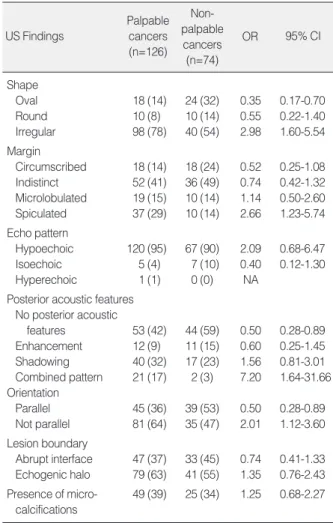
Table 2. US findings of palpable and nonpalpable breast can- cers
US Findings
Palpable cancers (n=126)
Non- palpable
cancers (n=74)
OR 95% CI
Shape
Oval 18 (14) 24 (32) 0.35 0.17-0.70
Round 10 (8) 10 (14) 0.55 0.22-1.40
Irregular 98 (78) 40 (54) 2.98 1.60-5.54 Margin
Circumscribed 18 (14) 18 (24) 0.52 0.25-1.08 Indistinct 52 (41) 36 (49) 0.74 0.42-1.32 Microlobulated 19 (15) 10 (14) 1.14 0.50-2.60 Spiculated 37 (29) 10 (14) 2.66 1.23-5.74 Echo pattern
Hypoechoic 120 (95) 67 (90) 2.09 0.68-6.47
Isoechoic 5 (4) 7 (10) 0.40 0.12-1.30
Hyperechoic 1 (1) 0 (0) NA
Posterior acoustic features No posterior acoustic
features 53 (42) 44 (59) 0.50 0.28-0.89 Enhancement 12 (9) 11 (15) 0.60 0.25-1.45 Shadowing 40 (32) 17 (23) 1.56 0.81-3.01 Combined pattern 21 (17) 2 (3) 7.20 1.64-31.66 Orientation
Parallel 45 (36) 39 (53) 0.50 0.28-0.89 Not parallel 81 (64) 35 (47) 2.01 1.12-3.60 Lesion boundary
Abrupt interface 47 (37) 33 (45) 0.74 0.41-1.33 Echogenic halo 79 (63) 41 (55) 1.35 0.76-2.43 Presence of micro- 49 (39) 25 (34) 1.25 0.68-2.27
calcifications
US=Ultrasonography; OR=crude odd ratio; CI=confidence interval;
NA=not applicable. Data are numbers (percentages) of cancers.
tumor size of palpable cancers was 32.41±73.43 mm and of nonpalpable cancers 17.89±13.48 mm and this
was significantly different (p<0.05).
Table 2 summarizes the US findings of palpable and
Fig 1. 64-yr-old woman with a palpable cancer. US shows a spiculated, irregular shaped, hypoechoic mass (arrows) that is not parallel to the skin, and which has a combined pattern of posterior acoustic feature. On pathologic examination, the mass proved to be a 12 mm sized invasive ductal carcinoma.
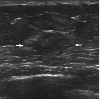
Fig 3. 48-yr-old woman with a nonpalpable cancer. US shows a microlobulated, oval, isoechoic mass (arrows) that is parallel to the skin. The mass has no posterior acoustic feature. On patho- logic examination, the mass proved to be an invasive ductal carcinoma with low grade DCIS of size 6 mm.
Fig 2. 71-yr-old woman with a palpable cancer. US demonstrates a microlobulated, irregular shaped, hypoechoic mass (arrows) that is not parallel to the skin. This mass has a combined pattern of posterior acoustic feature. On pathologic examination, the mass proved to be a mixed invasive ductal and lobular carcinoma of size 22 mm.
Fig 4. 52-yr-old woman with a nonpalpable breast cancer. US shows a circumscribed, oval, hypoechoic mass (arrows) lying parallel to skin. On pathologic examination, the mass proved to be an invasive ductal carcinoma with high grade DCIS of size 12 mm.
nonpalpable breast cancers. In terms of lesion character- istics by US, shape, margin, posterior acoustic features, and orientation were significantly different between the two study groups (p<0.05). An “irregular”shape was more frequent for palpable cancers (98/126, 78% vs 40/
74, 54%; OR=2.98, 95% CI=1.60-5.54) (Fig 1, 2), whereas an “oval”shape was more common for nonpalpable can- cers (24/74, 32% vs 18/126, 14%; OR=0.35, 95% CI=0.17- 0.70) (Fig 3, 4). In lesion margins, a “spiculated”margin was significantly more commonly observed in palpable cancers (37/126, 29% vs 10/74, 14%; OR=2.66, 95% CI=
1.23-5.74) (Fig 1). With respect to posterior acoustic fea- tures, a “combined pattern”was more frequent for pal- pable cancers (21/126, 17% vs 2/74, 3%; OR=7.20, 95%
CI=1.64-31.66) (Fig 1, 2), whereas “no posterior acoustic features”was more common for nonpalpable cancers (44/
74, 59% vs 53/126, 42%; OR=0.50, 95% CI=0.28-0.89) (Fig 3). A “not-parallel”orientation to skin was more common for palpable cancers (81/126, 64% vs 35/74, 47%; OR=2.01, 95% CI=1.12-3.60) (Fig 1, 2) and a “paral- lel”orientation was more common in nonpalpable can- cers (39/74, 53% vs 45/126, 36%; OR=0.50, 95% CI=0.28- 0.89) (Fig 3, 4). An “echogenic halo”(Fig 1, 2) and the
“presence of microcalcifications”were more frequent in palpable than in nonpalpable cancers, but this was with- out significance (p>0.05).
DISCUSSION
The most important roles of US in breast imaging are;
the characterization of masses that have been incom- pletely assessed by mammography, the characterization of palpable masses that are obscured by dense tissue during mammography, and screening for occult breast cancers in dense breasts by mammography. Breast US has been successfully used to differentiate benign and malignant breast lesions and to detect occult breast can- cers in dense breasts.(15-22) Our question was that breast US would correlate with clinical findings and prognosis.
There are a few reports to show correlation between tumor type or grade and US findings in breast cancers.
(25, 26) Moon et al.(26) reported that DCIS cases have
less often typical malignant US findings, thus, radiolo- gists or sonographer may misunderstand the lesion as benign. In this study, we investigated the correlation of clinical palpability with breast US findings. It has been widely reported that palpable and nonpalpable breast cancers have different clinicopathological findings and prognoses.(1-5, 7) Skinner et al.(6) demonstrated that palpability is correlated with pathologic tumor size, mitotic grade, nuclear grade, lymphovascular invasion, nodal positivity, and the lack of an extensive intraductal component, multifocality, and multicentricity. They sug- gested that palpable cancers inherently differ from non- palpable cancers, and that they have a less diffuse growth pattern, higher metastatic potential, higher proliferative activity, more nuclear abnormalities, and a poorer pro- gnosis. Moreover, the prognosis of nonpalpable cancers is better and their recurrence rate lower than those of palpable cancer.(2-5) Our results also demonstrate pal- pable breast cancers have a larger primary tumor size, a higher tumor stage, and are more frequently associat- ed with lymph node metastasis.
In the present study, we evaluated differences between palpable and nonpalpable cancers in terms of their US characteristics; shape, margin, echo pattern, posterior acoustic features, orientation, lesion boundary, and pre- sence of microcalcifications. Among these US charac- teristics, shape, margin, posterior acoustic feature, and orientation differed between the two study groups. An
“irregular”shape (OR=2.98), “spiculated”margin (OR
=2.66), “combined pattern”of posterior acoustic feature (OR=7.20) and “not-parallel”orientation (OR=2.01) were more frequent for palpable breast cancers; moreover, these are typical breast cancer US findings (22). Patho- logic examinations show that palpable cancers are larger and have a higher stage than nonpalpable cancers, and that various internal components, such as, necrosis or hemorrhage, are more likely in palpable cancers. These typical US findings are caused by an enlarging tumor, tumor invasion to surrounding tissue, and various inter- nal components. An “irregular”shape, a “spiculated”
margin, and a “not-parallel”orientation are related with a stellate configuration, which is associated with desmo-
plastic reaction indicating tumor-associated fibrosis.
Patients with abundant desmoplastic reaction show more frequent lymphovascular invasion, lymph node metas- tasis, and angiogenesis than those with a poor desmo- plastic reaction, and thus a poorer prognosis.(27) A “com- bined pattern”of posterior acoustic feature means a combination of shadowing and enhancement. Shadow- ing is the result of sound beam attenuation by desmo- plastic host response to breast cancer.(22) Enhancement is caused by highly cellular tumors and necrotic can- cers.(22) Thus, a “combined pattern”of posterior acoustic feature in palpable cancers might be related with the presence of various cellular components, desmoplastic reaction, necrosis, and hemorrhage caused by angiogen- esis. Thus, the different breast US characteristics of pal- pable and nonpalpable breast cancers might be related with pathologic differences and in turn be related with prognosis.
There are many reports to describe the usefulness of US in nonpalpable breast lesions for detection of malig- nancy and imaging-guided procedure.(15, 17-21, 28) However, there is no report to show that distinctive US findings for nonpalpable breast cancers when compared with palpable breast cancers. Our results demonstrated nonpalpable breast cancers had more often typical benign US findings; an “oval”shape, a “circumscribed”margin,
“no posterior acoustic features”, a “parallel”orientation, and an “abrupt”interface. Among these findings, an
“oval”shape, “no posterior acoustic features”, and a “par- allel”orientation were significantly different between palpable and nonpalpable breast cancers. Therefore, physicians must consider these results before assessment of nonpalpable breast lesions on US and it may be nec- essary of more strict application of US criteria to non- palpable breast lesions. If a nonpalpable lesion has a slight malignant feature on US, biopsy or aspiration should be considered.
The present study has several limitations. First, the study is limited by its retrospective nature. We included patients who underwent breast cancer surgery and breast US, and the total number of patients was small. Thus, a further prospective study in a larger population may be
warranted. Second, the characteristics of palpable and nonpalpable cancers were compared by breast US, but these different US characteristics were not correlated with pathologic findings. Third, we included both inva- sive carcinomas and DCIS cases, thus, the study popu- lation was heterogeneous and it could produce biased results. In the 200 breast cancers, 167 were invasive carcinomas and 33 were DCIS cases. We hope further study would be performed in separate groups of histo- logical tumor types.
CONCLUSION
In conclusion, the present study shows that the char- acteristics of palpable and nonpalpable cancers are quite different by breast US. Further study would be needed for investigation of the correlations between breast US findings and prognostic estimates in a large population.
REFERENCES
1. Bassett LW, Liu TH, Giuliano AE, Gold RH. The prevalence of carcinoma in palpable vs impalpable, mammographically detected lesions. AJR Am J Roentgenol 1991;157:21-4.
2. Pagana TJ, Lubbe WJ, Schwartz SM, Sprechini GD. A comparison of palpable and nonpalpable breast cancers. Arch Surg 1989;124:26-8.
3. Perdue P, Page D, Nellestein M, Salem C, Galbo C, Ghosh B. Early detection of breast carcinoma: a comparison of palpable and nonpal- pable lesions. Surgery 1992;111:656-9.
4. Silverstein MJ, Gamagami P, Masetti R, Legmann MD, Craig PH, Gierson ED. Results from a multidisciplinary breast center. Analysis of disease discovered. Surg Oncol Clin N Am 1997;6:301-14.
5. Silverstein MJ, Gierson ED, Waisman JR, Colburn WJ, Gamagami P.
Predicting axillary node positivity in patients with invasive carcinoma of the breast by using a combination of T category and palpability. J Am Coll Surg 1995;180:700-4.
6. Skinner KA, Silberman H, Sposto R, Silverstein MJ. Palpable breast cancers are inherently different from nonpalpable breast cancers. Ann Surg Oncol 2001;8:705-10.
7. Tafra L, Essner R, Brenner RJ, Giuliano AE. Nonpalpable versus palpable invasive breast tumors treated with breast-conserving surgical management. Am Surg 1996;62:395-9.
8. Burrell HC, Sibbering DM, Wilson AR, Pinder SE, Evans AJ, Yeoman LJ, et al. Screening interval breast cancers: mammographic features and prognosis factors. Radiology 1996;199:811-7.
9. Hollingsworth AB, Taylor LD, Rhodes DC. Establishing a histologic basis for false-negative mammograms. Am J Surg 1993;166:643-7.
10. Laya MB, Larson EB, Taplin SH, White E. Effect of estrogen replace- ment therapy on the specificity and sensitivity of screening mam- mography. J Natl Cancer Inst 1996;88:643-9.
11. Kerlikowske K, Grady D, Barclay J, Sickles EA, Ernster V. Effect of age, breast density, and family history on the sensitivity of first screening mammography. JAMA 1996;276:33-8.
12. Oza AM, Boyd NF. Mammographic parenchymal patterns: a marker of breast cancer risk. Epidemiol Rev 1993;15:196-208.
13. Saarenmaa I, Salminen T, Geiger U, Heikkinen P, Hyvarinen S, Isola J, et al. The effect of age and density of the breast on the sensitivity of breast cancer diagnostic by mammography and ultasonography.
Breast Cancer Res Treat 2001;67:117-23.
14. Saftlas AF, Wolfe JN, Hoover RN, Brinton LA, Schairer C, Salane M, et al. Mammographic parenchymal patterns as indicators of breast cancer risk. Am J Epidemiol 1989;129:518-26.
15. Berg WA, Gilbreath PL. Multicentric and multifocal cancer: whole- breast US in preoperative evaluation. Radiology 2000;214:59-66.
16. Buchberger W, DeKoekkoek-Doll P, Springer P, Obrist P, Dunser M. Incidental findings on sonography of the breast: clinical signifi- cance and diagnostic workup. AJR Am J Roentgenol 1999;173:921-7.
17. Gordon PB, Goldenberg SL. Malignant breast masses detected only by ultrasound. A retrospective review. Cancer 1995;76:626-30.
18. Crystal P, Strano SD, Shcharynski S, Koretz MJ. Using sonography to screen women with mammographically dense breasts. AJR Am J Roentgenol 2003;181:177-82.
19. Kaplan SS. Clinical utility of bilateral whole-breast US in the eval- uation of women with dense breast tissue. Radiology 2001;221:641-9.
20. Kolb TM, Lichy J, Newhouse JH. Occult cancer in women with
dense breasts: detection with screening US--diagnostic yield and tumor characteristics. Radiology 1998;207:191-9.
21. Leconte I, Feger C, Galant C, Berliere M, Berg BV, D’Hoore W, et al. Mammography and subsequent whole-breast sonography of non- palpable breast cancers: the importance of radiologic breast density.
AJR Am J Roentgenol 2003;180:1675-9.
22. Stavros AT, Thickman D, Rapp CL, Dennis MA, Parker SH, Sisney GA. Solid breast nodules: use of sonography to distinguish between benign and malignant lesions. Radiology 1995;196:123-34.
23. American College of Radiology. BI-RADS Committee. Breast ultrasound (1st ed). In: American College of Radiology, BI-RADS Committee, editors. ACR BI-RADS breast imaging and reporting data system: breast imaging atlas. Reston, VA: American College of Radiology; 2003.
24. Thurfjell MG, Lindgren A, Thurfjell E. Nonpalpable breast cancer:
mammographic appearance as predictor of histologic type. Radiology 2002;222:165-70.
25. Lamb PM, Perry NM, Vinnicombe SJ, Wells CA. Correlation bet- ween ultrasound characteristics, mammographic findings and his- tological grade in patients with invasive ductal carcinoma of the breast. Clin Radiol 2000;55:40-4.
26. Moon WK, Myung JS, Lee YJ, Park IA, Noh DY, Im JG. US of ductal carcinoma in situ. Radiographics 2002;22:269-80.
27. Colpaert C, Vermeulen P, van Beest P, Goovaerts G, Weyler J, Van Dam P, et al. Intratumoral hypoxia resulting in the presence of a fibrotic focus is an independent predictor of early distant relapse in lymph node-negative breast cancer patients. Histopathology 2001;
39:416-25.
28. Chiou SY, Chou YH, Chiou HJ, Wang HK, Tiu CM, Tseng LM, et al. Sonographic features of nonpalpable breast cancer: a study based on ultrasound-guided wire-localized surgical biopsies. Ultrasound Med Biol 2006;32:1299-306.