Copyright © 2013 The Korean Society for Bone and Mineral Research
This is an Open Access article distributed under the terms of the Creative Commons Attribution Non-Commercial Li- cense (http://creativecommons.org/licenses/by-nc/3.0/) which permits unrestricted non-commercial use, distribu- tion, and reproduction in any medium, provided the original work is properly cited.
Analysis of Clinical Features of Hip Fracture Patients with or without Prior Osteoporotic Spinal
Compression Fractures
Gang Deuk Kim1, Yeung Jin Kim2, Soo Uk Chae2, Deok Hwa Choi3
Departments of 1Radiology, 2Orthopeadic Surgery, and 3Anesthesiology and Pain Medicine, School of Medicine, Wonkwang University Hospital, Iksan, Korea
Background: To analyze and compare the clinical characteristics including bone mineral density (BMD) in a group who had operation of hip fracture with or without prior osteo- porotic spinal compression fractures. Methods: Two hundred forty patients who had undergone operation of hip fractures were evaluated, 127 patients who had with prior osteoporotic spinal compression fractures were in group I, and 113 patients without pri- or spinal fractures were in group II. In each group, we measured age, gender, body mass index (BMI, kg/m2), BMD (mg/cm3), type of hip fractures, concomitant diseases, presence of secondary hip fracture and history of percutaneous vertebroplasty. Results: The mean age of group I was 79.4 years (male/female: 28/99) and that of group II was 77.6 years (male/female: 37/76). The mean BMI of group I was 21.3 kg/m2 and that in group II was 22.0 kg/m2. The mean BMD and T-score of group I were 41.1 mg/cm3 and -4.45 and those in group II were 51.0 mg/cm3 and -4.17 (P<0.05). The numbers of patients of neck and intertrochanter fracture of group I were 31 and 96 patients and those in group II were 61 and 52 patients. Sixty in group I and 45 in group II patients had concomitant diseases.
Thirteen patients had undergone percutaneous vertebroplasty and 18 patients (7.5%) had second hip fractures. Conclusions: The hip fracture patients who had with prior os- teoporotic spinal compression fractures had lower BMD compared to the hip fracture patients without previous spinal compression fractures.
Key Words: Bone density, Hip fractures, Spinal fractures
INTRODUCTION
Recent studies reported increasing risks of hip, spine, and others among people who had previous clinically diagnosed fractures, or radiographic evidence of ver- tebral fractures.[1,2] Also, spinal radiographs identifying of prevalent vertebral deformities may be useful additional measurement to classify further a women’s risk of future fracture.[1] Women with preexisting vertebral fractures had approxi- mately 4 times higher risk of subsequent vertebral fractures and 2.3 times greater risk of subsequent hip fractures than those without prior fractures.[2]
Percutaneous vertebroplasty is one of the most commonly performed proce- dures for symptomatic osteoporotic spinal fractures.[3,4] But, subsequent fracture of spine is a potential complication of vertebroplasty.[3,5] There are many reports Corresponding author
Soo Uk Chae
Department of Orthopedic Surgery, School of Medicine, Wonkwang University, 27 Uiryowon-ro, Gunsan 573-713, Korea Tel: +82-63-472-5100
Fax: +82-63-472-5688 E-mail: [email protected] Received: January 11, 2013 Revised: April 10, 2013 Accepted: April 10, 2013
No potential conflict of interest relevant to this article was reported.
This paper was supported by Wonkwang University in 2013.
Original Article
of subsequent vertebral fracture while there are only a few reports of non-vertebral fracture, especially, hip fractures af- ter percutanous vertebroplasty in osteoporotic spinal com - pression fracture. This present study aim to analyze and compare the clinical characteristics of bone mineral density (BMD) in a group of hip fracture patients whether or not pri- or osteoporotic spinal compression fractures by cohort ret- rospective study. Additionally, whether or not of secondary hip fracture and history of percutaneous vertebroplasty in both groups were investigated.
METHODS
We reviewed 240 patients over 65 years of age among 436 cases who had undergone operation of intertrochan- teric and neck of hip fractures in one hospital by retrospec- tive study from June 2006 to June 2012. We excluded pa- tients who had hip fracture by high velocity injury such as a motor vehicle accident or falling down from height, those with under 65 years of age, unexamined BMD, and previ- ous spine fusion operation. We checked BMD and simple thoracolumbar radiograph of whole patients at the time of injury. The patients were divided into prior osteoporotic spinal compression fractures in group I (127 patients, male/
female: 28/99) and without compression fractures in group II (113 patients, male/female: 37/76). In each group, we measured age and gender, body mass index (BMI, kg/m2) with weight and height, type of hip fractures, concomitant diseases, and presence of secondary hip fracture and un- dergone percutaneous vertebroplasty. The fractures were classified as hip fractures in intertrochanteric and femoral neck fractures. The definition of second hip fracture was patients who have suffered a hip fracture with a subse- quent fracture of the contralateral hip. The included con- comitant diseases were hypertension, diabetes mellitus, cardiac disease, and senile dementia.
Assessment of prior radiographic vertebral fractures has been by a radiologist and two orthopedic surgeons. Six points digitations on each vertebral body of the thoracic and lumbar lateral radiograph for quantitative morphome- try were done as described previously, so that the anterior, middle, and posterior heights could be accurately mea- sured.[6,7] Baseline prevalent deformities were defined us- ing a modified Melton/Eastell vertebral height ratio crite- ria.[6] BMD was measured utilizing peripheral-quantitative
computed tomography (P-QCT; Somatom sensation 16, Si- mens, Erlangen, Germany) of thoracolumbar vertebrae that enables separate analyses of cortical, trabecular and total bone density including T-score.
Quantitative variables (age, BMI, BMD and T-score) were compared between groups by an independent T-test and difference in the categorical variables (sex, hip fracture type and concomitant disease) were compared using the chi-square test. The Mann-Whitney U test for continuous variables was used. SPSS version 20.0 (SPSS Inc., Chicago, IL, USA) was used for the statistical analyses. A P value of less than 0.05 was considered statistically significant.
RESULTS
The mean age of group I was 79.4 years (range, 65 to 95 years) and group II was 77.6 years (range, 65 to 93 years). The mean BMI of group I was 21.32±4.2 kg/m2 (mean weight/
mean height: 51.8 kg/155.9 cm) and group II was 22.03±
3.4 kg/m2 (mean weight/ mean height: 55.9 kg/158.9 cm).
The mean BMD and T-score of group I were 41.12±21.9 mg/cm3 and -4.45±0.8 and those in group II were 51.00±
20.3 mg/cm3 and -4.17±0.7. The age of group I was rela- tively older than those of group II (P=0.124), but the BMIs had no significant difference between groups (P=0.293).
The BMD of group I was significantly lower than those of group II (P<0.001). Also, the T-score of group I was signifi- cantly lower than those of group II (P=0.007) (Table 1). The numbers of patient of neck and intertrochanter fracture of group I were 31 (24.4%) and 96 (75.6%) patients and those
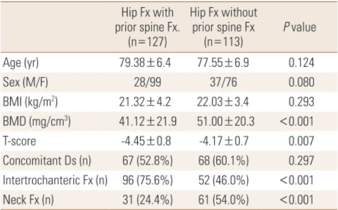
Table 1. Demographic data Hip Fx with prior spine Fx.
(n=127)
Hip Fx without prior spine Fx
(n=113) P value
Age (yr) 79.38±6.4 77.55±6.9 0.124
Sex (M/F) 28/99 37/76 0.080
BMI (kg/m2) 21.32±4.2 22.03±3.4 0.293
BMD (mg/cm3) 41.12±21.9 51.00±20.3 <0.001
T-score -4.45±0.8 -4.17±0.7 0.007
Concomitant Ds (n) 67 (52.8%) 68 (60.1%) 0.297 Intertrochanteric Fx (n) 96 (75.6%) 52 (46.0%) <0.001 Neck Fx (n) 31 (24.4%) 61 (54.0%) <0.001 Data are expressed as means±SD. Statistically significant at the 0.05 level.
Fx, fracture; BMI, body mass index; BMD, bone mineral density; Ds, dis- ease.
in group II were 61 (54.0%) and 52 (46.0%) patients, retro- spectively, which have a statistical significance (P<0.001).
Twenty-four patients (64.9%) among 37 male patients in group II had the type of neck fracture. Sixty (47.2%) in group I and 45 (39.8%) in group II patients had concomitant dis- eases, hypertension was the most common concomitant disease 46 in group I and 34 in group II patients.
Only 13 patients (5.4%) among 240 patients had under- gone percutaneous vertebroplasty (males/females: 2/11) and the mean age was 78.5 years (Table 2). Twelve in group I and six in group II patients had operation due to second hip fracture and the mean age of them was 82.7 years (males/females: 6/12). The age of patients of second hip fracture was significantly older than those of unilateral hip fracture patients (P=0.006) (Table 3).
DISCUSSION
The incidence of osteoporotic fracture is rising due to in- creasing of people living to an older age. Prior fractures have been documented consistently to predict the risk of subsequent incident fractures.[1,2,7] Many studies had demonstrated that prevalent vertebral fractures are risk factors for incident hip fracture and that predicts future vertebral and hip fractures.[1,7-9] In large meta-analyses, the pooled estimates for the risk of a subsequent hip frac- ture were found to be approximately twofold in the pres- ence of vertebral fracture.[2,10] However, only a few re- ports discussed about non-vertebral fracture, especially, hip fractures after percutanous vertebroplasty in osteopo- rotic spinal compression fracture. Mazzantini et al.[11] re-
ported in patients with osteoporosis treated with pecuta- neous vertebroplasty, the incidence of new vertebral frac- tures was 27.8% and non-vertebral fractures occurred in only 7% (only one at the hip) in as few as 3.5% of the pa- tients if exclude rib fractures after 39 months. In our study, only 13 patients (5.4%) had undergone percutaneous ver- tebroplasty in 240 patients who had undergone operation of hip fractures, retrospectively. The incidence of second hip fracture has been reported to be about 5% -10% and is expected to increase with the aging of the population in the future.[12,13] It has previously been reported that age and female sex are important risk factor for hip fracture.
[14,15] However, some reports have indicated that there are no differences in age or sex between unilateral and second hip fractures.[12,16] Mitani et al.[12] reported that the mean age at the time of initial fracture in the second fracture group tended to be slightly older than the mean age of the unilateral group. In our study, 18 patients (7.5%) had operated of second hip fractures in mean age 82.7 years. The age of patients of second hip fracture was sig- nificant older than those of unilateral hip fracture patients.
Female patients were 12, and male patients were 6 among 18 patients of second hip fracture.
Bone strength has been described as a reflection of the integration of bone density and bone quality.[17] BMD is strongly associated with fracture risk. Therefore, an impor- tant clinical goal is to prevent the bone loss that leads to the first fracture, because this bone loss is largely irrevers- ible by the time the first fracture occur. Across the range of BMD T-score, spine fracture burden had up to a 12-fold im- pact on the predicted vertebral fracture risk but only ap- proximately twofold impact on predict non-vertebral frac- Table 2. Comparison of between hip fracture with or without verte-
broplasty
Hip Fx with vertebroplasty
(n=13)
Hip Fx without vertebroplasty
(n=114) P value
Age (yr) 78.50±6.5 79.52±6.4 0.535
Sex (M/F) 2/11 26/88 1
BMI (kg/m2) 21.25±2.8 21.32±6.4 0.963
BMD (mg/cm3) 42.98±19.1 40.18±22.4 0.700
T-score -4.38±0.6 -4.46±0.8 0.694
Concomitant Ds (n) 6 47 0.308
Data are expressed as means±SD. Statistically significant at the 0.05 level.
Fx, fracture; BMI, body mass index; BMD, bone mineral density; Ds, dis- ease.
Table 3. Comparison of between unilateral hip fracture and second hip fracture
Unilateral hip fracture (n=222)
Second hip fracture
(n=18) P value
Age (yr) 78.18±6.6 82.72±6.7 0.006
Sex (M/F) 59/163 6/12 0.583
BMI (kg/m2) 21.57±3.9 22.35±2.2 0.586
BMD (mg/cm3) 46.14±22.1 41.18±16.4 0.353
T-score -4.30±0.8 -4.53±0.4 0.081
Concomitant Ds (n) 98 7 0.806
Data are expressed as means±SD. Statistically significant at the 0.05 level.
BMI, body mass index; BMD, bone mineral density; Ds, disease.
ture risk.[2] Siris et al.[8] stated that at any given BMD T- score, the risk of incident vertebral, non-vertebral, and any fracture depended heavily on prevalent radiographic ver- tebral fracture status. Most women with a history of low trauma risk fractures have low BMD, and women with both low BMD and prior fractures had a very high risk of further fractures. In our study, the BMD and T-score of multiple fractures group was significant lower than hip fractures group.
Our study has limitations of a relatively small number of patients. The main reason for small number was the rate of diagnosis osteoporosis of the hip fracture patients in early period (2006-2009: 51.5%) less than in late period (2010- 2012: 82.5%) of our study period. Many studies have re- ported low rates of osteoporosis treatment after hip frac- ture.[18,19] This result suggests that the orthopedic sur- geon can perform a leading role in the prevention of fur- ther fractures. We agree that the orthopedic surgeons can serve a pivotal role in optimizing treatment, not only of the fracture, but also of the underlying disease.[20] Sec- ond, we only considered operated hip fracture patients with or without prior osteoporotic spinal compression frac- tures by retrospective study, but the long term prospective study of patients treated with percutaneous vertebrao- plasty is needed.
In summary, the hip fracture patients who had with prior osteoporotic spinal compression fractures had lower BMD and T-score compared to the hip fracture patients without previous spinal compression fractures. These multiple os- teoporotic fracture patients should be clinically evaluated and treatment for osteoporosis.
REFERENCES
1. Black DM, Arden NK, Palermo L, et al. Prevalent vertebral deformities predict hip fractures and new vertebral defor- mities but not wrist fractures. Study of Osteoporotic Frac- tures Research Group. J Bone Miner Res 1999;14:821-8.
2. Klotzbuecher CM, Ross PD, Landsman PB, et al. Patients with prior fractures have an increased risk of future frac- tures: a summary of the literature and statistical synthesis.
J Bone Miner Res 2000;15:721-39.
3. Li YA, Lin CL, Chang MC, et al. Subsequent vertebral frac- ture after vertebroplasty: incidence and analysis of risk factors. Spine (Phila Pa 1976) 2012;37:179-83.
4. Hardouin P, Grados F, Cotten A, et al. Should percutaneous vertebroplasty be used to treat osteoporotic fractures? An update. Joint Bone Spine 2001;68:216-21.
5. Fribourg D, Tang C, Sra P, et al. Incidence of subsequent vertebral fracture after kyphoplasty. Spine (Phila Pa 1976) 2004;29:2270-6.
6. Black DM, Palermo L, Nevitt MC, et al. Comparison of methods for defining prevalent vertebral deformities: the Study of Osteoporotic Fractures. J Bone Miner Res 1995;
10:890-902.
7. Schousboe JT, Fink HA, Lui LY, et al. Association between prior non-spine non-hip fractures or prevalent radiograph- ic vertebral deformities known to be at least 10 years old and incident hip fracture. J Bone Miner Res 2006;21:1557- 64.
8. Siris ES, Genant HK, Laster AJ, et al. Enhanced prediction of fracture risk combining vertebral fracture status and BMD. Osteoporos Int 2007;18:761-70.
9. Melton LJ 3rd, Atkinson EJ, Cooper C, et al. Vertebral frac- tures predict subsequent fractures. Osteoporos Int 1999;
10:214-21.
10. Haentjens P, Autier P, Collins J, et al. Colles fracture, spine fracture, and subsequent risk of hip fracture in men and women. A meta-analysis. J Bone Joint Surg Am 2003;85- A:1936-43.
11. Mazzantini M, Carpeggiani P, d'Ascanio A, et al. Long-term prospective study of osteoporotic patients treated with percutaneous vertebroplasty after fragility fractures. Os- teoporos Int 2011;22:1599-607.
12. Mitani S, Shimizu M, Abo M, et al. Risk factors for second hip fractures among elderly patients. J Orthop Sci 2010;
15:192-7.
13. Schrøder HM, Petersen KK, Erlandsen M. Occurrence and incidence of the second hip fracture. Clin Orthop Relat Res 1993;289:166-9.
14. Cummings SR, Nevitt MC, Browner WS, et al. Risk factors for hip fracture in white women. Study of Osteoporotic Fractures Research Group. N Engl J Med 1995;332:767-73.
15. Hinton RY, Lennox DW, Ebert FR, et al. Relative rates of fracture of the hip in the United States. Geographic, sex, and age variations. J Bone Joint Surg Am 1995;77:695- 702.
16. Finsen V, Benum P. The second hip fracture. An epidemio- logic study. Acta Orthop Scand 1986;57:431-3.
17. NIH Consensus Development Panel on Osteoporosis Pre-
vention Diagnosis and Therapy. Osteoporosis prevention, diagnosis, and therapy. JAMA 2001;285:785-95.
18. Kim SR, Ha YC, Park YG, et al. Orthopedic surgeon's aware- ness can improve osteoporosis treatment following hip fracture: a prospective cohort study. J Korean Med Sci 2011;
26:1501-7.
19. Kaufman JD, Bolander ME, Bunta AD, et al. Barriers and
solutions to osteoporosis care in patients with a hip frac- ture. J Bone Joint Surg Am 2003;85-A:1837-43.
20. Bouxsein ML, Kaufman J, Tosi L, et al. Recommendations for optimal care of the fragility fracture patient to reduce the risk of future fracture. J Am Acad Orthop Surg 2004;
12:385-95.