70
ISSN 1975-4612 Copyright ⓒ 2009 Korean Society of Echocardiography www.kse-jcu.org
I
Innttrroodduuccttiioonn
Coronary arteriovenous fistula (CAVF) is an infrequent vascular anomaly that establishes a direct link between an epicardial coronary artery and a cardiac chamber, major vessels, or other vascular structures. We describe a case of 76-year-old woman who develop huge coronary artery aneurysm arising from Left main coronary artery and tortuous fistula form proximal right coronary artery drained to aneurysm.
C Caassee
A 76 year-old female patient presented with exertional dyspnea and effort angina for about 1month. She had a history of hypertension 2 years ago and medication of cal- cium channel blocker once daily. On physical examination, her blood pressure was 140/95 mmHg and chest auscultation revealed a continuous murmur graded IV/VI on her right second intercostal space along the right upper sternal border and crackles on both lower lung field. Electrocardiography revealed atrial fibrillation with rapid ventricular rhythm and left ventricular hypertrophy by voltage. Chest X-ray showed severe cardiomegaly and edematous change in both lower lung fields.
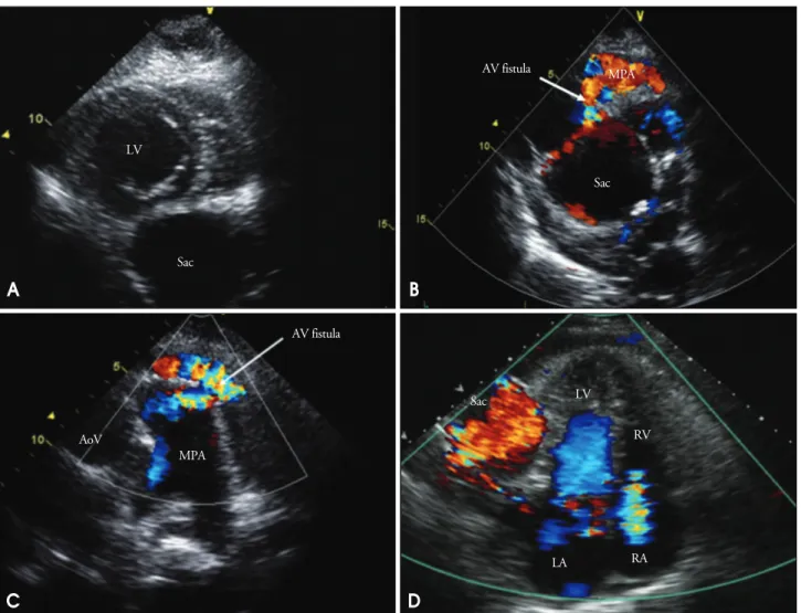
Echocardiography revealed huge saccular aneurysm in posterolateral side of left ventricle (Fig. 1A). Flow communi- cation between aneurysmal sac and main pulmonary trunk was observed (Fig. 1B). Color Doppler with continuous wave revealed arteriovenous fistula with huge aneurysm which drained to main pulmonary artery (Fig. 1C). Left ventricular ejection fraction was 45% and global hypokinesia was noted. Left ventricle was compressed by multiple fusiform aneurysms (maximal diameter of largest one: about 6.7 cm) in apical 4 chamber view (Fig. 1D). Moderate pulmonary hypertension (systolic pulmonary arterial pressure=
46.1 mmHg) and small amount pericardial effusion was noted. Cardiac computed tomography angiography (CTA) showed markedly enlarged tortuous anonymous vessel from left main coronary artery along course of left circumflex artery and several small tortuous anonymous vessels from proximal right coronary artery (Fig. 2). We performed coro- nary angiography for further evaluation of coronary artery anomaly. In coronary angiography, left main coronary artery was dilated and communicated to aneurysmal sac. Left ant- erior descending artery (LAD) was not visible. Instead, distal branch from left circumflex artery supplied left anterior
Received: December 31, 2008 Accepted: March 5, 2009
Address for Correspondence: Jong Chun Park, Department of Cardiology, The Heart Center, Chonnam National University Hospital, Cardiovascular Research Institute, Chonnam National University Medical School, 671 Jebong-ro, Dong-gu, Gwangju 501-757, Korea Tel: +82-62-220-6244, Fax: +82-62-225-6260, E- mail: [email protected]
A
A C Ca as se e o of f C Co or ro on na ar ry y A Ar rt te er riio ov ve en no ou us s F Fiis st tu ul la a A
As ss so oc ciia at te ed d w wiit th h G Giia an nt t C Co or ro on na ar ry y A Ar rt te er ry y A
An ne eu ur ry ys sm m
S
Soooo--HHyyuunn KKiimm,, MMDD,, JJuumm--SSuukk KKoo,, MMDD,, HHyyuunn--JJuu YYoooonn,, MMDD,, SSaanngg--CChhuunn LLiimm,, NNRR,, S
Sooookk--HHeeee CChhoo,, NNRR,, HHeeyy--SSooookk KKiimm,, NNRR,, JJii--SSuunn LLeeee,, NNRR aanndd JJoonngg CChhuunn PPaarrkk,, MMDD
Department of Cardiology, The Heart Center, Chonnam National University Hospital, Cardiovascular Research Institute, Chonnam National University Medical School, Gwangju, Korea
C
CAASSEE RREEPPOORRTT J Cardiovasc Ultrasound 2009;17(2):70-72
Coronary arteriovenous fistula is a more prevalent, hemodynamically significant congenital malformation. Both coronary arteries arise normally from their aortic sinuses, but the branches of fistula communicate directly with cardiac chamber, pulmonary trunk, coronary sinus, superior vena cava, or pulmonary vein. Fistula associated with coronary aneurysm is an uncommon finding. We report a rare case of 76-year-old female patient who had a coronary arteriovenous fistula with giant coronary artery aneurysm. This case is clearly diagnosed by echocardiography, three-dimensional computed tomography (3D- CT), and coronary angiography (CAG).
K
KEEYY WWOORRDDSS:Coronary arteriovenous fistula·Coronary artery aneurysm.
onlineⓒMLComm
Coronary Fistula with Aneursysm|Soo-Hyun Kim, et al.
71 descending artery territory (Fig. 3). The pulmonary to syste-
mic flow ratio was 1.7 by echocardiography. Reversible per- fusion defect in left anterior descending artery and right
coronary artery territory was identified in myocardial single- photon emission computed tomography (M-SPECT) which represented myocardial ischemia result from coronary steal.
We recommended surgical treatment to patient. But pati- ent refused surgery. After intensive medical treatment, patient showed improved clinical status and uneventful follow up.
D
Diissccuussssiioonn
The incidence of the CAVF has been reported to be 0.1- 0.2% in patients who undergo coronary artery angiography1) and 0.002% for general population.2)In about 50-55% of coronary arteriovenous fistulas, the right coronary artery provides the fistulous origin, and about 40% drain to the right ventricle.2)Generally, the symptoms develop depending on the amount of the shunt or the presence of coronary steal phenomenon of the fistulas, which tend to present in young adults with angina, exertional dyspnea, syncope, palpitations, B
A
D C
Fig. 1. A: Echocardiogram in parasternal short axis view show extrinsic cardiac compression in left ventricle side due to multiple fusiform aneurysms (maximal diameter of largest one: about 6.7 cm). B: Modified apical 2 chamber view showed a fistula from aneurismal sac to main pulmonary artery. C:
Parasternal short axis window in aortic valve level revealed a arteriovenous fistula from coronary artery to main pulmonary trunk. D: Apical 4 chamber view showed mild regurgitation of tricuspid valve. Sac: aneurysmal sac, LV: left ventricle, AoV: aortic valve, MPA: main pulmonary artery, AV:
arteriovenous, LA: left atrium, RV: right ventricle, RA: right atrium.
Fig. 2. A 64-detector row cardiac computed tomography with 3D reconstruction showed several small tortous anonymous vessels from proximal right coronary artery (A) and enlarged tortous anonymous vessel from left main coronary artery along course of left circulflex artery (B).
Arrow indicates RCA and LCX. Sac: aneurysmal sac, RCA: right coronary artery, LCX: left circumflex artery.
LV
LV
LA RA
RV Sac
Sac AV fistula
AV fistula
AoV
MPA
RCA
LCX Sac
Sac
MPA
Sac
A B
Journal of Cardiovascular Ultrasound 17|June 2009
72
myocardial ischemia and infarction, and to manifest in adults >40 years old with congestive heart failure, athero- sclerosis, and cardiac arrhythmias.1-2)Though some patients may have symptoms shortly after birth, however most of them are asymptomatic.1)Angina pectoris due to blood shun- ting and perfusion away from the myocardium (coronary steal phenomenon) occur. Steal phenomenon may be a cause of developing coronary artery aneurysmal change.1-2)Symp- toms of congestive heart failure can result when drainage occurs into the right system resulting in congestion and pulmonary hypertension. The predominant physical finding is a continuous murmur, representing the systolic-diastolic flow in large fistulas. A detailed physical examination combined with an adequate image studies such as echocar- diography, computed tomography may provide accurate diagnosis and avoid painful, invasive examinations.3-4) The decision to treat a coronary fistula is recommended in the presence of symptoms, a marker of functional significance in the absence of concomitant disease, fistula location, its size, and the resulting shunt volume.2)Anti-anginal therapy addressing the demand and supply mismatch induced by the coronary steal phenomenon may be effective for symptom relief.5-7)Of further clinical importance is recommended in endocarditis and antibiotic prophylaxis is recommended in CAVF patients. Surgical intervention is generally reserved for single, large, symptomatic fistula (with angina, cardiac
decompression or complications) and circulatory overload warrants surgical treatment even in asymptomatic patients.7) The main differences between our case and others are that giant aneurysm is detected in very elderly patient and in atypical location.
R
Reeffeerreenncceess
1. Shiga Y, Tsuchiya Y, Yahiro E, Kodama S, Kotaki Y, Shimoji E, Fukuda N, Morito N, Urata M, Saito N, Niimura H, Mihara H, Yamanouchi Y, Urata H. Left main coronary trunk connecting into right atrium with an aneurysmal coronary artery fistula. Int J Cardiol 2008;
123:e28-30.
2. Maleszka A, Kleikamp G, Minami K, Peterschröder A, Körfer R. Giant coronary arteriovenous fistula. A case report and review of the literature. Z Kardiol 2005;94:38-43.
3. Androulakis A, Chrysohoou C, Barbetseas J, Brili S, Kakavas A, Maragiannis D, Kallikazaros I, Stefanadis C. Arteriovenous connection between the aorta and the coronary sinus through a giant fistulous right coronary artery. Hellenic J Cardiol 2008;49:48-51.
4. Rangasetty UC, Ahmad M. Giant coronary artery fistula with aneurysm and multiple openings: A two-dimensional echocardiographic evaluation.
Echocardiography 2006;23:611-3.
5. Bobos D, Chatzis AC, Giannopoulos NM, Tsoutsinos A, Antoniadis A, Cokkinos D, Sarris GE. Successful surgical repair of a giant arteriovenous fistula of the coronary arteries. J Card Surg 2006;21:269-70.
6. Saxena P, Konstantinov IE, Burstow D, Tam R. Surgical repair of a large coronary artery aneurysm with arteriovenous fistula. J Thorac Cardiovasc Surg 2006;131:1167-8.
7. Olgunturk R, Kula S, Tunaoglu FS. Transcatheter closure of a rare form of coronary arteriovenous fistula (circumflex artery to coronary sinus). Int J Cardiol 2006;113:261-3.
Fig. 3. Coronary angiographic finding revealed left main coronary was dilated and communicated to aneurismal sac (A and B). And left anterior descending artery (LAD) was not visible. Small tortous anonymous vessel from proximal right coronary artery was drained to aneurysmal sac through a fistula formation (C).
A B C