ISSN 2234-3806 • eISSN 2234-3814
https://doi.org/10.3343/alm.2021.41.1.16
Clinical Utility of Mac-2 Binding Protein Glycosylation Isomer in Chronic Liver Diseases
Nobuharu Tamaki , M.D., Ph.D.
1,2, Masayuki Kurosaki , M.D., Ph.D.
1, Rohit Loomba , M.D., M.H.S.
2, and Namiki Izumi , M.D., Ph.D.
11Department of Gastroenterology and Hepatology, Musashino Red Cross Hospital, Tokyo, Japan; 2NAFLD Research Center, Division of Medicine, University of California, San Diego, La Jolla, California, USA
An accurate evaluation of liver fibrosis is clinically important in chronic liver diseases. Mac-2 binding protein glycosylation isomer (M2BPGi) is a novel serum marker for liver fibrosis. In this review, we discuss the role of M2BPGi in diagnosing liver fibrosis in chronic hepatitis B and C, chronic hepatitis C after sustained virologic response (SVR), and nonalcoholic fatty liver disease (NAFLD). M2BPGi predicts not only liver fibrosis but also the hepatocel- lular carcinoma (HCC) development and prognosis in patients with chronic hepatitis B and C, chronic hepatitis C after SVR, NAFLD, and other chronic liver diseases. M2BPGi can also be used to evaluate liver function and prognosis in patients with cirrhosis. M2B- PGi levels vary depending on the etiology and the presence or absence of treatment. There- fore, the threshold of M2BPGi for diagnosing liver fibrosis and predicting HCC develop- ment has to be adjusted according to the background and treatment status.
Key Words: Mac-2 binding protein glycosylation isomer, Liver fibrosis, Threshold, Hepato-
cellular carcinoma, Chronic hepatitis C, Chronic hepatitis B, Sustained virologic response, Nonalcoholic fatty liver disease
Received: April 21, 2020 Revision received: June 15, 2020 Accepted: July 29, 2020 Corresponding author:
Namiki Izumi, M.D., Ph.D.
Department of Gastroenterology and Hepatology, Musashino Red Cross Hospital, 1-26-1 Kyonan-cho, Musashino-shi, Tokyo 180-8610, Japan
Tel: +81-422-32-3111 Fax: +81-422-32-9551
E-mail: [email protected]
© Korean Society for Laboratory Medicine This is an Open Access article distributed under the terms of the Creative Commons Attribution Non-Commercial License (https://creativecom- mons.org/licenses/by-nc/4.0) which permits unrestricted non-commercial use, distribution, and reproduction in any medium, provided the original work is properly cited.
INTRODUCTION
Liver fibrosis correlates with hepatocarcinogenesis and progno- sis in chronic liver diseases; hence, an accurate evaluation of liver fibrosis is extremely important [1]. Although liver biopsy is currently the gold standard for liver fibrosis evaluation, it has many drawbacks, such as risk of complications and difficulty in repeated evaluation [2]. Therefore, various methods for the eval- uation of noninvasive liver fibrosis have been developed recently, including elastography using magnetic resonance imaging or ul- trasonography [3-11]. Although elastography has a high diag- nostic ability for not only liver fibrosis but also liver steatosis [12, 13] and is widely used, it has a number of drawbacks, such as the requirement of expensive equipment, limited available facili-
ties, and equipment incompatibility. Blood tests are also widely used to evaluate liver fibrosis and prognosis in patients with chro- nic hepatitis, and a two-step screening strategy to detect advanced fibrosis patients in a large population has been suggested [10, 14-17]. In the first-line screening, serum biomarkers are used to exclude patients with low risk of advanced fibrosis, and, in the second-line screening, patients with advanced fibrosis are identified by elastography. Recent advances in the non-invasive assessment of liver fibrosis based on serum biomarkers and im- aging have been summarized in reviews [10, 18].
In Japan, serum Mac-2 binding protein (M2BP) glycosylation isomer (M2BPGi) was identified in 2013 and clinically applied as a diagnostic marker for liver fibrosis; it is now widely used, mainly in Asia [19-21]. The clinical use of M2BPGi has rapidly
2017-03-16 https://crossmark-cdn.crossref.org/widget/v2.0/logos/CROSSMARK_Color_square.svg
increased in recent years, because it can be easily measured in the serum, and M2BPGi measurement has been used to assess liver fibrosis and carcinogenesis risk in chronic liver diseases [20, 22-26].
In this review, we summarize the current knowledge on the utility of M2BPGi in diagnosing liver fibrosis in chronic hepatitis B and C, nonalcoholic fatty liver disease (NAFLD), and other liver diseases, and as a carcinogenic risk factor.
M2BPGi and its Characteristics
M2BP, a secreted glycoprotein present in the extracellular ma- trix, is associated with cell adhesion and correlates with liver fi- brosis [27]. Recent advances in glycoproteomics have revealed that specific glycan structures of M2BP change as liver fibrosis progresses [28]. The concept of M2BPGi measurement involves
the evaluation of liver fibrosis by measuring M2BP with an al- tered glycan structure. The change in the M2BP glycan struc- ture was detected using the lectin Wisteria floribunda agglutinin (WFA) and was found to be correlated with the progression of fi- brosis [19]. Thus, it was demonstrated that WFA-positive M2BP [WFA
+-M2BP (M2BPGi)] detected by sandwich immunoassay with WFA and anti-M2BP antibody is clinically correlated with liver fibrosis. The sandwich immunoassay is automated using the HISCL-2000 system (Sysmex Co., Hyogo, Japan), and M2BPGi can be measured in 17 minutes using 10 μL of serum [29]. The measured values of WFA
+-M2BP conjugated to WFA were in- dexed with the obtained values using the following equation:
cutoff index (COI)=([M2BPGi]
sample– [M2BPGi]
NC)/
([M2BPGi]
PC– [M2BPGi]
NC),
where [M2BPGi]
sampleis the M2BPGi level in the serum sample,
Table 1. Thresholds of M2BPGi in liver fibrosis
Reference Etiology Mean value of M2BPGi Threshold for diagnosing fibrosis stage
F0 F1 F2 F3 F4 ≥F2 ≥F3 F4
Yamasaki, et al. [31] HCV 1.3 2.2 3.3 5.2
Tamaki, et al. [32] HCV 0.81 1.82 2.31 7.5
Ura, et al. [33] HCV 1.6 3.86 3.53 3.12 2.14 2.17
Huang, et al. [34] HCV 2.23 3.45 3.48 3.77 1.61 1.42 2.67
Fujita, et al. [35] HCV 1.26 1.81 4.03 7.86 2.19
Inoue, et al. [36] HCV 2.3 6.9
Nakamura, et al. [37] HCV 1.7 (F1–2) 5.1 (F3-4)
Xu, et al. [38] HCV 0.88 1.70 (F2–3) 5.68 0.95 1.35
0.88–2.23 1.81–3.86 2.3–3.53 3.12–7.86
Ishii, et al. [52] HBV 0.9 1.4 1.6 3.1 1.4 1.4 1.9
Ichikawa, et al. [53] HBV 0.75 1.14 1.03 1.64 0.94 1.26 1.26
Yeh, et al. [54] HBV 0.64 1.36 1.65 2.7 1.35 1.54 1.67
Jekarl, et al. [55] HBV 0.68 0.87 1.65 0.7 0.7
Mak, et al. [56] HBV 0.26 0.34 0.57 1.21 0.25 0.45 0.96
Wei, et al.* [57] HBV 0.88 1.17 (F2–3) 1.92 1.12 1.83
Jun, et al. [58] HBV 0.80 (F1–3) 2.67
0.26–0.9 0.34–1.36 0.57–1.65 1.21–3.1
Abe, et al. [71] NAFLD 0.57 0.7 1.02 1.57 2.96 0.94 1.46
Ogawa, et al. [72] NAFLD 0.43 0.62 0.92 1.12 2.94 0.83 0.83 1.26
Nishikawa, et al. [73] NAFLD 0.7 0.7 1.2 1.6 1.1 1.6
Atsukawa, et al. [74] NAFLD 0.71 1.17 1.36 1.98 1.23 1.37
Alkhouri, et al. [75] NAFLD 0.66 (F0–1) 1.2 (F2–3) 2.4
0.62–0.71 0.7–1.17 1.2–1.57 1.6–2.96
*Fibrosis stage determined using Fibroscan.
Abbreviations: M2BPGi, Mac-2 binding protein glycosylation isomer; HCV, hepatitis C virus infection; HBV, hepatitis B virus infection; NAFLD, nonalcoholic fatty liver disease.
PC is the positive control, and NC is the negative control. The PC was supplied as a calibration solution preliminarily standard- ized to yield a COI value of 1.0.
The pathophysiological role of M2BPGi is not completely elu- cidated. Hepatic stellate cells (HSCs) are the source of M2BPGi, and the M2BPGi secreted from HSCs induces Mac-2 expres- sion in Kupffer cells, which in turn activates HSCs and increases alpha-smooth muscle actin expression [30]. These findings in- dicated that M2BPGi plays an important role in the progression of liver fibrosis and M2BPGi levels are associated with the fibro- sis stage.
M2BPGi Level Predicts Liver Fibrosis and Carcinogenesis in Chronic Hepatitis C
M2BPGi was developed as a biomarker using the serum of chronic hepatitis C patients and is now widely used for diagnos- ing liver fibrosis in chronic hepatitis C [19, 31-38]. Numerous studies have compared the diagnostic accuracy of M2BPGi with that of liver biopsy in identifying liver fibrosis stage. The mean COI values of M2BPGi in histological fibrosis stages 1, 2, 3, and 4 are 0.88–2.23, 1.81–3.86, 2.3–3.53, and 3.12–7.86, respec- tively (Table 1) [31-38]. In all cases, M2BPGi levels significantly
increased as liver fibrosis progressed, which confirmed the util- ity of M2BPGi for diagnosing liver fibrosis in chronic hepatitis C.
Liver fibrosis is a risk factor for carcinogenesis in chronic hepati- tis C. Therefore, M2BPGi predicts the development of hepato- cellular carcinoma (HCC). In studies on carcinogenesis, M2B- PGi ≥4.0 indicated a high risk of HCC development (Table 2) [31, 32].
Another advantage of M2BPGi is that it can be measured eas- ily and repeatedly. An increase in M2BPGi levels over time was associated with HCC risk [32, 39]. High M2BPGi levels also cor- relate with the prognosis of chronic hepatitis C [36]. A meta-anal- ysis confirmed the utility of M2BPGi in diagnosing fibrosis and predicting HCC risk [21].
Utility of M2BPGi in Chronic Hepatitis C After Sustained Virologic Response (SVR)
In recent studies, several patients achieved SVR with direct-act- ing antiviral (DAA) treatment [40-43]; hence, we discuss the role of M2BPGi in relation to SVR. M2BPGi not only strongly corre- lates with liver fibrosis but also weakly correlates with inflamma- tion and alanine transaminase (ALT) [34, 36, 44]. Therefore, M2BPGi decreases rapidly during DAA treatment according to
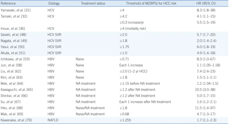
Table 2. Thresholds of M2BPGi in HCC development
Reference Etiology Treatment status Threshold of M2BPGi for HCC risk HR (95% CI)
Yamasaki, et al. [31] HCV ≥4 8.3 (1.8–38)
Tamaki, et al. [32] HCV ≥4.2 4.1 (1.1–15)
≥0.3 increase/yr 5.5 (1.5–19)
Inoue, et al. [36] HCV ≥4 (mortality risk)
Sasaki, et al. [48] HCV SVR ≥2.0 5.7 (1.7–20)
Nagata, et al. [49] HCV SVR ≥1.8 2.0 (1.4–2.4)
Yasui, et al. [50] HCV SVR ≥1.75 6.0 (1.8–19)
Akuta, et al. [51] HCV SVR ≥1.0 4.9 (1.4–18)
Ichikawa, et al. [53] HBV Naive ≥0.71 8.3 (1.0–67)
Jun, et al. [58] HBV Naive Each 1 increase 1.1 (1.05–1.18)
Liu, et al. [62] HBV Naive ≥2.0 (1–2 yr HCC) 7.4 (2.4–23)
Kim, et al. [63] HBV Naive ≥1.8 1.5 (1.1–2.1)
Mak, et al. [64] HBV NA treatment ≥1.15 before NA treatment 1.2 (1.04–1.5)
Kawaguchi, et al. [65] HBV NA treatment ≥1.2 after NA treatment 10.5 (3.0–38)
Shinkai, et al. [66] HBV NA treatment ≥1.2 after NA treatment 5.0 (1.7–15)
Su, et al. [67] HBV NA treatment Each 1 increase after NA treatment 1.6 (1.2–2.1)
Heo, et al. [68] HBV Naive/NA treatment ≥1.8 11.5 (1.4–97)
Mak, et al. [69] HBV Naive/NA treatment ≥0.68 4.7 (1.3–17)
Kawanaka, et al. [79] NAFLD ≥1.255 1.7 (1.1–2.3)
Abbreviations: M2BPGi, Mac-2 binding protein glycosylation isomer; HCC, hepatocellular carcinoma; SVR, sustained virologic response; NA, nucleotide/nu- cleoside analogue; NAFLD, nonalcoholic fatty liver disease; HCV, hepatitis C virus infection; HBV, hepatitis B virus infection; HR, hazard ratio.
the improvement of inflammation or ALT level [45, 46]. M2BPGi also increases during acute liver injury [47]. The threshold of M2BPGi for predicting HCC development after SVR should be adjusted because of the improved inflammation. In studies ex- amining the risk of HCC development after SVR, M2BPGi at SVR of 1.0–2.0, which was lower than that during continuous infec- tion with hepatitis C virus, indicated a carcinogenic risk (Table 2) [48-51].
Utility of M2BPGi in Chronic Hepatitis B
M2BPGi levels in patients with chronic hepatitis B increase as liver fibrosis progresses. The mean M2BPGi COI values in histo- logical fibrosis stages 1, 2, 3, and 4 are 0.26–0.9, 0.34–1.36, 0.57–1.65, and 1.21–3.1, respectively (Table 1) [52–58]. M2B- PGi was significantly lower in patients with chronic hepatitis B than in those with chronic hepatitis C [59]. Therefore, it is nec- essary to adjust the threshold of M2BPGi for liver fibrosis diag- nosis, considering its etiology.
In cross-sectional studies examining the association of M2B- PGi with HCC, M2BPGi level was significantly higher in patients with HCC than in those without HCC [60, 61]. When the pres- ence or absence of HCC was evaluated after the fibrosis stages were adjusted, no difference in M2BPGi levels was found [58], which was considered to reflect its carcinogenic potential due to fibrosis progression. Longitudinal observational studies have re- ported that patients with M2BPGi level ≥0.71−2.0 have a high risk of HCC development (Table 2) [53, 58, 62-69]. In consider- ing the risk of carcinogenesis, the presence or absence of treat- ment should be considered. M2BPGi level decreases with nu- cleotide/nucleoside analogue (NA) treatment [53, 56]. There- fore, it is necessary to reduce the threshold when evaluating M2BPGi level during NA treatment, because M2BPGi level de- creases with improvement in fibrosis and inflammation. M2B- PGi level ≥1.2 during NA treatment is associated with carcino- genesis and prognosis of chronic hepatitis B [60, 61], and the threshold tends to be lower than in treatment-naive cases. On the contrary, another study showed that the M2BPGi level dur- ing NA treatment is not associated with carcinogenesis [70], and further verification is needed in this regard.
Utility of M2BPGi in NAFLD
The utility of M2BPGi for diagnosing liver fibrosis in NAFLD has been reported. The mean M2BPGi COI values in histological fi- brosis stages 1, 2, 3, and 4 were 0.62–0.71, 0.7–1.17, 1.2–1.57, and 1.6–2.96, respectively, and M2BPGi levels increased with the progression of liver fibrosis (Table 1) [71–75]. M2BPGi level
has a higher diagnostic accuracy for fibrosis than FIB-4 or NAFLD fibrosis score [71]. The threshold of M2BPGi for predicting F3–4 according to these reports was 0.83–1.23 and that for predict- ing F4 was 1.26–1.46 [71-75]. In a report comparing the thresh- old of M2BPGi for diagnosing liver fibrosis between chronic hep- atitis C and NAFLD, the threshold for predicting F3–4 in NAFLD was lower than that for predicting F3–4 in chronic hepatitis C;
hence, it is necessary to consider the underlying liver disease when interpreting the M2BPGi results for the diagnosis of liver fibrosis [76]. Although FIB-4 is used for diagnosing liver fibrosis in NAFLD, its diagnostic accuracy is not enough, unless the thresh- old is adjusted according to age [77]. In a study comparing M2- BPGi level and FIB-4, the optimal threshold of FIB-4 increased with age, whereas that of M2BPGi remained unchanged; this finding indicated that M2BPGi has a high potential for advanced fibrosis screening in large populations [78]. In a report examin- ing its association with carcinogenesis (an M2BPGi level of 1.255 as the threshold), a higher level indicated an increased risk of subsequent carcinogenesis [79]. Other reports have also sug- gested that M2BPGi level is associated with carcinogenesis and liver-related complications [80]. Recently, it was reported that M2BPGi level is useful for predicting advanced fibrosis in health checkups [81].
Utility of M2BPGi in Cirrhosis
M2BPGi level increases when compensated cirrhosis develops to decompensated cirrhosis [82–84]. Therefore, in patients with cirrhosis, high M2BPGi levels indicate a poor prognosis [82, 85,
Fig. 1. Clinical utility of M2BPGi in chronic liver diseases. The M2BPGi levels increase as the disease progresses from minimal liv- er fibrosis to decompensated cirrhosis. M2BPGi can be used to as- sess disease status, such as liver fibrosis, HCC risk, HCC recur- rence risk, liver function, and prognosis of chronic liver diseases, with the progression of the disease.
Abbreviations: M2BPGi, Mac-2 binding protein glycosylation isomer; HCC, hepatocellular carcinoma.
86]. M2BPGi is reportedly useful as a predictive marker for liver failure and complications after hepatectomy or transcatheter ar- terial chemoembolization [82, 87-91]. Thus, M2BPGi level re- flects not only liver fibrosis but also liver function. M2BPGi level is also associated with recurrence and prognosis after hepatec- tomy and can be used for follow-up after HCC therapy [92-94].
Sarcopenia has recently attracted attention as a complication of cirrhosis. M2BPGi level is correlated with muscle mass and is useful as a predictive marker for sarcopenia [95, 96]. Thus, M2BPGi level is useful in patients with cirrhosis as it correlates with cirrhosis complications and prognosis.
M2BPGi level is useful for diagnosing liver fibrosis in autoim- mune hepatitis [97, 98], primary biliary cholangitis [99, 100], biliary atresia [101, 102], and primary sclerosing cholangitis [103], in addition to viral hepatitis and NAFLD; but relevant studies are limited in number and need validation.
Utility of M2BPGi in Chronic Liver Diseases
The clinical utility of M2BPGi in chronic liver diseases is shown in Fig. 1. M2BPGi levels increase as the disease progresses.
M2BPGi can be used to assess disease status, such as that of liver fibrosis [31-38, 52-58, 71-75], HCC risk [31, 32, 36, 48- 51, 53, 58, 62-69, 79, 104], HCC recurrence risk [92-94], liver function [82, 87-91], and prognosis of chronic liver diseases [82-86], with progression of the disease. In one relevant meta- analysis, the sensitivities and specificities for predicting signifi- cant fibrosis (≥F2), advanced fibrosis (≥F3), and cirrhosis were 0.690, 0.764, and 0.818 and 0.778, 0.758, and 0.839, respec- tively [21]. The hazard ratios for HCC development and overall survival were 5.946 and 1.068, respectively. The study included different etiologies, and the results indicated that the diagnostic accuracy differs depending on the etiology [21]. In the future, a meta-analysis needs to be performed separately for each etiol- ogy. Nearly all studies cited in this article reported the utility of M2BPGi; however, publication bias may exist, and further inves- tigation is required. Furthermore, the clinical utility of M2BPGi in patients with alcoholic liver diseases or liver transplantation has not been sufficiently investigated and requires further inves- tigation [105].
CONCLUSIONS
M2BPGi is useful for diagnosing liver fibrosis in chronic hepatitis B and C, chronic hepatitis C after SVR, and NAFLD and for pre- dicting HCC risk in these diseases. It is also useful for evaluating liver function in patients with cirrhosis, predicting complications
and prognosis. It should be noted that M2BPGi levels vary de- pending on the etiology of the disease and the presence or ab- sence of treatment.
ACKNOWLEDGEMENTS
None.
AUTHOR CONTRIBUTIONS
Study conception: NT, MK, NI; manuscript drafting: NT; clinical revision: MK, RL, NI; supervision: MK, RL, NI; funding acquisi- tion: NT, NI.
CONFLICTS OF INTEREST
None declared.
RESEARCH FUNDING
This study was supported by a grant-in-aid from Japan Agency for Medical Research and Development (grant number: JP19- fk0210025h0003, URL: http://www.amed.go.jp/en/). Namiki Izumi receives funding support from Japan Agency for Medical Re- search and Development (grant number: JP19fk0210025 h 0003, URL: http://www.amed.go.jp/en/). Nobuharu Tamaki receives funding support from the Uehara Memorial Foundation.
ORCID
Nobuharu Tamaki https://orcid.org/0000-0003-4634-6616 Masayuki Kurosaki https://orcid.org/0000-0001-7016-8931 Rohit Loomba https://orcid.org/0000-0002-4845-9991 Namiki Izumi https://orcid.org/0000-0002-0055-8229
REFERENCES
1. Dienstag JL. The role of liver biopsy in chronic hepatitis C. Hepatology 2002;36(S1):S152-60.
2. Gebo KA, Herlong HF, Torbenson MS, Jenckes MW, Chander G, Gha- nem KG, et al. Role of liver biopsy in management of chronic hepatitis C: a systematic review. Hepatology 2002;36(S1):S161-72.
3. Yin M, Talwalkar JA, Glaser KJ, Manduca A, Grimm RC, Rossman PJ, et al. Assessment of hepatic fibrosis with magnetic resonance elastog- raphy. Clin Gastroenterol Hepatol 2007;5:1207-1213.e2.
4. Loomba R, Wolfson T, Ang B, Hooker J, Behling C, Peterson M, et al.
Magnetic resonance elastography predicts advanced fibrosis in pa- tients with nonalcoholic fatty liver disease: a prospective study. Hepa- tology 2014;60:1920-8.
5. Tamaki N, Higuchi M, Kurosaki M, Kirino S, Osawa L, Watakabe K, et al. Risk assessment of hepatocellular carcinoma development by mag- netic resonance elastography in chronic hepatitis C patients who achi- eved sustained virological responses by direct-acting antivirals. J Viral Hepat 2019;26:893-9.
6. Higuchi M, Tamaki N, Kurosaki M, Watakabe K, Osawa L, Wang W, et al. Prediction of hepatocellular carcinoma after sustained virological responses using magnetic resonance elastography. Clin Gastroenterol Hepatol 2019;17:2616-8.
7. Castera L, Forns X, Alberti A. Non-invasive evaluation of liver fibrosis using transient elastography. J Hepatol 2008;48:835-47.
8. Tamaki N, Kurosaki M, Matsuda S, Nakata T, Muraoka M, Suzuki Y, et al. Prospective comparison of real-time tissue elastography and serum fibrosis markers for the estimation of liver fibrosis in chronic hepatitis C patients. Hepatol Res 2014;44:720-7.
9. Yada N, Tamaki N, Koizumi Y, Hirooka M, Nakashima O, Hiasa Y, et al. Diagnosis of fibrosis and activity by a combined use of strain and shear wave imaging in patients with liver disease. Dig Dis 2017;35:515- 20.
10. Loomba R and Adams LA. Advances in non-invasive assessment of hepatic fibrosis. Gut 2020;69:1343-52.
11. Ajmera VH, Liu A, Singh S, Yachoa G, Ramey M, Bhargava M, et al.
Clinical utility of an increase in magnetic resonance elastography in predicting fibrosis progression in nonalcoholic fatty liver disease. Hep- atology 2020;71:849-60.
12. Loomba R, Neuschwander-Tetri BA, Sanyal A, Chalasani N, Diehl AM, Terrault N, et al. Multicenter validation of association between decline in MRI-PDFF and histologic response in nonalcoholic steatohepatitis.
Hepatology 2020;21:31121.
13. Tamaki N, Koizumi Y, Hirooka M, Yada N, Takada H, Nakashima O, et al. Novel quantitative assessment system of liver steatosis using a new- ly developed attenuation measurement method. Hepatol Res 2018;48:
821-8.
14. Sterling RK, Lissen E, Clumeck N, Sola R, Correa MC, Montaner J, et al. Development of a simple noninvasive index to predict significant fi- brosis in patients with HIV/HCV coinfection. Hepatology 2006;43:1317- 25.
15. Tamaki N, Kurosaki M, Tanaka K, Suzuki Y, Hoshioka Y, Kato T, et al.
Noninvasive estimation of fibrosis progression overtime using the FIB- 4 index in chronic hepatitis C. J Viral Hepat 2013;20:72-6.
16. Tamaki N, Kurosaki M, Matsuda S, Muraoka M, Yasui Y, Suzuki S, et al. Non-invasive prediction of hepatocellular carcinoma development using serum fibrosis marker in chronic hepatitis C patients. J Gastro- enterol 2014;49:1495-503.
17. Takahashi Y, Kurosaki M, Tamaki N, Yasui Y, Hosokawa T, Tsuchiya K, et al. Non-alcoholic fatty liver disease fibrosis score and FIB-4 scoring system could identify patients at risk of systemic complications. Hepa- tol Res 2015;45:667-75.
18. Castera L, Friedrich-Rust M, Loomba R. Noninvasive assessment of liver disease in patients with nonalcoholic fatty liver disease. Gastroen- terology 2019;156:1264-81.e4.
19. Kuno A, Ikehara Y, Tanaka Y, Ito K, Matsuda A, Sekiya S, et al. A se- rum “sweet-doughnut” protein facilitates fibrosis evaluation and thera- py assessment in patients with viral hepatitis. Sci Rep 2013;3:1065.
20. Shirabe K, Bekki Y, Gantumur D, Araki K, Ishii N, Kuno A, et al. Mac-2 binding protein glycan isomer (M2BPGi) is a new serum biomarker for assessing liver fibrosis: more than a biomarker of liver fibrosis. J Gas- troenterol 2018;53:819-26.
21. Ito K, Murotani K, Nakade Y, Inoue T, Nakao H, Sumida Y, et al. Se- rum Wisteria floribunda agglutinin-positive Mac-2-binding protein lev-
els and liver fibrosis: a meta-analysis. J Gastroenterol Hepatol 2017;
32:1922-30.
22. Moon HW, Park M, Hur M, Kim H, Choe WH, Yun YM. Usefulness of enhanced liver fibrosis, glycosylation isomer of Mac-2 binding protein, galectin-3, and soluble suppression of tumorigenicity 2 for assessing liver fibrosis in chronic liver diseases. Ann Lab Med 2018;38:331-7.
23. Toshima T, Shirabe K, Ikegami T, Yoshizumi T, Kuno A, Togayachi A, et al. A novel serum marker, glycosylated Wisteria floribunda aggluti- nin-positive Mac-2 binding protein (WFA(+)-M2BP), for assessing liver fibrosis. J Gastroenterol 2015;50:76-84.
24. Baudi I, Inoue T, Tanaka Y. Novel biomarkers of hepatitis B and hepa- tocellular carcinoma: clinical significance of HBcrAg and M2BPGi. Int J Mol Sci 2020;21:949.
25. Yoneda M, Imajo K, Takahashi H, Ogawa Y, Eguchi Y, Sumida Y, et al.
Clinical strategy of diagnosing and following patients with nonalcoholic fatty liver disease based on invasive and noninvasive methods. J Gas- troenterol 2018;53:181-96.
26. Yasui Y, Abe T, Kurosaki M, Matsunaga K, Higuchi M, Tamaki N, et al.
Non-invasive liver fibrosis assessment correlates with collagen and elas- tic fiber quantity in patients with hepatitis C virus infection. Hepatol Res 2019;49:33-41.
27. Rosenberg I, Cherayil BJ, Isselbacher KJ, Pillai S. Mac-2-binding gly- coproteins. Putative ligands for a cytosolic beta-galactoside lectin. J Biol Chem 1991;266:18731-6.
28. Narimatsu H. Development of M2BPGi: a novel fibrosis serum glyco- biomarker for chronic hepatitis/cirrhosis diagnostics. Expert Rev Pro- teomics 2015;12:683-93.
29. Kuno A, Sato T, Shimazaki H, Unno S, Saitou K, Kiyohara K, et al. Re- construction of a robust glycodiagnostic agent supported by multiple lectin-assisted glycan profiling. Proteomics Clin Appl 2013;7:642-7.
30. Bekki Y, Yoshizumi T, Shimoda S, Itoh S, Harimoto N, Ikegami T, et al.
Hepatic stellate cells secreting WFA+-M2BP: its role in biological inter- actions with Kupffer cells. J Gastroenterol Hepatol 2017;32:1387-93.
31. Yamasaki K, Tateyama M, Abiru S, Komori A, Nagaoka S, Saeki A, et al. Elevated serum levels of Wisteria floribunda agglutinin-positive hu- man Mac-2 binding protein predict the development of hepatocellular carcinoma in hepatitis C patients. Hepatology 2014;60:1563-70.
32. Tamaki N, Kurosaki M, Kuno A, Korenaga M, Togayachi A, Gotoh M, et al. Wisteria floribunda agglutinin positive human Mac-2-binding pro- tein as a predictor of hepatocellular carcinoma development in chronic hepatitis C patients. Hepatol Res 2015;45:E82-8.
33. Ura K, Furusyo N, Ogawa E, Hayashi T, Mukae H, Shimizu M, et al.
Serum WFA(+) -M2BP is a non-invasive liver fibrosis marker that can predict the efficacy of direct-acting anti-viral-based triple therapy for chronic hepatitis C. Aliment Pharmacol Ther 2016;43:114-24.
34. Huang CI, Huang CF, Yeh ML, Lin YH, Liang PC, Hsieh MH, et al. Se- rum Wisteria floribunda agglutinin-positive Mac-2-binding protein ex- pression predicts disease severity in chronic hepatitis C patients. Kaoh- siung J Med Sci 2017;33:394-9.
35. Fujita K, Kuroda N, Morishita A, Oura K, Tadokoro T, Nomura T, et al.
Fibrosis staging using direct serum biomarkers is influenced by hepa- titis activity grading in hepatitis C virus infection. J Clin Med 2018;7:267.
36. Inoue T, Tsuzuki Y, Iio E, Shinkai N, Matsunami K, Fujiwara K, et al.
Clinical evaluation of hepatocarcinogenesis and outcome using a novel glycobiomarker Wisteria floribunda agglutinin-positive Mac-2 binding protein (WFA+-M2BP) in chronic hepatitis C with advanced fibrosis.
Jpn J Infect Dis 2018;71:177-83.
37. Nakamura M, Kanda T, Jiang X, Haga Y, Takahashi K, Wu S, et al. Se- rum microRNA-122 and Wisteria floribunda agglutinin-positive Mac-2 binding protein are useful tools for liquid biopsy of the patients with hep-
atitis B virus and advanced liver fibrosis. PLoS One 2017;12:e0177302.
38. Xu H, Kong W, Liu L, Chi X, Wang X, Wu R, et al. Accuracy of M2BP- Gi, compared with Fibro Scan®, in analysis of liver fibrosis in patients with hepatitis C. BMC Gastroenterol 2017;17:62.
39. Lin YJ, Chang CL, Chen LC, Hu HH, Liu J, Korenaga M, et al. A glyco- marker for short-term prediction of hepatocellular carcinoma: a longi- tudinal study with serial measurements. Clin Transl Gastroenterol 2018;
9:183.
40. Tsuji K, Kurosaki M, Itakura J, Mori N, Takaki S, Hasebe C, et al. Real- world efficacy and safety of ledipasvir and sofosbuvir in patients with hepatitis C virus genotype 1 infection: a nationwide multicenter study by the Japanese Red Cross Liver Study Group. J Gastroenterol 2018;
53:1142-50.
41. Izumi N, Takehara T, Chayama K, Yatsuhashi H, Takaguchi K, Ide T, et al. Sofosbuvir-velpatasvir plus ribavirin in Japanese patients with geno- type 1 or 2 hepatitis C who failed direct-acting antivirals. Hepatol Int 2018;12:356-67.
42. Fujii H, Kimura H, Kurosaki M, Hasebe C, Akahane T, Yagisawa H, et al. Efficacy of daclatasvir plus asunaprevir in patients with hepatitis C virus infection undergoing and not undergoing hemodialysis. Hepatol Res 2018;48:746-56.
43. Akahane T, Kurosaki M, Itakura J, Tsuji K, Joko K, Kimura H, et al. Re- al-world efficacy and safety of sofosbuvir + ribavirin for hepatitis C gen- otype 2: A nationwide multicenter study by the Japanese Red Cross Liver Study Group. Hepatol Res 2019;49:264-70.
44. Sato S, Genda T, Ichida T, Amano N, Sato S, Murata A, et al. Predic- tion of hepatocellular carcinoma development after hepatitis C virus eradication using serum Wisteria floribunda agglutinin-positive Mac- 2-Binding protein. Int J Mol Sci 2016;17:2143.
45. Nagata H, Nakagawa M, Nishimura-Sakurai Y, Asano Y, Tsunoda T, Miyoshi M, et al. Serial measurement of Wisteria floribunda agglutinin positive Mac-2-binding protein is useful for predicting liver fibrosis and the development of hepatocellular carcinoma in chronic hepatitis C patients treated with IFN-based and IFN-free therapy. Hepatol Int 2016;
10:956-64.
46. Miyaki E, Imamura M, Hiraga N, Murakami E, Kawaoka T, Tsuge M, et al. Daclatasvir and asunaprevir treatment improves liver function pa- rameters and reduces liver fibrosis markers in chronic hepatitis C pa- tients. Hepatol Res 2016;46:758-64.
47. Morio K, Imamura M, Daijo K, Teraoka Y, Honda F, Nakamura Y, et al.
Wisteria floribunda agglutinin positive Mac-2-binding protein level in- creases in patients with acute liver injury. J Gastroenterol 2017;52:
1252-7.
48. Sasaki R, Yamasaki K, Abiru S, Komori A, Nagaoka S, Saeki A, et al.
Serum Wisteria floribunda agglutinin-positive Mac-2 binding protein values predict the development of hepatocellular carcinoma among patients with chronic hepatitis C after sustained virological response.
PLoS One 2015;10:e0129053.
49. Nagata H, Nakagawa M, Asahina Y, Sato A, Asano Y, Tsunoda T, et al.
Effect of interferon-based and -free therapy on early occurrence and recurrence of hepatocellular carcinoma in chronic hepatitis C. J Hepa- tol 2017;67:933-9.
50. Yasui Y, Kurosaki M, Komiyama Y, Takada H, Tamaki N, Watakabe K, et al. Wisteria floribunda agglutinin-positive Mac-2 binding protein pre- dicts early occurrence of hepatocellular carcinoma after sustained vi- rologic response by direct-acting antivirals for hepatitis C virus. Hepa- tol Res 2018;48:1131-9.
51. Akuta N, Suzuki F, Sezaki H, Kobayashi M, Fujiyama S, Kawamura Y, et al. Complex association of virus- and host-related factors with hepa- tocellular carcinoma rate following hepatitis C virus clearance. J Clin
Microbiol 2019;57:01463-18.
52. Ishii A, Nishikawa H, Enomoto H, Iwata Y, Kishino K, Shimono Y, et al.
Clinical implications of serum Wisteria floribunda agglutinin-positive Mac-2-binding protein in treatment-naive chronic hepatitis B. Hepatol Res 2017;47:204-15.
53. Ichikawa Y, Joshita S, Umemura T, Shobugawa Y, Usami Y, Shibata S, et al. Serum Wisteria floribunda agglutinin-positive human Mac-2 bind- ing protein may predict liver fibrosis and progression to hepatocellular carcinoma in patients with chronic hepatitis B virus infection. Hepatol Res 2017;47:226-33.
54. Yeh ML, Huang CF, Huang CI, Dai CY, Lin IH, Liang PC, et al. Wisteria floribunda agglutinin-positive Mac-2-binding protein in the prediction of disease severity in chronic hepatitis B patients. PLoS One 2019;14:
e0220663.
55. Jekarl DW, Choi H, Lee S, Kwon JH, Lee SW, Yu H, et al. Diagnosis of liver fibrosis with Wisteria floribunda agglutinin-positive Mac-2 binding protein (WFA-M2BP) among chronic hepatitis B patients. Ann Lab Med 2018;38:348-54.
56. Mak LY, Wong DK, Cheung KS, Seto WK, Lai CL, Yuen MF. Role of se- rum M2BPGi levels on diagnosing significant liver fibrosis and cirrho- sis in treated patients with chronic hepatitis B virus infection. Clin Transl Gastroenterol 2018;9:163.
57. Wei B, Feng S, Chen E, Li D, Wang T, Gou Y, et al. M2BPGi as a po- tential diagnostic tool of cirrhosis in Chinese patients with hepatitis B virus infection. J Clin Lab Anal 2018;32:24.
58. Jun T, Hsu YC, Ogawa S, Huang YT, Yeh ML, Tseng CH, et al. Mac-2 binding protein glycosylation isomer as a hepatocellular carcinoma mark- er in patients with chronic hepatitis B or C infection. Hepatol Commun 2019;3:493-503.
59. Nishikawa H, Enomoto H, Iwata Y, Kishino K, Shimono Y, Hasegawa K, et al. Serum Wisteria floribunda agglutinin-positive Mac-2-binding pro- tein for patients with chronic hepatitis B and C: a comparative study. J Viral Hepat 2016;23:977-84.
60. Chuaypen N, Chittmittraprap S, Pinjaroen N, Sirichindakul B, Poovo- rawan Y, Tanaka Y, et al. Serum Wisteria floribunda agglutinin-positive Mac-2 binding protein level as a diagnostic marker of hepatitis B virus- related hepatocellular carcinoma. Hepatol Res 2018;48:872-81.
61. Cheung KS, Seto WK, Wong DK, Mak LY, Lai CL, Yuen MF. Wisteria floribunda agglutinin-positive human Mac-2 binding protein predicts liver cancer development in chronic hepatitis B patients under antiviral treatment. Oncotarget 2017;8:47507-17.
62. Liu J, Hu HH, Lee MH, Korenaga M, Jen CL, Batrla-Utermann R, et al.
Serum levels of M2BPGi as short-term predictors of hepatocellular car- cinoma in untreated chronic hepatitis B patients. Sci Rep 2017;7:14352.
63. Kim SU, Heo JY, Kim BK, Park JY, Kim DY, Han KH, et al. Wisteria flo- ribunda agglutinin-positive human Mac-2 binding protein predicts the risk of HBV-related liver cancer development. Liver Int 2017;37:879- 87.
64. Mak LY, Ko M, To E, Wong DK, Ma JH, Hui TL, et al. Serum Mac-2-bind- ing protein glycosylation isomer and risk of hepatocellular carcinoma in entecavir-treated chronic hepatitis B patients. J Gastroenterol Hepa- tol 2019;34:1817-23.
65. Kawaguchi K, Honda M, Ohta H, Terashima T, Shimakami T, Arai K, et al. Serum Wisteria floribunda agglutinin-positive Mac-2 binding protein predicts hepatocellular carcinoma incidence and recurrence in nucleo- s(t)ide analogue therapy for chronic hepatitis B. J Gastroenterol 2018;
53:740-51.
66. Shinkai N, Nojima M, Iio E, Matsunami K, Toyoda H, Murakami S, et al. High levels of serum Mac-2-binding protein glycosylation isomer (M2BPGi) predict the development of hepatocellular carcinoma in hep-
atitis B patients treated with nucleot(s)ide analogues. J Gastroenterol 2018;53:883-9.
67. Su TH, Peng CY, Tseng TC, Yang HC, Liu CJ, Liu CH, et al. Serum Mac- 2-Binding protein glycosylation isomer at virological remission predicts hepatocellular carcinoma and death in chronic hepatitis B-related cir- rhosis. J Infect Dis 2020;221:589-97.
68. Heo JY, Kim SU, Kim BK, Park JY, Kim DY, Ahn SH, et al. Use of Wis- teria floribunda agglutinin-positive human Mac-2 binding protein in assessing risk of hepatocellular carcinoma due to hepatitis B virus. Med- icine (Baltimore) 2016;95:e3328.
69. Mak LY, To WP, Wong DK, Fung J, Liu F, Seto WK, et al. Serum Mac-2 binding protein glycosylation isomer level predicts hepatocellular car- cinoma development in E-negative chronic hepatitis B patients. World J Gastroenterol 2019;25:1398-408.
70. Hsu YC, Jun T, Huang YT, Yeh ML, Lee CL, Ogawa S, et al. Serum M2BPGi level and risk of hepatocellular carcinoma after oral anti-viral therapy in patients with chronic hepatitis B. Aliment Pharmacol Ther 2018;48:1128-37.
71. Abe M, Miyake T, Kuno A, Imai Y, Sawai Y, Hino K, et al. Association between Wisteria floribunda agglutinin-positive Mac-2 binding protein and the fibrosis stage of non-alcoholic fatty liver disease. J Gastroen- terol 2015;50:776-84.
72. Ogawa Y, Honda Y, Kessoku T, Tomeno W, Imajo K, Yoneda M, et al.
Wisteria floribunda agglutinin-positive Mac-2-binding protein and type 4 collagen 7S: useful markers for the diagnosis of significant fibrosis in patients with non-alcoholic fatty liver disease. J Gastroenterol Hepatol 2018;33:1795-803.
73. Nishikawa H, Enomoto H, Iwata Y, Kishino K, Shimono Y, Hasegawa K, et al. Clinical significance of serum Wisteria floribunda agglutinin posi- tive Mac-2-binding protein level in non-alcoholic steatohepatitis. Hepa- tol Res 2016;46:1194-202.
74. Atsukawa M, Tsubota A, Okubo T, Arai T, Nakagawa A, Itokawa N, et al. Serum Wisteria floribunda agglutinin-positive Mac-2 binding protein more reliably distinguishes liver fibrosis stages in non-alcoholic fatty liver disease than serum Mac-2 binding protein. Hepatol Res 2018;48:
424-32.
75. Alkhouri N, Johnson C, Adams L, Kitajima S, Tsuruno C, Colpitts TL, et al. Serum Wisteria floribunda agglutinin-positive Mac-2-binding protein levels predict the presence of fibrotic nonalcoholic steatohepatitis (NASH) and NASH cirrhosis. PLoS One 2018;13:e0202226.
76. Shigefuku R, Takahashi H, Nakano H, Watanabe T, Matsunaga K, Mat- sumoto N, et al. Correlations of hepatic hemodynamics, liver function, and fibrosis markers in nonalcoholic fatty liver disease: comparison with chronic hepatitis related to hepatitis C virus. Int J Mol Sci 2016;17:
1545.
77. Ishiba H, Sumida Y, Tanaka S, Yoneda M, Hyogo H, Ono M, et al. The novel cutoff points for the FIB4 index categorized by age increase the diagnostic accuracy in NAFLD: a multi-center study. J Gastroenterol 2018;53:1216-24.
78. Tamaki N, Higuchi M, Kurosaki M, Kirino S, Osawa L, Watakabe K, et al. Wisteria floribunda agglutinin-positive mac-2 binding protein as an age-independent fibrosis marker in nonalcoholic fatty liver disease. Sci Rep 2019;9:10109.
79. Kawanaka M, Tomiyama Y, Hyogo H, Koda M, Shima T, Tobita H, et al. Wisteria floribunda agglutinin-positive Mac-2 binding protein pre- dicts the development of hepatocellular carcinoma in patients with non- alcoholic fatty liver disease. Hepatol Res 2018;48:521-8.
80. Kanno M, Kawaguchi K, Honda M, Horii R, Takatori H, Shimakami T, et al. Serum aldo-keto reductase family 1 member B10 predicts ad- vanced liver fibrosis and fatal complications of nonalcoholic steato-
hepatitis. J Gastroenterol 2019;54:549-57.
81. Nah EH, Cho S, Kim S, Kim HS, Cho HI. Diagnostic performance of Mac-2 binding protein glycosylation isomer (M2BPGi) in screening liv- er fibrosis in health checkups. J Clin Lab Anal 2020:e23316.
82. Hanai T, Shiraki M, Ohnishi S, Miyazaki T, Ideta T, Kochi T, et al. Im- pact of serum glycosylated Wisteria floribunda agglutinin positive Mac- 2 binding protein levels on liver functional reserves and mortality in patients with liver cirrhosis. Hepatol Res 2015;45:1083-90.
83. Uojima H, Hidaka H, Tanaka Y, Inoue T, Onoue M, Wada N, et al. Wis- teria floribunda agglutinin-positive human Mac-2 binding protein in decompensated cirrhosis. J Gastroenterol Hepatol 2018;33:1889-96.
84. Xu WP, Wang ZR, Zou X, Zhao C, Wang R, Shi PM, et al. Serum Wis- teria floribunda agglutinin-positive Mac-2-binding protein evaluates liv- er function and predicts prognosis in liver cirrhosis. J Dig Dis 2018;19:
242-53.
85. Hayashi T, Tamaki N, Kurosaki M, Wang W, Okada M, Higuchi M, et al. Use of the serum Wisteria floribunda agglutinin-positive Mac2 bind- ing protein as a marker of gastroesophageal varices and liver-related events in chronic hepatitis C patients. Diagnostics (Basel) 2020;10:
173.
86. Lin J, Ko CJ, Hung YJ, Lin PY, Lin KH, Hsieh CE, et al. Prognostic role of serum Wisteria floribunda agglutinin-positive Mac-2 binding protein level in early stage hepatocellular carcinoma. Sci Rep 2020;10:5651.
87. Imai D, Maeda T, Wang H, Sanefuji K, Kayashima H, Yoshiya S, et al.
Elevation of Mac-2 binding protein glycosylation isomer after hepatec- tomy is associated with post-hepatectomy liver failure, total Pringle time, and renal dysfunction. Ann Gastroenterol Surg 2019;3:515-22.
88. Ishii N, Harimoto N, Araki K, Muranushi R, Hoshino K, Hagiwara K, et al. Preoperative Mac-2 binding protein glycosylation isomer level pre- dicts postoperative ascites in patients with hepatic resection for hepa- tocellular carcinoma. Hepatol Res 2019;49:1398-405.
89. Okuda Y, Taura K, Yoshino K, Ikeno Y, Nishio T, Yamamoto G, et al.
Usefulness of Mac-2 binding protein glycosylation isomer for predic- tion of posthepatectomy liver failure in patients with hepatocellular car- cinoma. Ann Surg 2017;265:1201-8.
90. Eso Y, Takai A, Takahashi K, Ueda Y, Taura K, Marusawa H, et al. Com- bination of Mac-2 binding protein glycosylation isomer and Up-To-sev- en criteria as a useful predictor for child-Pugh grade deterioration after transarterial chemoembolization for hepatocellular carcinoma. Cancers (Basel) 2019;11:405.
91. Totani H, Kusumoto S, Tanaka Y, Suzuki N, Hagiwara S, Kinoshita S, et al. The value of serum Wisteria floribunda agglutinin-positive human Mac-2-binding protein as a predictive marker for hepatitis C virus-re- lated complications after systemic chemotherapy. Int J Hematol 2016;
104:384-91.
92. Fujiyoshi M, Kuno A, Gotoh M, Fukai M, Yokoo H, Kamachi H, et al.
Clinicopathological characteristics and diagnostic performance of Wis- teria floribunda agglutinin positive Mac-2-binding protein as a preop- erative serum marker of liver fibrosis in hepatocellular carcinoma. J Gastroenterol 2015;50:1134-44.
93. Toyoda H, Kumada T, Tada T, Kaneoka Y, Maeda A, Korenaga M, et al.
Serum WFA+-M2BP levels as a prognostic factor in patients with early hepatocellular carcinoma undergoing curative resection. Liver Int 2016;
36:293-301.
94. Kim HS, Kim SU, Kim BK, Park JY, Kim DY, Ahn SH, et al. Serum Wis- teria floribunda agglutinin-positive human Mac-2 binding protein level predicts recurrence of hepatitis B virus-related hepatocellular carcino- ma after curative resection. Clin Mol Hepatol 2020;26:33-44.
95. Sung JH, Uojima H, Hidaka H, Tanaka Y, Wada N, Kubota K, et al. Risk factors for loss of skeletal muscle mass in patients with cirrhosis. Hep-
atol Res 2019;49:550-8.
96. Nishikawa H, Enomoto H, Yoh K, Iwata Y, Sakai Y, Kishino K, et al. Sig- nificant correlation between grip strength and m2bpgi in patients with chronic liver Diseases. J Clin Med 2019;8:1359.
97. Nishikawa H, Enomoto H, Iwata Y, Hasegawa K, Nakano C, Takata R, et al. Clinical significance of serum Wisteria floribunda agglutinin posi- tive Mac-2-binding protein level and high-sensitivity C-reactive protein concentration in autoimmune hepatitis. Hepatol Res 2016;46:613-21.
98. Migita K, Horai Y, Kozuru H, Koga T, Abiru S, Yamasaki K, et al. Serum cytokine profiles and Mac-2 binding protein glycosylation isomer (M2- BPGi) level in patients with autoimmune hepatitis. Medicine (Baltimore) 2018;97:e13450.
99. Umemura T, Joshita S, Sekiguchi T, Usami Y, Shibata S, Kimura T, et al. Serum Wisteria floribunda agglutinin-positive Mac-2-binding protein level predicts liver fibrosis and prognosis in primary biliary cirrhosis.
Am J Gastroenterol 2015;110:857-64.
100. Nishikawa H, Enomoto H, Iwata Y, Hasegawa K, Nakano C, Takata R, et al. Impact of serum Wisteria floribunda agglutinin positive Mac-2-bin- ding protein and serum interferon-gamma-inducible protein-10 in pri- mary biliary cirrhosis. Hepatol Res 2016;46:575-83.
101. Ueno T, Kodama T, Noguchi Y, Saka R, Takama Y, Tazuke Y, et al. Clini-
cal implications of serum Mac-2-binding protein (M2BPGi) during reg- ular follow-up of patients with biliary atresia. Pediatr Surg Int 2018;34:
1065-71.
102. Yamada N, Katano T, Hirata Y, Okada N, Sanada Y, Ihara Y, et al. Se- rum Mac-2 binding protein glycosylation isomer predicts the activation of hepatic stellate cells after liver transplantation. J Gastroenterol Hep- atol 2019;34:418-24.
103. Umetsu S, Inui A, Sogo T, Komatsu H, Fujisawa T. Usefulness of se- rum Wisteria floribunda agglutinin-positive Mac-2 binding protein in children with primary sclerosing cholangitis. Hepatol Res 2018;48:
355-63.
104. Osawa L, Tamaki N, Kurosaki M, Kirino S, Watakabe K, Wang W, et al.
Wisteria floribunda agglutinin-positive Mac-2 binding protein but not α-fetoprotein as a long-term hepatocellular carcinoma predictor. Int J Mol Sci 2020;21:3640.
105. Uchiyama H, Shirabe K, Bekki Y, Toshima T, Harimoto N, Ikegami T, et al. Peritransplant kinetics of Mac-2-binding protein glycosylation iso- mer levels in living donor liver transplantation: its implication of post- transplant small-for-size syndrome. Transl Gastroenterol Hepatol 2019;
4:41.