Radiofrequency Ablation
of Hepatocellular Carcinoma (≤ 5 cm) with Saline-Perfused Electrodes: Factors Affecting Local Tumor Progression
5 cm 이하의 간암에서 식염수 주입방식 전극을 이용한 고주파 소작술: 국소 재발에 영향을 미치는 인자
Dong Ho Kim, MD1 , Dong Jin Chung, MD1* , Se Hyun Cho, MD2 , Joon-Yeol Han, MD2
Departments of 1Radiology, 2Gastroenterology, Yeouido St. Mary’s Hospital, The Catholic University of Korea, Seoul, Korea
Purpose We aimed to assess local tumor progression (LTP) rate and associated prognostic fac- tors in 92 patients who underwent radiofrequency ablation (RFA) using saline-perfused elec- trodes to treat hepatocellular carcinoma (HCC) (≤ 5 cm).
Materials and Methods Total 92 patients with 148 HCCs were treated with RFA using saline- perfused electrodes, from 2009 to 2015. We retrospectively evaluated technical success, tech- nique efficacy, and LTP rates. Potential prognostic factors for LTP were perivascular tumor, sub- phrenic tumor, artificial ascites, tumor size (≥ 2 cm), and previous treatment of transarterial chemoembolization. Analysis was performed by lesion, rather than by person.
Results During follow-up period from 1 to 97.4 months, total cumulative LTP rates were 7.9%, 11.4%, and 14.6% at 1, 3, and 5 years, respectively. These values were significantly higher in the perivascular (35.1%; p = 0.009) and subphrenic group (38.9%; p = 0.002) at 5-year. We did not observe any significant difference in LTP according to other prognostic factors (p > 0.05).
Conclusion RFA with saline-perfused electrode is a safe and effective treatment modality for HCC (≤ 5 cm), with lower LTP rates. Nevertheless, perivascular and subphrenic HCCs demon- strated higher LTP rate than other sites. It is imperative to note that perivascular and subphren- ic location of HCC are associated with a high risk of local recurrence, despite the use of saline- perfused electrodes.
Index terms Radiofrequency Ablation; Ablation Techniques; Hepatocellular Carcinoma;
Saline Solution; Neoplasm Recurrence, Local
Received June 6, 2019 Revised July 25, 2019 Accepted September 14, 2019
*Corresponding author Dong Jin Chung, MD Department of Radiology, Yeouido St. Mary’s Hospital, The Catholic University of Korea, 10 63-ro, Yeongdeungpo-gu, Seoul 07345, Korea.
Tel 82-2-3779-1327 Fax 82-2-780-9114
E-mail [email protected] This is an Open Access article distributed under the terms of the Creative Commons Attribu- tion Non-Commercial License (https://creativecommons.org/
licenses/by-nc/4.0) which permits unrestricted non-commercial use, distribution, and reproduc- tion in any medium, provided the original work is properly cited.
ORCID iDs Dong Ho Kim https://
orcid.org/0000-0002-5926-5156 Dong Jin Chung
https://
orcid.org/0000-0002-3250-142X Se Hyun Cho
https://
orcid.org/0000-0002-3627-1588 Joon-Yeol Han
https://
orcid.org/0000-0003-1750-008X
INTRODUCTION
Radiofrequency ablation (RFA) is a safe and effective therapeutic modality for patients with early-stage hepatocellular carcinoma (HCC) (< 3 cm) according to Barcelona Clinic Liver Can- cer (BCLC) guidelines (1-6). However, local tumor progression (LTP) rate, which is a common and a significant prognostic factor (7) for HCC treated with RFA, ranged from 17% to 35% af- ter a mean of 16–25.7 months of follow-up (8-10). The known risk factors of LTP include large tumor size (11-14), an insufficient ablative margin (13, 15), peritumoral blood vessels (13, 15), poor histologic grade (16), and viable tumor cells attached to the electrode used in conven- tional RFA (17). Saline-perfused electrodes, which increase energy diffusion by injecting sa- line into the target tumor and reduce impedance by preventing its overheating and charring, significantly increase the size of the ablative volume than do conventional electrodes (18). Sa- line-perfused electrodes have been reported to be safe and used successfully for RFA in HCC patients in clinical practice (19). However, no prior study, to the best of our knowledge, has evaluated the prognostic factors for LTP after saline-perfused RFA of HCC via multivariate analysis.
Through this retrospective single-institution study, we aimed to assess the technical suc- cess, technique efficacy, and LTP rate and analyze prognostic factors for LTP when using sa- line-perfused electrodes to treat patients with HCCs measuring ≤ 5 cm.
MATERIALS AND METHODS STUDY POPULATION
This retrospective study was approved by the Institutional Review Board, and the require- ment for informed consent was waived (IRB No. SC16RISI0053). Patients were selected based on the following inclusion criteria: presence of three or fewer HCCs (< 3 cm) or a single HCC (≤ 5 cm) (including both viable HCC and retained lipiodol after chemoembolization) and no evidence of tumor vascular invasion or extrahepatic metastasis. The indication for RFA was based on BCLC staging system. Patients were excluded from RFA if they had more than four HCC nodules, a lesion larger than 5 cm in diameter, evidence of vascular or ductal invasion, extrahepatic metastasis, or coagulopathy (platelet count < 60 × 103/µL or international nor- malized ratio > 1.5). We selected 92 consecutive patients with 148 HCC nodules, between Oc- tober 2009 and December 2015, who underwent RFA with saline-perfused electrodes.
RADIOFREQUENCY ABLATION SYSTEM
All RFA procedures were performed percutaneously, using ultrasound guidance, by a sin- gle radiologist who had 10 years of experience in RFA, under general anesthesia in the oper- ating room. We used a 17-gauge internally cooled saline-perfused electrode RFA system with a 2-cm or 3-cm active tip (RF medical electrode; RF Medical, Seoul, Korea) for all patients. In- ternal cooling was ensured using chilled 0.9% isotonic saline diffused into the internal lu- men of the electrode with a peristaltic pump to maintain a perfusion rate of 100–110 mL/min for cooling and 1–1.3 mL/min for infusion. RF current was emitted for 7–12 minutes by a 100–
200 W generator that was set to deliver energy via the automatic impedance control method.
RF energy of 120–140 W was applied until there was a marked increase in impedance. Alter- natively, higher RF energy levels of 140–180 W were applied according to the operator’s expe- rience. In cases with tumor size equal to or larger than 2 cm, a multiple overlapping tech- nique using two electrodes with a multichannel generator was typically selected since it could provide a larger ablation zone in a shorter time. However, in cases with tumor size of less than 2 cm, one electrode with or without an overlapping technique was used according to the operator’s preference. The endpoint of ablation was complete ablation of both the visi- ble tumor and margins of at least 0.5–1.0 cm in the normal liver parenchyma surrounding the tumor. RFA of both viable HCC and retained iodized oil was performed, in cases when re- tained iodized oil was conspicuous on sonography. In a number of patients, artificial ascites or a pleural effusion was created to visualize the lesion or to avoid thermal injury to the adja- cent diaphragm. After ablation, we cauterized the electrode path while retracting the elec- trode to minimize bleeding and tract seeding.
FOLLOW-UP
All patients underwent contrast-enhanced multiphase CT (pre-contrast, arterial, portal, and delayed phase) immediately after RFA to assess technical success and immediate com- plications. If residual unablated tumor was detected near the ablated lesion, additional RFA was performed to treat the residual tumor.
Technical success was defined as complete ablation of the target tumor with ablative mar- gin observed on immediate post-RFA CT, as determined at the time of the procedure. Techni- cal failure was defined as failure of the ablation zone to completely encompass the tumor ob- served on CT or MR imaging performed prior to RFA. In cases of technical failure, tumors were treated with optimal second-line treatment such as additional RFA and transarterial chemoembolization (TACE) according to treatment guideline, liver function, and general condition of the patient. Technique efficacy was defined as complete ablation of the macro- scopic tumor as shown on follow-up imaging 1 month after the ablation (20). Patients classi- fied under the technique efficacy criterion at the 1-month follow-up imaging study under- went follow-up contrast-enhanced multiphasic liver CT or MR imaging and measurement of serum alpha fetoprotein (AFP) levels every 3 months during the first year and every 3–6 months during the second year.
The primary endpoint of this study was LTP, which was defined as a newly developed nod- ular or infiltrative enhancement at the margin of the ablated region at any follow-up assess- ment after 1 month. Major complications were defined as clinical events that required addi- tional therapeutic treatment or prolonged hospitalization such as hepatic/portal vein clot or hepatic bleeding, including readmission after discharge, and permanent adverse effects in- cluding substantial morbidity, disability, or death.
Tumors were categorized into subgroups based on pre-established prognostic factors. We selected the following factors to categorize the same: potential prognostic predictors of LTP were perivascular tumor, subphrenic tumor, artificial ascites, tumor size in the largest di- mension (≥ 2 cm), and previous TACE treatment. Perivascular tumor, subphrenic tumor, and tumor size were determined based on CT findings obtained before RFA. Perivascular tu- mor was defined as an index tumor with any contact with first- or second degree branches of
a portal or hepatic vein with axial diameter greater than or equal to 3 mm (21). The sub- phrenic tumor in our study was defined as a tumor that abutted the diaphragm on CT axial or coronal images. Artificial ascites was induced in some tumors adjacent to extrahepatic or- gans using a solution of 5% dextrose in water before performing RFA. Data were analyzed considering the lesion, rather than the individual. The rates of technical success, technique efficacy, LTP, and major complications were compared between subgroups.
STATISTICAL ANALYSIS
Student’s t test was used to compare pairs of independent continuous variables. Fisher’s exact test was used to compare categorical variables. LTP-free rates were calculated using the Kaplan–Meier method. Univariate and multivariate Cox proportional hazards regressions were performed to determine significant prognostic factors for LTP. All variables with p < 0.2 were included in the multivariate analysis, and stepwise Cox proportional hazards regression was used to evaluate their value as independent prognostic factors. In stepwise regression, p <
0.05 was considered to indicate a significant difference. All statistical analyses were per- formed using the Statistical Package for the Social Sciences (version 24.0; IBM Corp., Ar- monk, NY, USA).
RESULTS
BASELINE CHARACTERISTICS
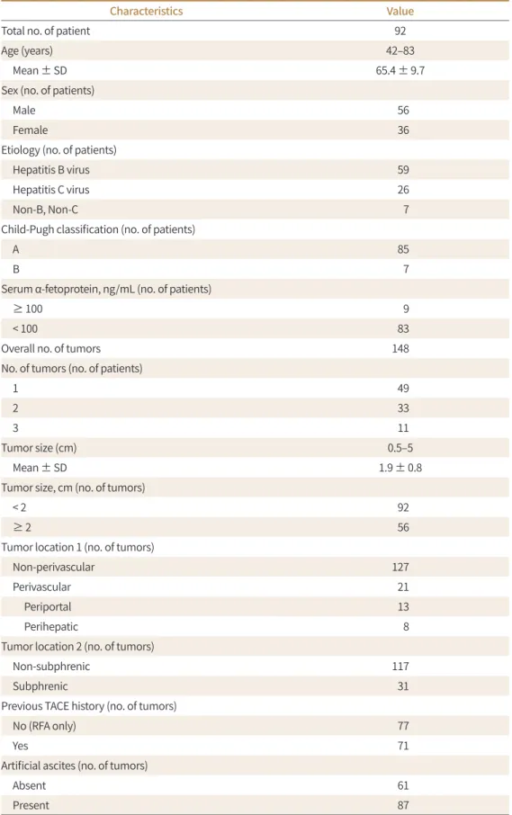
The baseline characteristics of all patients are summarized in Table 1. There were no sig- nificant differences between treatment groups regarding patient age, sex, positivity for hepa- titis B, liver function (Child–Pugh classification), and serum AFP levels (p > 0.05).
LOCAL TUMOR CONTROL
Technical success–After the initial RFA sessions, technical success was achieved for 145 of 148 tumors (98.0%). After initial RFA there were three tumors with residual unablated tumors at the ablated margin on immediately assessed multiphase CT. Additional RFA was performed immediately to treat the residual unablated tumor of three tumors, to ensure complete abla- tion on the follow-up images. These three tumors were included in subsequent analyses of therapeutic outcomes.
Technique efficacy–Complete ablation was achieved in 148 tumors based on follow-up CT obtained at 1 month. No newly developed nodular or infiltrative enhancement at the margin of the ablation zone was demonstrated. Therefore, the primary technique efficacy rate was 100% (148 of 148).
LIP–During follow-up for 1–97.4 months after RFA (median, 38.3 months), LTP was identi- fied in 21 of 148 tumors (14.6%) (Figs. 1, 2). LTP was specifically identified in 10 of 92 tumors (10.9%) (< 2 cm) and 11 of 56 tumors (19.6%) (≥ 2 cm); in 14 of 117 tumors (12.0%) in the non- subphrenic group and 9 of 31 tumors (29.0%) in the subphrenic group; in 14 of 127 tumors (11.0%) in the non-perivascular group and 7 of 21 tumors (33.3%) in the perivascular group;
in 7 of 77 tumors (9.1%) in the no TACE history group and 14 of 71 tumors (19.7%) in the pre- vious TACE history group; and in 8 of 61 tumors (13.1%) without artificial ascites and 13 of 87
Table 1. Baseline Patient and Tumor Characteristics
Characteristics Value
Total no. of patient 92
Age (years) 42–83
Mean ± SD 65.4 ± 9.7
Sex (no. of patients)
Male 56
Female 36
Etiology (no. of patients)
Hepatitis B virus 59
Hepatitis C virus 26
Non-B, Non-C 7
Child-Pugh classification (no. of patients)
A 85
B 7
Serum α-fetoprotein, ng/mL (no. of patients)
≥ 100 9
< 100 83
Overall no. of tumors 148
No. of tumors (no. of patients)
1 49
2 33
3 11
Tumor size (cm) 0.5–5
Mean ± SD 1.9 ± 0.8
Tumor size, cm (no. of tumors)
< 2 92
≥ 2 56
Tumor location 1 (no. of tumors)
Non-perivascular 127
Perivascular 21
Periportal 13
Perihepatic 8
Tumor location 2 (no. of tumors)
Non-subphrenic 117
Subphrenic 31
Previous TACE history (no. of tumors)
No (RFA only) 77
Yes 71
Artificial ascites (no. of tumors)
Absent 61
Present 87
RFA = radiofrequency ablation, SD = standard deviation, TACE = transarterial chemoembolization
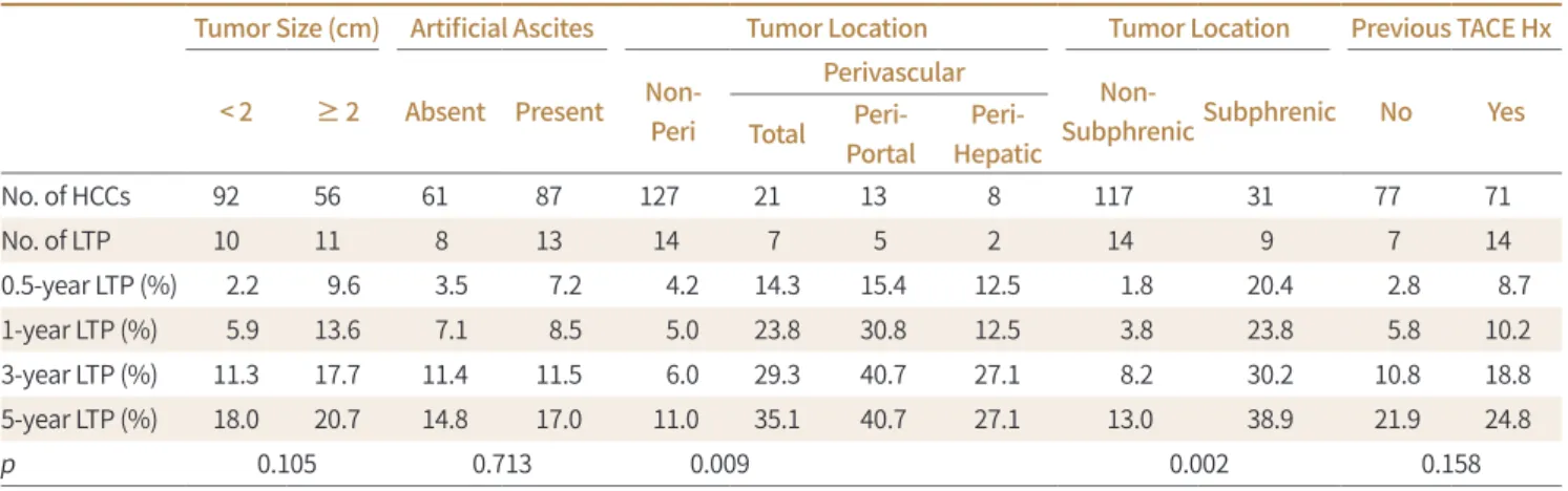
tumors (14.9%) with artificial ascites. Perivascular and subphrenic tumor locations were sig- nificant factors for the development of LTP (p = 0.013 and p = 0.04, respectively). The 1-, 3-, and 5-year cumulative LTP rates for all 148 tumors were 7.9%, 11.4%, and 14.6%, respectively.
Subgroup analyses revealed that the corresponding rates were 5.0%, 6.0%, and 11.0% for non-perivascular tumors and 23.8%, 29.3%, and 35.1% for perivascular tumors (p = 0.009).
We noted the following of peritumoral vessel types: portal vein in 5 instances and hepatic vein in 2 instances in the LTP group. Periportal location of HCC was significantly associated with LTP (p = 0.005), contrary to the perihepatic venous location of HCC (p = 0.305) (Table 2, Fig. 3).
Subphrenic location was also significantly associated with LTP (p = 0.04). The LTP rates were 3.8%, 8.2%, and 13.0% for non-subphrenic tumors and 23.8%, 30.2%, and 38.9% for subphren- Fig. 1. 73-year-old woman with perivascular hepatocellular carcinoma who underwent RFA using saline- perfused electrodes.
A. Axial CT image obtained during arterial phase shows a 2.5 cm hypervascular mass (arrow) in segment IV.
The index tumor is proximal to the left portal vein.
B. Axial CT image obtained 15 months after RFA shows the ablation zone (arrow) without any evidence of local tumor progression. A peritumoral portal vein shows the maintenance of vascular patency without thrombosis formation.
RFA = radiofrequency ablation
Fig. 2. 57-year-old man with perivascular hepatocellular carcinoma who underwent RFA using saline-per- fused electrodes.
A. Axial CT image obtained during arterial phase shows a 1.6 cm hypervascular mass (arrow) in segment IV.
The index tumor is located close to the left portal vein.
B. Axial CT image obtained 12 months after RFA demonstrates focal arterial enhancement at ablation zone, suggesting local tumor progression (black arrow). This mass is in direct contact with dilated intrahepatic bile duct (white arrow), suggestive of biliary stricture caused by RFA.
RFA = radiofrequency ablation
A B
A B
ic tumors, respectively (p = 0.002) (Table 2, Fig. 3). Perivascular and subphrenic locations were significant factors for LTP in univariate analysis (p = 0.013 and p = 0.04, respectively) and remained independent significant prognostic factors in multivariate analysis (p = 0.006 and p = 0.018, respectively) (Table 3). Tumor size, presence of artificial ascites and previous TACE history did not significantly affect LTP.
MAJOR COMPLICATIONS
The overall major complication rate after RFA was 1.4% (2/148 tumors; colonic bleeding, and portal vein thrombosis, respectively). One patient demonstrated colonic bleeding adja- cent to the ablated region, which was subsequently treated with partial embolization after superior mesenteric angiography. The bleeding persisted despite the treatment and resulted in death. Another patient had portal vein thrombosis at left main portal vein on follow-up imaging 1 year after the ablation. The case of portal vein thrombosis was successfully treated using anti-thrombolytic treatment.
Table 2. Results of the Subgroups Analysis for LTP. The Cumulative Rates of LTPs were Compared between Two Groups by Using a Log-Rank Test Tumor Size (cm) Artificial Ascites Tumor Location Tumor Location Previous TACE Hx
< 2 ≥ 2 Absent Present Non- Peri
Perivascular
Non-
SubphrenicSubphrenic No Yes Total Peri-
Portal
Peri- Hepatic
No. of HCCs 92 56 61 87 127 21 13 8 117 31 77 71
No. of LTP 10 11 8 13 14 7 5 2 14 9 7 14
0.5-year LTP (%) 2.2 9.6 3.5 7.2 4.2 14.3 15.4 12.5 1.8 20.4 2.8 8.7
1-year LTP (%) 5.9 13.6 7.1 8.5 5.0 23.8 30.8 12.5 3.8 23.8 5.8 10.2
3-year LTP (%) 11.3 17.7 11.4 11.5 6.0 29.3 40.7 27.1 8.2 30.2 10.8 18.8
5-year LTP (%) 18.0 20.7 14.8 17.0 11.0 35.1 40.7 27.1 13.0 38.9 21.9 24.8
p 0.105 0.713 0.009 0.002 0.158
HCC = hepatocellular carcinoma, Hx = history, LTP = local tumor progression, TACE = transarterial chemoembolization
Fig. 3. Kaplan-Meier curves show LTP rates.
A-C. Data for all 148 tumors (A) for non-perivascular and perivascular tumors (p = 0.009) (B) and for non-subphrenic and subphrenic tumors (p = 0.002) (C).
LTP = local tumor progression
0.4 0.3 0.2 0.1 0.0
1.0 0.8 0.6 0.4 0.2 0.0
1.0 0.8 0.6 0.4 0.2 0.0
LTP rates LTP rates LTP rates
0 20 40 60 80 100 0 20 40 60 80 100 0 20 40 60 80 100
Follow up (month) Follow up (month) Follow up (month)
A B Non-perivascular tumors C
Perivascular tumors Non-Subphrenic tumors
Subphrenic tumors
DISCUSSION
In this study, RFA using saline-perfused electrodes ensured a high initial technical success rate (98%), with 100% technique efficacy. During a median follow-up duration of 38.3 months (range 1–97.4 months), the cumulative LTP rates were 7.9%, 11.4% and 14.6% for 1, 3, and 5 years, respectively. These findings were comparable to those reported in previous studies, which used conventional RFA to treat HCC and reported 5-year LTP rates of 14.5%–20.9% (22, 23) Our results indicated that RFA using saline-perfused electrodes ensures successful local tu- mor control in patients with HCC.
Our results also demonstrated that perivascular and subphrenic locations were significant predictive factors for LTP development (hazard ratio = 3.16; 95% confidence interval: 1.27–
7.86; p = 0.013, hazard ratio = 3.54; 95% confidence interval: 1.14–8.42; p = 0.004, respectively) when using saline-perfused RFA to treat HCC (< 5 cm). Multivariate analysis revealed that perivascular and subphrenic tumor locations were significant factors independently associ- ated with LTP. Interestingly, the location of the periportal tumor was highly associated with LTP development, whereas perihepatic venous tumor location was not. Subgroup analysis of perivascular HCC with saline-perfused RFA, according to the type of peritumoral vessels, re- vealed that periportal location of HCC (40.7%) showed a higher LTP rate at 5-year period than did perihepatic location of HCC (27.1%) (p = 0.005). Lee et al. (24) recently reported increased risk of LTP when performing RFA for periportal HCC than when performing RFA for perihe- patic venous HCC. Although not fully understood, this unfavorable tumor control mecha- nism of periportal HCCs was also observed in our study. Our results suggest that presence of a hepatic vessel near the tumor and tumors abutting the diaphragm were more important prog- nostic indicators than tumor size, presence of artificial ascites, or previous TACE treatment.
Similarly, Lu et al. (21) reported poor and incomplete treatment (including incomplete ab- lation and local tumor recurrence within 6 months) in 23% and local tumor recurrence be- yond 6 months after RFA in 26% of perivascular intrahepatic tumors including metastatic le- sions after RFA using a conventional electrode. In the previous study, mean tumor size was 2.4 cm, and mean follow-up period was 11.3 months. They suggested that local tumor recur- Table 3. Prognostic Factors for LTP after Successful RFA Treatment
Factor
LTP
Univariate Analysis Multivariate Analysis
HR (95% CI) p HR (95% CI)* p
Index tumor size (cm) 2.00 (0.85–4.72) 0.112 1.03 (0.40–2.66) 0.953 Artificial ascites 1.18 (0.49–2.85) 0.714
Perivascular HCC 3.16 (1.27–7.86) 0.013 2.11 (0.40–11.08) 0.006
Subphrenic HCC 3.54 (1.14–8.42) 0.004 2.28 (0.63–9.97) 0.018
Previous TACE 0.53 (0.21–1.31) 0.165 0.66 (0.26–1.72) 0.400
The cox proportional hazard model was used for univariate and multivariate analysis. Data are HR (95% CI) values.
*Muxltivariate analysis was performed, including covariates with a p value < 0.2 in univariate analysis.
CI = confidence interval, HCC = hepatocellular carcinoma, HR = Hazard ratio, LTP = local tumor progression, RFA = radiofrequency ablation, TACE = transarterial chemoembolization
rence was significantly related to the presence of a large vessel adjacent to intrahepatic tu- mor due to promotion of the heat-sink effect generated by such vessels. Our technical suc- cess rate with saline-perfused RFA in the perivascular group was higher than that achieved by Lu et al. (21) using a conventional electrode. However, direct comparison was difficult giv- en that baseline characteristics including tumor size of our study population were different from those of Lu et al. (21) study.
Kang et al. (25) recently reported therapeutic outcomes (LTP and long-term survival) using conventional RFA for single HCC (< 3 cm). Their cumulative LTP rates were 16% and 26% at 3 and 5 years in perivascular HCC, respectively. Despite the efforts to ensure complete ablation via saline-perfused electrodes, our results indicated that the LTP of perivascular HCC (29.3%
and 35.1% at 3 and 5 years, respectively) were higher than those reported by Kang et al. (25), with the limitation of direct comparison. We present one hypothesis to explain this. HCC is a hypervascular tumor with or without the presence of microscopic invasion around the pe- ripheral portion of the index tumor. Increased intratumoral pressure associated with heating during RFA procedure could facilitate the spread of tumor cells around the ablation zone (26).
Intratumoral pressure might be further increased by using perfused saline from electrodes;
therefore, the tumor spread might be promoted in perivascular HCC. Considering all LTP of perivascular tumors demonstrated early recurrence (median 7 months, range 3.1–20.7 months) in the current study, aggressive tumor characteristics or incomplete treatment may be attributed to LTP. Furthermore, our study included several recurrent HCCs with retained iodized oil, which might cause incomplete treatment. Incomplete ablation, although unde- tected on immediate follow-up CT, may have occurred and regrowth of untreated tumor could have manifested as an early LTP. Based on this assumption, saline-perfused RFA was relatively inefficient in completely controlling local tumors in the perivascular HCC in our study.
The LTP rate was 38.9% at 5 years for the subphrenic group in our study. We observed that LTP rate was significantly higher in the subphrenic group compared to the non-subphrenic group (p = 0.002) when using saline-perfused electrodes. Characteristically, subphrenic HCCs are considered to be more difficult to treat with percutaneous RFA than are non-subphrenic HCCs, considering the poor sonic window due to the presence of overlying lung and ribs, the difficulty in placing electrodes, and inability to obtain enough ablative margin along the he- patic capsules (11, 27, 28). Therefore, it is our understanding that the percutaneous RFA of subphrenic HCCs may result in poor local tumor control. A previous study by Song et al. (28) reported comparable outcomes for subphrenic HCC in terms of LTP, while using conven- tional electrodes. The LTP rate was 46.6% at the 5-year follow-up time point. There could be one possible reason for the higher LTP rates in the subphrenic group than in the non-sub- phrenic group for RFA with saline-perfused electrodes. Although RFA using saline-perfused electrodes provided a larger ablation zone than did RFA using conventional electrodes, the operator’s concern of thermal injury to the adjacent diaphragm might lead to incomplete ab- lation due to a poor sonic window during the ablation. Nevertheless, our study demonstrated that subphrenic HCC could safely be treated using a percutaneous RFA with saline-perfused electrode without significant complication. Recent American Association for the Study of Liver Diseases guidelines (29) reported that tumor size was a risk factor for a poorer outcome and RFA was an effective treatment modality in HCC (< 3 cm) in a systematic review. Contrary
to previous studies, we noted no significant correlation between LTP and tumor size. In our study, LTP was identified in 21 of 148 tumors ranging from 1.2 to 3.6 cm. Except two, the re- maining tumors were smaller than 3 cm in size (median = 2 cm). Considering this, the tumor size might be less important as a prognostic factor than the perivascular location of HCC and might demonstrate no significant difference in LTP noted in the current study.
Our study had several limitations. The principal limitation of this study was its retrospec- tive nature, followed by the single-centered, single arm, and non-randomized design. The lack of a control group was another weak point. Despite consecutively enrolling patients, we could not completely exclude the effect of selection bias on the study outcomes due to the preference of the attending physician. However, we tried to eliminate bias by establishing clear exclusion criteria before starting the analysis. Another limitation was that several re- current tumors after TACE were included in our study, making the analysis particularly diffi- cult. Further well-designed trials of RFA for HCC with saline-perfused electrodes are war- ranted to reach a definitive conclusion regarding LTP. To the best of our knowledge, however, no prior study has evaluated prognostic factors affecting LTP of RFA with saline-perfused elec- trodes to treat HCC in an objective and quantitative manner.
In conclusion, RFA with a saline-perfused electrode is safe and effective and can be applied to large tumor, instead of a conventional electrode, to treat HCC (≤ 5 cm), without a higher rate of LTP. Nevertheless, perivascular and subphrenic HCCs demonstrated higher LTP rates than did the other sites. We should note that the perivascular and subphrenic HCC locations were associated with a high risk of local recurrence, even after the use of saline-perfused elec- trodes.
Author Contributions
Conceptualization, K.D.H., C.D.J.; data curation, K.D.H., C.D.J.; formal analysis, K.D.H., C.D.J.; in- vestigation, all authors; project administration, K.D.H., C.D.J.; resources, C.D.J., C.S.H., H.J.; supervi- sion, C.D.J.; validation, K.D.H., C.D.J.; visualization, K.D.H.; writing—original draft, K.D.H.; and writ- ing—review & editing, C.D.J., C.S.H., H.J.
Conflicts of Interest
The authors have no potential conflicts of interest to disclose.
REFERENCES
1. Forner A, Llovet JM, Bruix J. Hepatocellular carcinoma. Lancet 2012;379:1245-1255
2. Curley SA, Izzo F, Ellis LM, Nicolas Vauthey J, Vallone P. Radiofrequency ablation of hepatocellular cancer in 110 patients with cirrhosis. Ann Surg 2000;232:381-391
3. Lencioni RA, Allgaier HP, Cioni D, Olschewski M, Deibert P, Crocetti L, et al. Small hepatocellular carcinoma in cirrhosis: randomized comparison of radio-frequency thermal ablation versus percutaneous ethanol in- jection. Radiology 2003;228:235-240
4. Shibata T, Shibata T, Maetani Y, Isoda H, Hiraoka M. Radiofrequency ablation for small hepatocellular carci- noma: prospective comparison of internally cooled electrode and expandable electrode. Radiology 2006;
238:346-353
5. Cho YK, Kim JK, Kim MY, Rhim H, Han JK. Systematic review of randomized trials for hepatocellular carci- noma treated with percutaneous ablation therapies. Hepatology 2009;49:453-459
6. McWilliams JP, Yamamoto S, Raman SS, Loh CT, Lee EW, Liu DM, et al. Percutaneous ablation of hepatocel- lular carcinoma: current status. J Vasc Interv Radiol 2010;21:S204-S213
7. Xu HX, Lu MD, Xie XY, Yin XY, Kuang M, Chen JW, et al. Prognostic factors for long-term outcome after percu-
taneous thermal ablation for hepatocellular carcinoma: a survival analysis of 137 consecutive patients.
Clin Radiol 2005;60:1018-1025
8. Vivarelli M, Guglielmi A, Ruzzenente A, Cucchetti A, Bellusci R, Cordiano C, et al. Surgical resection versus percutaneous radiofrequency ablation in the treatment of hepatocellular carcinoma on cirrhotic liver. Ann Surg 2004;240:102-107
9. Hong SN, Lee SY, Choi MS, Lee JH, Koh KC, Paik SW, et al. Comparing the outcomes of radiofrequency ab- lation and surgery in patients with a single small hepatocellular carcinoma and well-preserved hepatic function. J Clin Gastroenterol 2005;39:247-252
10. Montorsi M, Santambrogio R, Bianchi P, Donadon M, Moroni E, Spinelli A, et al. Survival and recurrences af- ter hepatic resection or radiofrequency for hepatocellular carcinoma in cirrhotic patients: a multivariate analysis. J Gastrointest Surg 2005;9:62-67; discussion 67-68
11. Nam SY, Rhim H, Kang TW, Lee MW, Kim YS, Choi D, et al. Percutaneous radiofrequency ablation for hepatic tumors abutting the diaphragm: clinical assessment of the heat-sink effect of artificial ascites. AJR Am J Roentgenol 2010;194:W227-W231
12. Hori T, Nagata K, Hasuike S, Onaga M, Motoda M, Moriuchi A, et al. Risk factors for the local recurrence of hepatocellular carcinoma after a single session of percutaneous radiofrequency ablation. J Gastroenterol 2003;38:977-981
13. Kim YS, Rhim H, Cho OK, Koh BH, Kim Y. Intrahepatic recurrence after percutaneous radiofrequency abla- tion of hepatocellular carcinoma: analysis of the pattern and risk factors. Eur J Radiol 2006;59:432-441 14. Komorizono Y, Oketani M, Sako K, Yamasaki N, Shibatou T, Maeda M, et al. Risk factors for local recurrence
of small hepatocellular carcinoma tumors after a single session, single application of percutaneous radio- frequency ablation. Cancer 2003;97:1253-1262
15. Nakazawa T, Kokubu S, Shibuya A, Ono K, Watanabe M, Hidaka H, et al. Radiofrequency ablation of hepa- tocellular carcinoma: correlation between local tumor progression after ablation and ablative margin. AJR Am J Roentgenol 2007;188:480-488
16. Kim SH, Lim HK, Choi D, Lee WJ, Kim SH, Kim MJ, et al. Percutaneous radiofrequency ablation of hepato- cellular carcinoma: effect of histologic grade on therapeutic results. AJR Am J Roentgenol 2006;186:S327- S333
17. Sofocleous CT, Nascimento RG, Petrovic LM, Klimstra DS, Gonen M, Brown KT, et al. Histopathologic and immunohistochemical features of tissue adherent to multitined electrodes after RF ablation of liver malig- nancies can help predict local tumor progression: initial results. Radiology 2008;249:364-374
18. Giorgio A, Tarantino L, De Stefano G, Scala V, Liorre G, Scarano F, et al. Percutaneous sonographically guid- ed saline-enhanced radiofrequency ablation of hepatocellular carcinoma. AJR Am J Roentgenol 2003;
181:479-484
19. Kim JH, Kim PN, Won HJ, Shin YM. Percutaneous radiofrequency ablation using internally cooled wet elec- trodes for the treatment of hepatocellular carcinoma. AJR Am J Roentgenol 2012;198:471-476
20. Ahmed M, Solbiati L, Brace CL, Breen DJ, Callstrom MR, Charboneau JW, et al. Image-guided tumor abla- tion: standardization of terminology and reporting criteria--a 10-year update. Radiology 2014;273:241-260 21. Lu DS, Raman SS, Limanond P, Aziz D, Economou J, Busuttil R, et al. Influence of large peritumoral vessels
on outcome of radiofrequency ablation of liver tumors. J Vasc Interv Radiol 2003;14:1267-1274
22. Kang TW, Lim HK, Lee MW, Kim YS, Rhim H, Lee WJ, et al. Long-term therapeutic outcomes of radiofre- quency ablation for subcapsular versus nonsubcapsular hepatocellular carcinoma: a propensity score matched study. Radiology 2016;280:300-312
23. Lee DH, Lee JM, Lee JY, Kim SH, Yoon JH, Kim YJ, et al. Radiofrequency ablation of hepatocellular carcino- ma as first-line treatment: long-term results and prognostic factors in 162 patients with cirrhosis. Radiology 2014;270:900-909
24. Lee S, Kang TW, Cha DI, Song KD, Lee MW, Rhim H, et al. Radiofrequency ablation vs. surgery for perivascu- lar hepatocellular carcinoma: propensity score analyses of long-term outcomes. J Hepatol 2018;69:70-78 25. Kang TW, Lim HK, Lee MW, Kim YS, Choi D, Rhim H. Perivascular versus nonperivascular small HCC treated
with percutaneous RF ablation: retrospective comparison of long-term therapeutic outcomes. Radiology 2014;270:888-899
26. Mori Y, Tamai H, Shingaki N, Moribata K, Shiraki T, Deguchi H, et al. Diffuse intrahepatic recurrence after percutaneous radiofrequency ablation for solitary and small hepatocellular carcinoma. Hepatol Int 2009;
3:509-515
27. Kang TW, Rhim H, Kim EY, Kim YS, Choi D, Lee WJ, et al. Percutaneous radiofrequency ablation for the he- patocellular carcinoma abutting the diaphragm: assessment of safety and therapeutic efficacy. Korean J Radiol 2009;10:34-42
28. Song KD, Lim HK, Rhim H, Lee MW, Kang TW, Paik YH, et al. Hepatic resection vs percutaneous radiofre- quency ablation of hepatocellular carcinoma abutting right diaphragm. World J Gastrointest Oncol 2019;
11:227-237
29. Heimbach JK, Kulik LM, Finn RS, Sirlin CB, Abecassis MM, Roberts LR, et al. AASLD guidelines for the treat- ment of hepatocellular carcinoma. Hepatology 2018;67:358-380
5 cm 이하의 간암에서 식염수 주입방식 전극을 이용한 고주파 소작술: 국소 재발에 영향을 미치는 인자
김동호1 · 정동진1* · 조세현2 · 한준열2
목적 5 cm 이하 크기의 간암을 가진 92명의 환자군에서 식염수 주입방식 전극을 이용한 고주 파 소작술의 국소 재발률과 예후인자를 평가하였다.
대상과 방법 2009년부터 2015년까지 간암으로 식염수 주입전극 고주파 소작술을 받은 92명 환자(148개 간암)를 대상으로 하였다. 후향적으로 기술적인 성공과 효능, 국소 재발률을 분석 하였다. 국소 재발의 가능한 예후인자로써 혈관주위종양, 횡경막하종양, 인공복수 유무, 2 cm 이상 크기, 이전 간동맥색전술 치료 여부를 평가하였다. 결과는 각각의 병변 별로 분석하였다.
결과 1~97.4개월의 추적관찰 기간 동안 누적 국소 재발률은 1년, 3년, 5년에서 각각 7.9%, 11.4%, 14.6%였다. 5년 누적 국소 재발률은 혈관주위간암과(35.1%; p = 0.009) 횡경막하간 암에서(38.9%; p = 0.002) 각각 비교군에 비해 유의미하게 높았다. 다른 예후인자들에서 국 소 재발률의 유의미한 차이는 없었다(p > 0.05).
결론 식염수 주입전극을 이용한 고주파 소작술은 5 cm 이하 크기의 간암을 국소 재발률 증가 없이 안전하고 효과적으로 치료할 수 있다. 그렇지만 식염수 주입전극으로도 혈관 주위와 횡 경막하 위치의 간암은 다른 부위에 비해 국소 재발률이 높으므로, 혈관주위간암과 횡경막하 간암은 식염수 주입전극을 이용한 고주파 소작술에서도 국소 재발의 위험성이 크다는 것을 유념하여야 한다.
가톨릭대학교 여의도성모병원 1영상의학과, 2소화기내과