십이지장 무공증을 동반한 복부 내장 전위증
- 1예 보고-
경북대학교 의과대학 외과학교실, 소아과학교실1
박진영․최병호1․장수일
이 논문의 요지는 1996 년 6월 21일 제주도에서 개 최된 제 12회 대한소아외과학회 춘계학술대회에서 구연되었음.
접수일 : 09 / 5 / 28 게재승인일 : 09 / 8 / 6
교신저자:박진영, 700-721 대구광역시 중구 삼덕 2가 50번지 경북대학병원 외과
Tel : 053)420-5612, Fax : 053)421-0510 E-mail: [email protected]
서 론
복부 내장 전위증은 복강 내 장기가 거울 상처럼 좌우가 바뀌어있는 질환으로 매우 드물며 태생학적 원인은 아직까지 정확하게 알려져 있지 않다. 복부 내장 전위증 환아에 서는 선천성 심장 질환이나 비장 기형이 빈 번하게 동반된다1-3. 선천성 장 폐색증을 동 반한 복부 내장 전위증은 복잡한 외과적 문 제를 야기시키며, 치료를 위한 올바른 결정 을 내리기 위해서는 수술 전에 정확히 동반 기형을 알아내야 하며 올바른 부위에 외과 적 절개를 시행하기 위해서 술 전 복부 초 음파 촬영이나 전산화 단층촬영 등이 요구 된다. 저자들은 십이지장 무공증을 동반한 복부 내장 전위증 환아 1예를 경험하였기에 문헌 고찰과 함께 보고하는 바이다.
증 례
환아는 생후 6일된 여아로 출생 후 3일째 부터 지속된 담즙성 구토를 주소로 본원으 로 전원 되었다. 환아는 재태기간 41주째 정 상 질식분만으로 출생하였으며, 출생 시 체 중은 3.42 Kg, Apgar 점수는 정상이었다. 산 전 진찰상 특별한 문제가 없었으며, 가족력 상 특이한 소견은 없었다. 입원 당시 생체징 후상 체온은 36.2 ℃, 맥박은 128회/min, 호 흡수 38회/min 이었다. 외관상 다운증후군을 의심할 만한 소견은 보이지 않았으며, 청진 상 심박동은 규칙적이었고 심잡음은 없었으 며 복부 이학적 검사상 복부 팽만은 없었다.
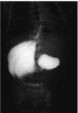
심 초음파상 특이소견은 없었으며 Infanto- gram 상에서 심장은 정상적인 위치에 놓여 있었으며, 역 쌍기포 상이 나타났으며 그 하 부에는 공기 음영이 관찰되지 않았다(Fig.
1). 타 병원에서 시행한 상부 위장관 조영술 에서 복강 내의 우측에 팽창된 위장이, 좌측 에 팽창된 십이지장이 보였다(Fig. 2). 술 전 에 시행한 복부 전산화 단층촬영에서 복강 내 좌측에 간장, 우측에 위장과 다비장 소견을 보였으며 좌측에 십이지장이 보였다(Fig. 3).
Fig. 2. Upper gastrointestinal series shows the stomach on the right side and the duodenum on the left side.
Fig. 3. Abdominal CT scan shows situs inversus abdominis with the stomach and polysplenia on the right side and the duodenum on the left side.
Fig. 1. Chest-abdomen plain radiography shows the reversed double bubble sign.
Note the normal location of the heart and the absence of gas in the rest of the abdomen.
모든 장기는 좌우가 바뀌어 있었다. 간장은 복강 내 중앙부위에서부터 좌측에 걸쳐져 있었고 십이지장은 좌측에 위치하였다. 십이 지장 제 1부위는 직경 3 cm로 팽창되어있었 으며 그 하방의 십이지장은 허탈 되어 있었 다. 십이지장 제 2부위 앞쪽에 틈을 동반한 불완전 환상 췌장이 보였으며 복강 내 우측 에 2개의 비장이 위치하였다. 장간막 기저부 는 불완전하게 고정되어있었으며 대부분의 소장은 복강 내 우측에 위치하였고 대장은 좌측에 위치하였다. 수술은 팽창된 십이지장 제 1 부위에 십이지장 절개을 시행하였다.
십이지장은 횡격막에 의해서 막혀져 있어서 횡격막절제술을 시행하였고, 불완전 환상 췌 장이 동반되어있어서 십이지장십이지장 문 합술을 측측으로 시행하였으며 충수절제술 을 시행하였다. 환아는 술 후 2주일째에 합 병증 없이 퇴원하여 현재 정상적인 성장을 하고 있다.
고 찰
복부 내장 전위증은 abdominal hetero- taxia 혹은 isolated levocardia로 불려지기도 하며, 특징적으로 복강 내 장기의 위치가 거 울상으로 바뀌지만 심장은 정상적인 위치에 있는 경우를 말한다. Aristotle이 동물에서 처음 복부 내장 전위증을 보고하였으며4,5, 17세기에 들어서 Fabricius 등이 인간에서 복부 내장 전위증을 처음 보고하였다1,5. 그 후로 복부 내장 전위증의 보고는 점점 증가 하고 있다.
복부 내장 전위증의 발생학적 원인은 아
Virchow 는 탯줄의 reversed spiral twist 의 중요성에 관해서 강조하였으며 Von Baer는 배아와 배꼽 소포(umbilical vesicle) 사이의 변경된 상관관계가 원인인자라고 주 장하였으며 Serre는 배꼽창자간막 동맥과 간장의 비정상적인 발달이 중요한 역할을 한다고 주장하였다5.
발생빈도는 출생 4,000-20,000 명당 1명꼴 로 발생하는 매우 드문 질환이며1,3 성별빈도 는 남녀에서 차이가 거의 없으나 남자에서 비장의 기형이 동반되는 경우가 흔하다고 한다6.
복부 내장 전위증 환아에서 동반되는 기 형은 주로 심장과 비장 기형이 많으며, 동반 된 심장기형은 다양하며 생명을 위협하는 심한 심장 기형이 많다. Ruben 등1(1983)은 복부 내장 전위증 환아의 60.3 %에서 선천 성 심장 질환이 동반되었고, 문헌 고찰상 27 명의 복부 기형을 동반한 복부 내장 전위증 환자 중 16명이 동반된 심장 기형으로 사망 하였다고 보고하였다. 수술적 치료가 필요한 복부 내장 전위증 환자의 경우에 술 전에 심 초음파를 시행하여 동반된 선천성 심장 기형의 상태를 정확하게 알아내야 한다.
이 등7(2006)은 45명의 복부 내장 전위증 환아 중 26명(58 %)에서 십이지장 무공증, 담도 폐쇄증 등의 복강 내 기형이 동반하였 다고 보고하였다. 복부 내장 전위증 환자에 서 십이지장 폐쇄증이 동반되는 경우는 매 우 드물어서 최근까지 문헌상에 약 20예 정 도만이 보고되었다3, 8-12. 복부 내장 전위증과 동반된 십이지장 폐쇄증의 원인은 환상 췌 장이 가장 많았으며, 십이지장 격막, pre-
duodenal portal vein, 십이지장 협착 및 무 공증 등이 보고되었으며8-12, 저자들의 경우 에는 횡격막과 불완전 환상 췌장에 의해서 십이지장 폐쇄증이 발생하였다.
복부 내장 전위증은 대부분 증상이 없어 서, 수술 중이나 부검 시에 우연히 발견되지 만 선천성 십이지장 폐쇄증을 동반한 경우 에는 신생아 시기에 증상이 발생한다. 진단 은 단순 복부 촬영상 역 쌍기포상으로 쉽게 내릴 수 있고 중심부에 천공을 동반한 십이 지장 격막의 경우에는 조영제를 이용한 상 부 위장관 촬영을 시행하여 확진할수도 있 다3.
복부 내장 전위증 환아의 합리적인 접근 은 수술 전에 가능한 한 많은 정보를 얻어 내는 것이 중요하다. 예를 들면 복부의 올바 른 부위에 외과적 절개를 시행하기 위하여 술 전에 복부 초음파나 복부 전산화 단층촬 영 등의 방사선적 촬영을 시행하여 복강 내 장기의 위치 변화를 인지해 내야하며 술 전 에 심 초음파를 시행하여 생명을 위협하는 동반된 선천성 심장기형을 알아내야 한다.
개복시에는 동반된 여러 기형을 알아내는 것이 중요하며 또한 충수돌기의 위치가 불 분명하기 때문에 충수 절제술을 반드시 시 행해야 한다. 이러한 외과적 원칙을 준수한 다면 복강 내 기형을 동반한 복부 내장 전 위증 환아에서 적절한 수술적 치료를 시행 할 수 있을 것이다.
참 고 문 헌
1. Ruben GD, Templeton JM Jr, Ziegler MM:
Situs Inversus: The Complex
Inducing Neonatal Intestinal Obstruction.
J Pediatr Surg 18:751-6, 1983
2. Chacko KA, Krishnaswami S, Sukumar IP, Cherian G: Isolated levocardia: two
cases with abdominal situs inversus, thoracic situs solitus, and normal circulation. Am Heart J 106:155-9, 1983
3. Nawaz A, Matta H, Hamchou M,Jacobez A, Trad O, Al Salem AH: Situs
inversus abdominis in association with congenital duodenal obstruction: a report of two cases and review of the literature.
Pediatr Surg Int 21:589-92, 2005
4. Fonkalsrud EW, Tompkins R, Clatworthy Hw Jr: Abdominal manifestations of situs
inversus in infants and children. Arch
Surg 92:791-5, 19665. Blegen HM: Surgery in Situs Inversus.
Ann Surg 129:244-59, 1949
6. Gray SW, Skandalakis JE: Embryology
for Surgeons, The Embryological Basis for the Treatment of Congenital Defects.
Philadelphia, WB Saunders, 1972, Pp 880-90
7. Lee SE, Kim HY, Jung SE, Lee SC, Park KW, Kim WK: Situs anomalies and
gastrointestinal abnormalities. J Pediatr
Surg 41:1237-42, 20068. Iuchtman M, Golan Y, Heldenberg D, Kessler FB: Situs inversus abdominis in
association with duodenal obstruction and intestinal hernia. Am J Perinatol 10:255-
7, 19939. Akel S, Halabi J, Shawis R: Abdominal
situs inversus with congenital duodenal stenosis: rare association. Eur J Pediatr
Surg 8:55-7, 199810. Mordehai J, Cohen Z, Kurzbart E, Mares AJ: Preduodenal portal vein causing
duodenal obstruction associated with situs
inversus, intestinal malrotation, and
polysplenia: A case report. J Pediatr Surg
37:E5, 2002Late presentation of a duodenal web in a patient with situs inversus and apple peel jejunal atresia. Pediatr Surg Int 20:301-3,
2004Laparoscopic duodenoduodenostomy in a newborn with situs inversus totalis. J Laparoendosc Adv Surg Tech A 18:654-6, 2008
Situs Inversus Abdominis Associated with Duodenal Atresia
- A Case Report-
Jinyoung Park, M.D., Byung Ho Choe, M.D.
1, Sooil Chang, M.D.
Department of Surgery, Department of Pediatric1, School of Medicine, Kyungpook National University,
Taegu, Korea
Situs inversus abdominis is a rare congenital condition commonly associated with serious cardiac and splenic malformations. The importance of recognizing the presence of situs inversus abdominis preoperatively is emphasized by the fact that the surgical incision is placed on the incorrect side of the abdomen. A 6 day-old girl was referred to our hospital because of bile stained vomiting. A plain radiography of abdomen and chest showed the heart to be normal position and a reversed "double-bubble" picture with no other gas shadow in the rest of the abdomen. Abdominal computed tomography scan revealed situs inversus with the stomach and polysplenia on the right side and the liver on the left side. A laparotomy confirmed the diagnosis of situs inversus with duodenal atresia. The obstruction was bypassed by constructing a side-to-side duodenoduodenostomy.
The postoperative course was uneventful.
(J Kor Assoc Pediatr Surg 15(1):52~57), 2009.
Index Words:Situs inversus abdominis, Duodenal atresia
Correspondence:Jinyoung Park, M.D., Department of Surgery, Kyungpook National University Hospital, 50 Samduk-2 Ga, Chung-gu, Taegu 700-721, Korea
Tel : 053)420-5612, Fax : 053)421-0510 E-mail: [email protected]