Hepatocellular Carcinoma Risk of Compensated Cirrhosis Patients with Elevated HBV DNA Levels according to Serum Aminotransferase Levels
Sometimes, hepatitis B virus (HBV)-related cirrhotic patients with normal aminotransferase levels are closely followed-up for the elevation of aminotransferase levels instead of prompt antiviral therapy (AVT). We analyzed the long-term hepatocellular carcinoma (HCC) risk according to the aminotransferase levels in a retrospective cohort of 1,468 treatment- naïve, HBV-related, compensated cirrhosis patients with elevated HBV DNA levels (≥ 2,000 IU/mL). Based on aminotransferase levels, patients were categorized into normal (< 40 U/L, n = 364) and elevated group (≥40 U/L, n = 1,104). During a median of 5.3 yr of follow-up (range: 1.0-8.2 yr), HCC developed in 296 (20%) patients. The 5-yr
cumulative HCC incidence rate was higher in patients with elevated aminotransferase level, but was not low in normal aminotransferase level (17% vs. 14%, P = 0.004). During the follow-up, 270/364 (74%) patients with normal aminotransferase levels experienced elevation of aminotransferase levels, and AVT was initiated in 1,258 (86%) patients. Less patients with normal aminotransferase levels received AVT (70% vs. 91%, P < 0.001) and median time to start AVT was longer (17.9 vs. 2.4 months, P < 0.001). AVT duration was an independent factor associated with HCC, and median duration of AVT was shorter (4.0 vs. 2.6 yr, P < 0.001) in patients with normal aminotransferase levels. The HCC risk of compensated cirrhosis patients with normal aminotransferase level is not low, and AVT duration is associated with lowered HCC risk, indicating that prompt AVT should be strongly considered even for those with normal aminotransferase levels.
Keywords: Liver Neoplasms; Antiviral Therapy; Aminotransferase; Treatment; Viruses Junggyu Lee,1* Dong Hyun Sinn,1*
Jung Hee Kim,1 Geum-Youn Gwak,1 Hye Seung Kim,2 Sin-Ho Jung,2 Yong-Han Paik,1 Moon Seok Choi,1 Joon Hyeok Lee,1 Kwang Cheol Koh,1 Byung Chul Yoo,1 and Seung Woon Paik1
1Department of Medicine, Samsung Medical Center, Sungkyunkwan University School of Medicine;
2Biostatistics and Clinical Epidemiology Center, Samsung Medical Center, Seoul, Korea
*Junggyu Lee and Dong Hyun Sinn contributed equally to this work.
Received: 14 May 2015 Accepted: 5 August 2015 Address for Correspondence:
Seung Woon Paik, MD
Department of Medicine, Samsung Medical Center, Sungkyunkwan University School of Medicine, 81 Irwon-ro, Gangnam-gu, Seoul 06351, Korea
Tel: +82.2-3410-3409, Fax: +82.2-3410-6983 E-mail: [email protected]
http://dx.doi.org/10.3346/jkms.2015.30.11.1618 • J Korean Med Sci 2015; 30: 1618-1624
INTRODUCTION
The hepatitis B virus (HBV) is a major public health problem worldwide, as well as in Korea (1,2). Chronic HBV infection can evolve into cirrhosis and/or hepatocellular carcinoma (HCC) (3), and HBV is the most important risk factor for HCC in HBV- endemic areas, including Korea (4). Unfortunately, there are no effective cures for HBV; currently available treatments, such as interferon and nucleos(t)ide analogues (NUCs), can suppress viral replication but cannot eradicate the virus (3,5). Therefore, the decision to treat should be based on assessing the risk and benefit of the treatment.
Guidelines put forth by the American Association for the Study of Liver Diseases (AASLD), the European Association for the Study of the Liver (EASL), the Asian Pacific Association for the Study of the Liver (APASL), and the Korean Association for the Study of the Liver (KASL) recommends different treatment stra- tegy according to the liver disease statues (6-9). For patients with chronic hepatitis, treatment is recommended when both serum HBV DNA levels and serum aminotransferase levels are elevat-
ed, however, for patients with compensated cirrhosis, treatment is recommended when serum HBV DNA levels are elevated, ir- respective of serum aminotransferase levels (6-9).
Serum aminotransferase level is a useful indicator to assess the risk of HCC and guide treatment initiation (9,10). However, serum alanine aminotransferase (ALT) levels can be elevated in patients with normal liver histology and can be normal in pa- tients with advanced fibrosis (11-13). Furthermore, cirrhosis is a major risk factor for HCC (14), and antiviral therapy (AVT) us- ing NUCs can effectively suppress HBV replications and have been shown to reduce the risk of HCC (15). Therefore, for com- pensated cirrhosis patients, AVT is recommended irrespective of serum ALT levels by AASLD, EASL, APASL, and KASL guide- lines (6-9). However, in Korea, the costs of AVT for HBV are cov- ered by National Health Insurance only if the patient’s serum aminotransferase levels are elevated above the upper limit of normal, even for compensated cirrhotic patients with elevated HBV DNA level. As cost for AVT is not reimbursed for patients with normal aminotransferase levels, many Korean patients with normal aminotransferase levels are closely monitored for
a change in serum aminotransferase levels, instead of prompt AVT. Limited data are available about the actual HCC risk in HBV-related compensated cirrhosis patients with elevated HBV DNA levels plus normal aminotransferase levels. Therefore, in this study, we analyzed the long-term risk as well as the risk fac- tors for HCC in cirrhotic patients with elevated HBV DNA levels plus normal aminotransferase levels, and compared to cirrhotic patients with elevated aminotransferase levels.
MATERIALS AND METHODS Study design, setting, and participants
This is a retrospective cohort study of chronic HBV-infected com- pensated cirrhosis patients who received care at Samsung Med- ical Center in Seoul, Korea. All patients who had their serum HBV DNA levels measured using the COBAS TaqMan HBV DNA Test (Roche Diagnostics, Branchburg, NJ) between 2006 (when serum HBV DNA testing with the COBAS TaqMan HBV DNA Test first began) and 2011 were screened for potential inclusion in the study. The time of this initial HBV DNA measurement was considered the baseline. We included patients who met all of following criteria: 1) aged ≥ 18 yr with chronic HBV infection, defined by the presence of hepatitis B surface antigen (HBsAg) in serum for more than 6 months or appropriate medical histo- ry; 2) the presence of one or more of the following clinical indi- cators of cirrhosis: thrombocytopenia (<150,000 platelets per µL), cirrhotic configuration of the liver (nodular liver surface or caudate lobe hypertrophy) and/or splenomegaly confirmed in imaging studies, or the presence of varices (abnormally enlarg- ed veins, detected by upper endoscopy or cross-sectional imag- es); 3) a baseline serum HBV DNA level ≥ 2,000 IU/mL; 4) no previous history of interferon or NUC treatment; 5) no previous history of HCC or HCC diagnosed within a year; 6) no evidence of decompensated liver cirrhosis as indicated by the presence (or history) of ascites, esophageal or gastric variceal bleeding, hepatic encephalopathy, or a Child-Pugh score ≥ 7; 7) no co- infection with hepatitis C virus or human immunodeficiency virus; 8) follow-up duration of more than a year. Ultimately, a total of 1,468 patients were included in the study.
Study variables
The primary outcome variable was the diagnosis of HCC dur- ing follow-up. The follow-up period was the time elapsed be- tween baseline HBV DNA measurement and the date of data analysis, which was the 30th of April, 2014. Follow-up assess- ments were performed on all patients every 3-6 months or more frequently as required, for a period of at least one year. Serum HBV DNA levels were measured in all patients during the entire follow-up period, and patient use of AVT during follow-up (in the form of NUC treatment) was recorded.
HCC (the primary outcome variable) was diagnosed either
by histological evaluation or clinical imaging (16). The follow- ing parameters were reviewed: age, sex, medical history, ultra- sonography and upper endoscopy results, baseline levels of se- rum platelet, hepatitis B e antigen (HBeAg), hepatitis B e anti- body (anti-HBe), alphafetoprotein (AFP) and HBV DNA, and other blood chemistry parameters at baseline, including aspar- tate aminotransferase (AST) and ALT levels. Normal amino- transferase was defined as when both AST and ALT level were below 40 U/L. Patients were divided into the two groups (nor- mal aminotransferase and elevated aminotransferase levels) based on serum aminotransferase levels at baseline. The initial lower limit of 12 IU/mL for HBV DNA detection was lowered to 9 IU/mL, but for this study, an HBV DNA level of 12 IU/mL was considered to represent an undetectable HBV DNA level. Com- plete virological response (CVR) was defined when serum HBV DNA became undetectable (< 12 IU/mL) after NUCs therapy.
Statistical analysis
Baseline and clinical characteristics were summarized with mean
± standard deviation (SD), median (quartile) or frequency (per- cent) as appropriate, and their distributions between groups were compared by two-sample t-test, Mann-Whitney U-test, or chi-square test as appropriate. The cumulative incidence rate of HCC was calculated and plotted by using the Kaplan-Meier method. Log-rank tests were used to examine differences of the incidence rate among the groups. Risk factor for HCC was as- sessed using Cox’s regression analysis. As the time to start AVT was not uniform, we used AVT duration (which is a time-de- pendent variable), instead of AVT (yes vs. no), in the Cox’s re- gression analyses. CVR was assessed as CVR duration, instead of CVR (yes vs. no), for the same reason. Independent risk fac- tor for HCC was assessed via multivariate Cox-regression analy- ses. As AVT duration and CVR duration have high co-linearity, they were separately assessed in the multivariate model. Statis- tical significance was declared when a P value < 0.05.
Ethics statement
The study protocol was reviewed and approved by the institu- tional review board at Samsung Medical Center (IRB No. 2014- 09-082). Because the study is based on the retrospective analy- sis of existing administrative and clinical data, the requirement of obtaining informed patient consent was waived by the board.
RESULTS
Baseline characteristics of study participants
Baseline characteristics of the analyzed patients are shown in Table 1. All enrolled patients had evidence of cirrhosis in the form of thrombocytopenia, cirrhotic configuration with or without splenomegaly, and/or varices. At baseline, 364 patients showed normal aminotransferase levels, while 1,104 patients showed
elevated aminotransferase levels. There were more female pa- tients with normal aminotransferase levels and fewer who were HBeAg (+). The normal aminotransferase levels group also show- ed higher albumin, lower bilirubin, higher platelet, lower AFP, and lower HBV DNA levels than patients with elevated amino- transferase levels (Table 1).
AVT during follow-up
During follow-up, a total of 1,258 patients (86%) started AVT af- ter a median of 4.4 months from enrollment in form of NUCs.
Entecavir was most frequently used drug (72%). CVR was no- ticed in 1,085 patients. More patients with elevated aminotrans- ferase levels received AVT (91% vs. 70%, P < 0.001). Median time to start AVT was shorter (2.4 months vs. 17.9 months, P < 0.001), and median duration of AVT was longer (4.0 yr vs. 2.6 yr, P < 0.001) (Table 1). Also, more patients with elevated aminotransferase levels achieved CVR (78% vs. 60%, P < 0.001), median time to CVR was shorter (15.1 months vs. 26.6 months, P < 0.001), and median duration of CVR was longer (2.8 yr vs. 1.6 yr, P < 0.001).
However, when analysis was limited for those who received AVT,
there was no significant difference of CVR rate between patients with elevated vs. normal aminotransferase levels (86%, 866/1,005 patients vs. 87%, 19/253 patients, P = 0.87).
HCC incidence rates and risk factors
Throughout follow-up (medium follow-up duration 5.2 yr; range 1.0-8.2 yr), HCC was newly diagnosed in 296 patients (20.2%).
Patients who developed HCC were older, more often male, and HBeAg (+). ALT levels was similar, but AST level was slightly higher in patients who developed HCC. Serum albumin and platelet level was lower while bilirubin, and AFP level was high- er. Baseline HBV DNA levels were similar, but AVT and CVR was less frequently seen in patients who developed HCC. AVT duration and CVR duration was shorter in patients who devel- oped HCC (Table 1).
The overall 5-yr cumulative HCC incidence rate was 16.4%.
Age, gender, HBeAg status, AFP levels, and baseline aminotrans- ferase levels (elevated vs. normal) were independent predictors for HCC (Table 2). AVT duration was also significant factors as- sociated with HCC. As AVT induces CVR, AVT showed high co-
Table 1. Comparison of characteristics
Characteristics ALL (n = 1,468) Normal (n = 364) Elevated (n = 1,104) P value No HCC (n = 1,172) HCC (n = 296) P value
Age (yr) 50.0 ± 9.0 49.4 ± 9.0 50.1 ± 9.0 0.20 49.0 ± 9.0 53.5 ± 8.2 < 0.001
Male 948 (65%) 205 (56%) 743 (67%) < 0.001 726 (62%) 222 (75%) < 0.001
Hepatitis B e antigen positive 827 (56%) 177 (49%) 650 (59%) 0.001 645 (55%) 182 (62%) 0.046
ALT (U/L) 47 (33-75) 28 (22-34) 58 (43-94) < 0.001 48 (33-80) 46 (34-62) 0.13
AST (U/L) 48 (35-72) 30 (25-34) 56 (44-84) < 0.001 47 (33-73) 50 (38-68) 0.041
Albumin (g/dL) 4.0 (3.7-4.3) 4.1 (3.9-4.3) 4.0 (3.6-4.2) < 0.001 4.0 (3.8-4.3) 3.9 (3.5-4.1) < 0.001 Bilirubin (mg/dL) 1.0 (0.7-1.3) 0.8 (0.7-1.1) 1.0 (0.8-1.4) < 0.001 1.0 (0.7-1.3) 1.1 (0.8-1.5) < 0.001 Platelet (× 103/L) 123 (96-145) 128 (104-150) 121 (93-143) < 0.001 125 (100-145) 82 (62-113) < 0.001 AFP (ng/mL) 7.3 (4.2-18.3) 4.6 (3.1-8.0) 9.0 (5.0-25.2) < 0.001 6.7 (3.9-17.1) 10.9 (6.2-21.6) < 0.001 Baseline HBV DNA (log10 IU/mL) 5.8 ± 1.4 4.7 (3.9-5.8) 6.0 (5.0-7.2) < 0.001 5.8 ± 1.4 5.7 ± 1.3 0.78
Antiviral therapy 1,258 (86%) 253 (70%) 1,005 (91%) < 0.001 1,020 (87%) 238 (80%) 0.004
Entecavir 905 (72%) 198 (78%) 707 (70%) 737 (72%) 168 (71%)
Others* 353 (28%) 55 (22%) 298 (30%) 283 (28%) 70 (29%)
Time to AVT (months) 4.4 (0.4-21.0) 17.9 (6.4-33.9) 2.4 (0.4-17.1) < 0.001 4.3 (0.4-20.9) 5.1 (0.4-21.9) 0.60 AVT duration (yr) 3.7 (1.8-5.5) 2.6 (0-4.5) 4.0 (2.4-5.8) < 0.001 4.1 (2.5-5.9) 1.9 (0.4-3.7) < 0.001
CVR† 1,085 (74%) 219 (60%) 866 (78%) < 0.001 919 (78%) 166 (56%) < 0.001
Time to CVR (months) 18.0 (7.2-35.0) 26.6 (14.4-43.9) 15.1 (6.5-30.5) < 0.001 18.0 (7.3-34.7) 17.5 (6.7-35.9) 0.62 CVR duration (yr) 2.5 (0-4.5) 1.6 (0-3.6) 2.8 (0.8-4.7) < 0.001 2.9 (1.1-5.0) 0.5 (0-2.4) < 0.001
*Other medications include lamivudine, telbivudine, clevudine and adefovir; †Complete virological response was defined when HBV DNA became undetectable ( < 12 IU/mL) af- ter AVT. ALT, Alanine aminotransferase; AST, Aspartate aminotransferase; HBV, hepatitis B virus; AVT, antiviral therapy; CVR, Complete virological response.
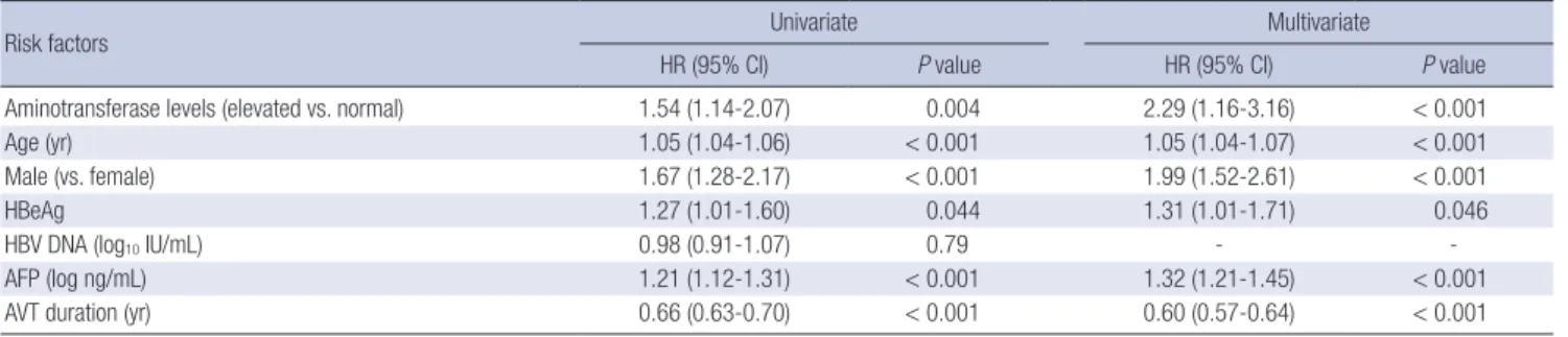
Table 2. Risk factors for hepatocellular carcinoma development
Risk factors Univariate Multivariate
HR (95% CI) P value HR (95% CI) P value
Aminotransferase levels (elevated vs. normal) 1.54 (1.14-2.07) 0.004 2.29 (1.16-3.16) < 0.001
Age (yr) 1.05 (1.04-1.06) < 0.001 1.05 (1.04-1.07) < 0.001
Male (vs. female) 1.67 (1.28-2.17) < 0.001 1.99 (1.52-2.61) < 0.001
HBeAg 1.27 (1.01-1.60) 0.044 1.31 (1.01-1.71) 0.046
HBV DNA (log10 IU/mL) 0.98 (0.91-1.07) 0.79 - -
AFP (log ng/mL) 1.21 (1.12-1.31) < 0.001 1.32 (1.21-1.45) < 0.001
AVT duration (yr) 0.66 (0.63-0.70) < 0.001 0.60 (0.57-0.64) < 0.001
HR, hazard ratio; CI, confidence interval; HBeAg, hepatitis b e antigen; HBV, hepatitis B virus; AFP, alphafetoprotein; AVT, antiviral therapy.
linearity with CVR. Therefore impact of CVR on HCC risk was separately assessed. In univariate analysis, CVR duration (year) was associated with HCC (hazard ratio, 0.62; 95% confidence interval, 0.58-0.66; P < 0.001). CVR was associated with HCC development even after adjusted for age, gender, HBeAg, HBV DNA levels and AFP levels (hazard ratio, 0.60; 95% confidence interval, 0.56-0.64; P < 0.001).
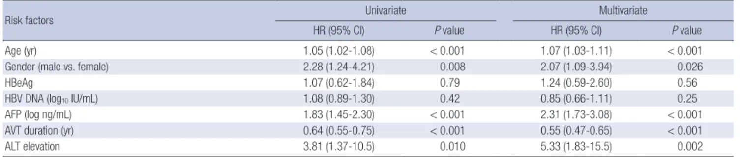
HCC risk according to aminotransferase levels
When compared according to the aminotransferase levels, the cumulative HCC incidence rate was higher in patients with ele- vated aminotransferase levels (3-, 5-, and 7-yr cumulative HCC incidence rate: 9%, 17%, and 28%) but HCC incidence rate was not low in patients with normal aminotransferase levels (3-, 5-, and 7-yr cumulative HCC incidence rate: 7%, 14%, and 18%, P = 0.004, Fig. 1).
Among 364 patients with normal aminotransferase levels at baseline, AVT was started in 253 patients (70%), and the 5-yr cu- mulative incidence rate of HCC for patients who received AVT
was lower than patients who did not (12% vs. 21%, P = 0.003, Fig. 2). Elevation of ALT levels above 40 U/L was also noticed in 270 of 364 (74%) patients after median of 17.4 months (range:
0.2-84.8 months) of follow-up, and the 5-yr cumulative incidence rate of those who experienced ALT elevation was higher than those without (18% vs. 4%, P = 0.005). Age, gender, AFP level, ALT elevation, and AVT duration were independent factors as- sociated with HCC risk in patients with normal aminotransfer- ase levels (Table 3). Among 1,104 patients with elevated amino- transferase levels, the 5-yr cumulative incidence rate of HCC for patients who started AVT was lower than patients who did not (15% vs. 38%, P < 0.001, Fig. 2).
HCC risk according to the use of AVT
During follow-up, 1,258/1,468 (86%) patients started AVT after
Cumulative incidence of hepatocellular carcinoma
Follow-up duration (yr)
No. at risk
Elevated 1,104 1,104 1,013 895 752 597 429 278 40 Normal 364 364 333 288 240 196 160 106 12
0 1 2 3 4 5 6 7 8 1.0
0.8
0.6
0.4
0.2
0
P = 0.004
Fig. 1. Cumulative incidence rate of hepatocellular carcinoma according to the serum aminotransferase levels. When the aminotransferase groups were compared, the cu- mulative HCC incidence rate was significantly higher in patients with elevated amino- transferase levels than normal aminotransferase levels (P = 0.004).
Fig. 2. Cumulative incidence rate of hepatocellular carcinoma according to the serum aminotransferase levels and use of antiviral therapy during follow-up. The 5-yr cumu- lative HCC incidence rate was highest in patients with elevated aminotransferase lev- els who did not received antiviral therapy (38%, yellow, group 1), followed by patients with normal aminotransferase levels who did not received antiviral therapy (21%, blue, group 2), patients with elevated aminotransferase levels who received antiviral thera- py (15%, purple, group 3), and patients with normal aminotransferase levels who re- ceived antiviral therapy (12%, green, group 4).
Cumulative incidence of hepatocellular carcinoma
Follow-up duration (yr)
No. at risk
Group 1 99 99 76 52 38 30 20 10 3
Group 2 111 111 93 69 50 38 24 14 3
Group 3 1,005 1,005 937 843 713 567 409 268 36
Group 4 253 253 240 219 191 158 136 92 10
0 1 2 3 4 5 6 7 8 1.0
0.8
0.6
0.4
0.2
0
P < 0.001
Table 3. Risk factors for hepatocellular carcinoma development in patients with normal aminotransferase levels
Risk factors Univariate Multivariate
HR (95% CI) P value HR (95% CI) P value
Age (yr) 1.05 (1.02-1.08) < 0.001 1.07 (1.03-1.11) < 0.001
Gender (male vs. female) 2.28 (1.24-4.21) 0.008 2.07 (1.09-3.94) 0.026
HBeAg 1.07 (0.62-1.84) 0.79 1.24 (0.59-2.60) 0.56
HBV DNA (log10 IU/mL) 1.08 (0.89-1.30) 0.42 0.85 (0.66-1.11) 0.25
AFP (log ng/mL) 1.83 (1.45-2.30) < 0.001 2.31 (1.73-3.08) < 0.001
AVT duration (yr) 0.64 (0.55-0.75) < 0.001 0.55 (0.47-0.65) < 0.001
ALT elevation 3.81 (1.37-10.5) 0.010 5.33 (1.83-15.5) 0.002
HR, hazard ratio; CI, confidence interval; HBeAg, hepatitis b e antigen; HBV, hepatitis B virus; AFP, alphafetoprotein; AVT, antiviral therapy; ALT, alanine aminotransferase.
a median of 4.4 months of follow-up. For those started AVT, the 5-yr cumulative incidence rate of HCC was 14%, and was higher in patients with elevated aminotransferase levels (15% vs. 12%, P = 0.001, Fig. 2). Among patients without AVT during follow- up, the 5-yr cumulative incidence rate of HCC was 29%, and the rate was higher in patients with elevated aminotransferase lev- els (40 U/L) than normal aminotransferase levels (38% vs. 21%, P = 0.005, Fig. 2).
DISCUSSION
In this retrospective cohort with treatment-naïve HBV-related compensated cirrhosis, we found that patients with elevated aminotransferase levels are at higher risk for HCC (17% at 5-yr) than patients with normal aminotransferase levels. However, the long-term HCC risk was not low in patients with normal aminotransferase levels (14% at 5-yr). During follow-up, many patients (86%) eventually started AVT. Those without AVT dur- ing follow-up showed higher HCC incidence rates (29% at 5-yr) than those with AVT (14% at 5-yr), and AVT duration was a sig- nificant factor associated with HCC. In overall, those with ele- vated aminotransferase levels started AVT more frequently and earlier than those with normal aminotransferase levels, which resulted in higher AVT duration, higher CVR rate, and higher CVR duration, yet, CVR rate was similar between those with el- evated and normal aminotransferase levels, when analysis re- stricted for those who received AVT. Among patients with nor- mal aminotransferase levels, many patients experienced eleva- tion of aminotransferase levels during follow-up, and AVT du- ration was independent factor associated with lower HCC risk, along with age, gender, AFP levels and ALT elevation during fol- low-up.
A strength of this study is that this is a ‘real-life’ cohort, with large numbers of patients, large cases of primary end-point (296 patients with HCC), and a long follow-up period (median 5.2 yr) that can demonstrate clinical course of these patients. This study has clearly shown that compensated cirrhosis patients are at risk for developing HCC, and those with normal amino- transferase levels at baseline are also at risk for HCC, although HCC risk is lower than patients with elevated aminotransferase levels. As HCC risk is considerably high for those with normal aminotransferase levels (14% at 5-yr), efforts to minimize the risk is needed. In this study, older age, male gender, higher AFP levels, and experiencing ALT elevation was associated with in- creased HCC risk, while AVT duration was associated with low- ered HCC risk. Although effective suppression of HBV replica- tion cannot completely eliminate the risk of HCC (17), NUC treatment has been shown to reduce the incidence of HCC (15, 18), and reverse cirrhosis (19). Therefore, this data suggest that increasing AVT duration by prompt AVT can be a way to decrease HCC risk in cirrhotic patients with normal aminotransferase
levels, and support the recommendation from AASLD, EASL, APASL, and KASL that AVT should be strongly considered for compensated cirrhosis patients with elevated HBV DNA levels, irrespective of serum aminotransferase levels (6-9). We observ- ed that time to start AVT was significantly longer for patients with normal aminotransferase levels. As this study is retrospec- tive study, we could not accurately assess the exact reason for initiating and not initiating AVT for individual patient, however, current reimbursement policy for NUCs in Korea (cost for AVT is not reimbursed for patients with normal aminotransferase levels) might be a reason. Revision of current reimbursement policy for HBV-related cirrhotic patients in Korea should be strong- ly considered.
As HCC risk was low (4% at 5-yr) in those with ‘persistently’
normal aminotransferase levels, one may choose close moni- toring over prompt AVT and treat for those who shows elevated aminotransferase levels. However, this approach needs careful consideration, as one cannot accurately know whether patients will remain having ‘persistently’ normal aminotransferase lev- els or not at baseline. In this study, those who experience eleva- tion of aminotransferase levels are at increased risk of HCC, and early initiation of NUC therapy can potentially prevent elevation of aminotransferase levels.
Our findings have several limitations. First, this study is a ret- rospective cohort study with several potential biases. AVT is well-known factor associated with HCC risk, yet AVT was initi- ated at variable time point during follow-up. We used time-de- pendent variables (AVT duration) instead of use of AVT (yes vs.
no) to minimize potential bias. However, in order to see defi- nitely whether prompt AVT can reduce development of HCC over watchful monitoring (and AVT after ALT elevation), ran- domized-controlled trials are needed. Therefore decision to initiate AVT should be individualized balancing the risk and benefit of the treatment until those data are available, yet, con- sidering potential benefit, efficacy and safety of NUC therapy, it may be unethical to perform such trial. Second, large propor- tion of patients who were classified as normal aminotransferase group experienced ALT elevation during follow-up at different time-point from enrollment. Many of those who were initially classified as normal aminotransferase group may actually be in group of patients with elevated aminotransferase level, as we classified patients according to single point value. Third, it should be noted that almost all Korean chronic hepatitis B patients are infected with HBV genotype C (20), which is known to progress more rapidly to HCC than other genotypes (9). Fourth, this study has been conducted in a tertiary referral center, which may have introduced selection bias that more severe cases may have been included. Fifth, we used an aminotransferase cutoff of 40 U/L, as this is the currently used aminotransferase cutoff used by National Health Insurance, however, many suggested lowered cutoff for normal ALT levels (11,12,21). In this study we tested
lowered ALT cutoff point (e.g., 30 IU/L for men and 19 IU/L for women) to define normal aminotransferase level, yet, the find- ing were similar and there was no significant difference in the HCC risk between patients with upper normal ALT levels (30-40 IU/L for men, 19-40 IU/L for women) or lower normal ALT lev- els (< 30 IU/L for men, < 19 IU/L for women) (data not shown).
In conclusion, the present data suggest that compensated cirrhotic patients with elevated serum HBV DNA levels are at risk for HCC (16% at 5-yr). The HCC risk was higher when ami- notransferase levels are elevated (17% at 5-yr), but patients with normal aminotransferase levels were not at low risk for HCC (14% at 5-yr). Intervention to decrease HCC risk is definitely needed for cirrhotic patients with elevated serum HBV DNA levels, both with elevated and normal aminotransferase levels.
AVT duration was significantly associated with lower HCC inci- dence, which suggests that increasing AVT duration by prompt AVT, instead of close monitoring for elevation of serum amino- transferase levels, can reduce the risk of HCC in cirrhotic pati- ents with elevated HBV DNA levels even for those with normal aminotransferase levels.
DISCLOSURE
The authors have no conflicts of interest to disclose.
AUTHOR CONTRIBUTION
Study design: Paik SW, Sinn DH. Data collection: Lee J, Kim JH.
Statistical analysis: Lee J, Sinn DH, Kim HS, Jung SH. Writing:
Lee J, Sinn DH, Jung SH. Critical review and revision: Kim JH, Gwak GY, Paik YH, Choi MS, Lee JH, Koh KC, Yoo BC, Paik SW.
Approval of the final manuscript and submission: all authors.
ORCID
Junggyu Lee http://orcid.org/0000-0002-7449-933X Dong Hyun Sinn http://orcid.org/0000-0002-7126-5554 Jung Hee Kim http://orcid.org/0000-0001-5435-6166 Geum-Youn Gwak http://orcid.org/0000-0002-6453-3450 Hye Seung Kim http://orcid.org/0000-0003-4666-7049 Sin-Ho Jung http://orcid.org/0000-0002-1473-7236 Yong-Han Paik http://orcid.org/0000-0002-3076-2327 Moon Seok Choi http://orcid.org/0000-0002-9690-9301 Joon Hyeok Lee http://orcid.org/0000-0003-3547-7434 Kwang Cheol Koh http://orcid.org/0000-0002-9146-450X Byung Chul Yoo http://orcid.org/0000-0002-2408-331X Seung Woon Paik http://orcid.org/0000-0002-6746-6652 REFERENCES
1. Yang BM, Kim DJ, Byun KS, Kim HS, Park JW, Shin S. The societal bur-
den of HBV-related disease: South Korea. Dig Dis Sci 2010; 55: 784-93.
2. Chae HB, Kim JH, Kim JK, Yim HJ. Current status of liver diseases in Ko- rea: hepatitis B. Korean J Hepatol 2009; 15: S13-24.
3. Trepo C, Chan HL, Lok A. Hepatitis B virus infection. Lancet 2014; 384:
2053-63.
4. Song IH, Kim KS. Current status of liver diseases in Korea: hepatocellu- lar carcinoma. Korean J Hepatol 2009; 15: S50-9.
5. Lim YS, Suh DJ. Current antiviral therapy for chronic hepatitis B. J Kore- an Med Sci 2004; 19: 489-94.
6. Lok AS, McMahon BJ. Chronic hepatitis B: update 2009. Hepatology 2009;
50: 661-2.
7. EASL clinical practice guidelines: Management of chronic hepatitis B virus infection. J Hepatol 2012; 57: 167-85.
8. Liaw YF, Kao JH, Piratvisuth T, Chan HL, Chien RN, Liu CJ, Gane E, Lo- carnini S, Lim SG, Han KH, et al. Asian-Pacific consensus statement on the management of chronic hepatitis B: a 2012 update. Hepatol Int 2012;
6: 531-61.
9. KASL Clinical Practice Guidelines: Management of chronic hepatitis B.
Clin Mol Hepatol 2012; 18: 109-62.
10. Chen CF, Lee WC, Yang HI, Chang HC, Jen CL, Iloeje UH, Su J, Hsiao CK, Wang LY, You SL, et al. Changes in serum levels of HBV DNA and alanine aminotransferase determine risk for hepatocellular carcinoma.
Gastroenterology 2011; 141: 1240-8, 8.e1-2.
11. Lee JK, Shim JH, Lee HC, Lee SH, Kim KM, Lim YS, Chung YH, Lee YS, Suh DJ. Estimation of the healthy upper limits for serum alanine amino- transferase in Asian populations with normal liver histology. Hepatolo- gy 2010; 51: 1577-83.
12. Park HN, Sinn DH, Gwak GY, Kim JE, Rhee SY, Eo SJ, Kim YJ, Choi MS, Lee JH, Koh KC, et al. Upper normal threshold of serum alanine amino- transferase in identifying individuals at risk for chronic liver disease. Liv- er Int 2012; 32: 937-44.
13. Lai M, Hyatt BJ, Nasser I, Curry M, Afdhal NH. The clinical significance of persistently normal ALT in chronic hepatitis B infection. J Hepatol 2007;
47: 760-7.
14. Bruix J, Sherman M, American Association for the Study of Liver Dis- eases. Management of hepatocellular carcinoma: an update. Hepatolo- gy 2011; 53: 1020-2.
15. Sung JJ, Tsoi KK, Wong VW, Li KC, Chan HL. Meta-analysis: Treatment of hepatitis B infection reduces risk of hepatocellular carcinoma. Aliment Pharmacol Ther 2008; 28: 1067-77.
16. Korean Liver Cancer Study Group, National Cancer Center (KR). Prac- tice guidelines for management of hepatocellular carcinoma 2009. Kore- an J Hepatol 2009; 15: 391-423.
17. Cho JY, Paik YH, Sohn W, Cho HC, Gwak GY, Choi MS, Lee JH, Koh KC, Paik SW, Yoo BC. Patients with chronic hepatitis B treated with oral an- tiviral therapy retain a higher risk for HCC compared with patients with inactive stage disease. Gut 2014; 63: 1943-50.
18. Papatheodoridis GV, Lampertico P, Manolakopoulos S, Lok A. Incidence of hepatocellular carcinoma in chronic hepatitis B patients receiving nucleos(t)ide therapy: a systematic review. J Hepatol 2010; 53: 348-56.
19. Marcellin P, Gane E, Buti M, Afdhal N, Sievert W, Jacobson IM, Wash- ington MK, Germanidis G, Flaherty JF, Schall RA, et al. Regression of cir- rhosis during treatment with tenofovir disoproxil fumarate for chronic hepatitis B: a 5-year open-label follow-up study. Lancet 2013; 381: 468- 75.
20. Bae SH, Yoon SK, Jang JW, Kim CW, Nam SW, Choi JY, Kim BS, Park YM, Suzuki S, Sugauchi F, et al. Hepatitis B virus genotype C prevails among chronic carriers of the virus in Korea. J Korean Med Sci 2005; 20:
816-20.
21. Prati D, Taioli E, Zanella A, Della Torre E, Butelli S, E DV, Vianello L, Za- nuso F, Mozzi F, Milani S, et al. Updated definitions of healthy ranges for serum alanine aminotransferase levels. Ann Intern Med 2002; 137: 1-10.