Urinary Nucleic Acid TSPAN13-to-S100A9 Ratio as a Diagnostic Marker in Prostate Cancer
The potential use of urinary nucleic acids as diagnostic markers in prostate cancer (PCa) was evaluated. Ninety-five urine samples and 234 prostate tissue samples from patients with PCa and benign prostatic hyperplasia (BPH) were analyzed. Micro-array analysis was used to identify candidate genes, which were verified by the two-gene expression ratio and validated in tissue mRNA and urinary nucleic acid cohorts. Real-time quantitative polymerase chain reaction (qPCR) was used to measure urinary nucleic acid levels and tissue mRNA expression. The TSPAN13-to-S100A9 ratio was selected to determine the diagnostic value of urinary nucleic acids in PCa (P = 0.037) and shown to be significantly higher in PCa than in BPH in the mRNA and nucleic acid cohort analyses (P < 0.001 and P = 0.013, respectively). Receiver operating characteristic (ROC) analysis showed that the area under the ROC curve was 0.898 and 0.676 in tissue mRNA cohort and urinary nucleic acid cohort, respectively. The TSPAN13-to-S100A9 ratio showed a strong potential as a diagnostic marker for PCa. The present results suggest that the analysis of urine
supernatant can be used as a simple diagnostic method for PCa that can be adapted to the clinical setting in the future.
Keywords: Prostatic Neoplasms; Cell-free Nucleic Acid; Urine; Two-gene Expression Ratio;
S100A9; TSPAN13; Diagnostic Marker Chunri Yan,1 Ye-Hwan Kim,1
Ho Won Kang,1 Sung Phil Seo,1
Pildu Jeong,1 Il-Seok Lee,1 Dongho Kim,2 Jung Min Kim,3 Yung Hyun Choi,4 Sung-Kwon Moon,5 Seok Joong Yun,1 and Wun-Jae Kim1
1Department of Urology, College of Medicine, Chungbuk National University, Cheongju;
2Bio-Medical Science Co. Ltd, Daejeon; 3Nucleic Acid Research Center, Inc., Daejeon Oriental Hospital of Daejeon University, Daejeon; 4Department of Biochemistry, Dong-Eui University, Busan;
5Department of Food and Biotechnology, Chung- Ang University, Seoul, Korea
Received: 29 May 2015 Accepted: 2 September 2015 Address for Correspondence:
Wun-Jae Kim, MD
Department of Urology, Chungbuk National University, College of Medicine and Institute for Tumor Research, 776 1Sunhwan-ro, Seowon-gu, Cheongju 28644, Korea Tel: +82.43-269-6371, Fax: +82.43-269-6144 E-mail: wjkim@chungbuk.ac.kr
Funding: This research was supported by the Basic Science Research Program through the National Research Foundation of Korea (NRF) funded by the Ministry of Science, ICT & Future Planning (No. NRF-2014R1A2A1A09006983) and by the Basic Science Research Program through the National Research Foundation of Korea (NRF) funded by the Ministry of Education, Science, and Technology (2013R1A1A2004740).
http://dx.doi.org/10.3346/jkms.2015.30.12.1784 • J Korean Med Sci 2015; 30: 1784-1792
INTRODUCTION
Prostate cancer (PCa) is the most commonly diagnosed malig- nancy in Western men (1), and the number of newly diagnosed PCa cases in Korea is increasing (2). The widespread use of prostate-specific antigen (PSA) testing led to a dramatic in- crease in the incidence and survival of PCa patients (3). How- ever, PSA is an excellent organ-specific but not cancer-specific marker, and its specificity for the detection of PCa is low (4).
Transrectal prostate biopsy, which is necessary to confirm the diagnosis of PCa, has a limited value because of its low detec- tion rate and uncomfortable nature of the procedure. Therefore, accurate, specialized and convenient diagnostic biomarkers for PCa are needed.
Cell-free nucleic acid-based biomarkers in body fluids have recently become a topic of interest (5). Fragmented DNAs and RNAs exist as circulating nucleic acids and can be isolated from body fluids such as blood, feces and urine (6-8). Circulating
(cell-free) nucleic acids (CNA) are released from apoptotic and necrotic cells, although their exact origin is poorly understood (9). Despite the fact that their source remains unclear, CNAs re- flect the molecular changes occurring in specific tissues (10).
Furthermore, they can be isolated from body fluids using non- invasive procedures, which make them superior as cancer bio- markers to tissue markers that are obtained by invasive proce- dures.
Despite their value as cancer biomarkers, the quantification of CNAs is difficult. Therefore, we adopted the two-gene ex- pression ratio method previously described by Ma et al. (11).
The two-gene expression ratio consists of the comparison of one up-regulated gene to one down-regulated gene. In our pre- vious study (10), urinary expression levels of S100A9 nucleic acids were significantly lower in PCa than in benign prostatic hyperplasia (BPH). Therefore, S100A9 was selected as the down-regulated gene for the two-gene expression ratio.
In the present study, the up-regulated gene for the two-gene
expression ratio was identified, and the mRNA expression lev- els were assessed. The ratio of up-regulated to down-regulated gene was assessed in PCa and BPH tissues to confirm our find- ing at the tissue mRNA level, and in urine samples from PCa and BPH to determine the value of urinary nucleic acids as di- agnostic biomarkers of PCa.
MATERIALS AND METHODS Study design
A schematic of the study design, which included four different stages, is shown in Fig. 1. Candidate genes were selected from tissue mRNA micro-array data and evaluated using the two- gene expression ratio method in urinary nucleic acids. The genes selected for two-gene expression ratio were validated in tissue mRNA and urinary nucleic acid cohorts.
Candidate gene selection from micro-array data
The gene expression profiles for GSE2618 (12), GSE6099 (13), GSE6608 (14), GSE6919 (14), and GSE23388 (15) were down- loaded from the Gene Expression Omnibus (GEO; http://www.
ncbi.nlm.nih.gov/geo/) database and differentially expressed genes (DEGs) were selected using Gene Spring GX 7.3 software (Agilent Technology, Santa Clara, CA, USA). The DEGs were identified using a t-test with a threshold P value < 0.05 (Ben- jamini and Hochberg False Discovery Rate) and a fold-change
≥ 2; PCa-associated genes were obtained from NCBI’s Entrez Gene (http://www.ncbi.nlm.nih.gov/gene/) and GeneCards (http://www.genecards.org/) databases.
Study population and samples
A total of 95 urine samples and 234 prostate tissue samples ob- tained from patients with PCa and BPH treated at our institute were used in the study. In detail, 12 PCa cases and 5 BPH con- trols were used in the two-gene expression ratio selection study;
and 129 PCa and 105 BPH tissue samples and 37 PCa and 31 BPH urine samples were used in the validation cohort (tissue mRNA validation cohort and urinary nucleic acid validation cohort) (Fig. 1). All of the urine samples were collected prior to surgery on the first morning and centrifuged at 25,000 rpm for 15 min, and the supernatants were stored at -20°C until use.
The tissue study included patients with PCa who underwent palliative transurethral resection (TUR) or radical prostatectomy, and patients with BPH who underwent TUR. All prostate tissue samples were macro-dissected within 15 min of surgical resec- tion. Each prostate specimen was confirmed by pathological analysis of fresh-frozen sections, and the remaining tissue was frozen in liquid nitrogen and stored at -80°C until use. The con- trols were matched by age; and subjects were screened to en- sure that their laboratory values were within the normal range and that they had no history of cancer. Controls with serum PSA levels ≥ 3 ng/mL underwent transrectal prostate biopsy before transurethral resection of the prostate (TURP) to rule out the presence of cancer. Patients who received neoadjuvant ther- apies, such as radiation therapy or androgen deprivation, were not included in the study. Gleason grades were measured in 12- core transrectal biopsies, TURP or radical prostatectomy speci- mens. Tumor stage was estimated from specimens obtained from radical prostatectomy or from magnetic resonance imag- ing, computed tomography, or bone scans.
Nucleic acid extraction from urine
Urinary nucleic acids were extracted using the QIAquickR gel extraction kit (Qiagen GmbH, Hilden, Germany). Each frozen urine sample (1 mL) was melted down at room temperature and treated with 500 µL of QG buffer (contained in QIAquickR gel extraction kit). After incubation for 10 min at 50°C, 500 µL of isopropanol was added and the sample was mixed. The sample was transferred onto a QIAquick column, which binds nucleic acids, and the column was placed into a 2 mL collection tube and centrifuged for 1 min at 13,000 rpm. The aqueous flow- through was discarded and the QIAquick column was placed into the same collection tube. After addition of 500 µL of QG buffer, the column was centrifuged for 1 min at 13,000 rpm, and bound nucleic acids were washed with 750 µL of PE buffer (contained in QIAquickR gel extraction kit) and centrifugation for 1 min. The aqueous flow-through was discarded and the Fig. 1. Study design and validation strategies. GEO, Gene Expression Omnibus; PCa,
prostate cancer; BPH, benign prostatic hyperplasia.
Candidate genes selection (GSE2618, GSE6099, GSE6608,
GSE6919, GSE23388)
Using the gene expression profilers from GEO
Two-gene expression ratio for diagnosis of PCa selected Two-gene expression ratio selection
· 12 PCa cases · 5 BPH controls
Tissue mRNA validation cohort · 129 PCa cases · 105 BPH controls
Urinary nucleic acid validation cohort · 37 PCa cases
· 31 BPH controls
QIAquick column was centrifuged for an additional 1 min at 13,000 rpm and placed into a clean 1.5 mL microcentrifuge tube, and nucleic acids were eluted by addition of 50 µL of EB buffer (contained in QIAquickR gel extraction kit) to the center of the QIAquick membrane and centrifugation for 1 min at 13,000 rpm.
The nucleic acids dissolved in EB buffer were stored at -20°C until use.
RNA extraction from tissues and cDNA synthesis
RNA was extracted from tissues using TRIzol reagent (Invitro- gen, Carlsbad, CA, USA) as described in a previous report (10).
The cDNA was synthesized from 1 μg of total RNA using the first strand cDNA synthesis kit (Amersham Biosciences Europe GmbH, Freiburg, Germany), according to the manufacturer’s protocol.
Real-time PCR
To quantify urinary nucleic acid and tissue mRNA expression, real-time PCR was performed using a Rotor Gene 6000 instru- ment (Corbett Research, Mortlake, Australia). The Real-time PCR were performed in micro-reaction tubes (Corbett Re- search) using SYBR Premix EX Taq (Takara Bio Inc., Otsu, Ja-
pan). The nucleotide sequences of gene-specific primers for re- al-time PCR are shown in Table 1. In the urine study, a standard plasmid was included in real-time PCR to produce a standard curve using copy number (with copy numbers of 105, 104, 103, 102, and 10) and threshold cycle (Ct) values. To construct stan- dard plasmids, 360 bps of each gene including the PCR ampli- fied target region were synthesized and ligated into pUC57 plasmid DNA (GenScript, Piscataway, NJ, USA). The synthe- sized target regions of each gene were confirmed by capillary sequencing. The Ct values of each gene from the real-time PCR run were plotted on the standard curve to calculate copy num- ber. For the relative quantification of gene expression, the com- parative Ct method was used to calculate the two-gene expres- sion ratio. The final two-gene expression ratio values expressing the ratio of up-regulated gene to down-regulated gene (∆Ct = Ct up-regulated gene-Ct down-regulated gene) were calculated using the following formula: 2-∆Ct, which allowed for the com- parison of the different samples of our study. Finally, the for- mula 2-[Ct up-regulated gene-Ct down regulated gene] was used for the calculation of the ratio of up-regulated to down-regulated gene in urinary nucleic acids. In tissue study, GAPDH was used as an endoge- nous RNA reference gene and the expression of each gene of in-
Table 1. Primer sequences used in the study
Target gene Primer sequences for the urinary nucleic acids study Primer sequences for the tissue mRNA study
ACSL3 Sense, 5´-CACAAGTGGATCCACAGGAC-3´
Antisense, 5´-TGGAATCCTTTCTGCCATCC-3´
AGR2 Sense, 5´-CCCATCTCTGACAGTTAGAG-3´
Antisense, 5´-AGAGCTGTATCTGCAGGTTC-3´
C19orf48 Sense, 5´-TGGGCTACCAAGGGCTTCC-3´
Antisense, 5´-CTCACGGTGGCGTATGGAC-3´
CANT1 Sense, 5´-GAATCGCAGTTATCGCAGAC-3´
Antisense, 5´-CTGACAGGGTCAGGTAGCC-3´
HPN Sense, 5´-GTCTGCAATGGCGCTGACTTC-3´
Antisense, 5´-TGGCAGGCATCAATGCCAC-3´
OR51E2 Sense, 5´-GTGGTCTTCATCGTAAGGAC-3´
Antisense, 5´-GCCAGGTCAATGGCTGCAA-3´
PCBP2 Sense, 5´-TGCTACCCAACTCAACTGAG-3´
Antisense, 5´-CCAACATGACCACGCAGATC-3´
RASD1 Sense, 5´-GGACGCCTACACGCCTAC-3´
Antisense, 5´-GATGTCGAGCTGGTAGACC-3´
SLC45A3 Sense, 5´-GATTCGGCACTCGAGCAGTC-3´
Antisense, 5´-ACTGTGGGACAGGCATGTGG-3´
SPON2 Sense, 5´-ATAGCTCCGACTACAGCATG-3´
Antisense, 5´-CGATCTCCTTCATCAGCGC-3´
SYT7 Sense, 5´-TATGGCTGATGTACAAGGAC-3´
Antisense, 5´-TATCGAAGGCGAAGGACTC-3´
VPS13B Sense, 5´-TCCTGCTCAGCATAAAGGTC-3´
Antisense, 5´-CACAATTGCAGGCACAAAGG-3´
TSPAN13 Sense, 5´-GGTTAGTCTGCTGCTAATTG-3´ Sense, 5´-CTCGAAATGACATCCAGAGAA-3´
Antisense, 5´-TCAGACCCACTAAAGCAATC-3´ Antisense, 5´-GTCACTTTTAACACAGCTAGC-3´
S100A9 Sense, 5´-GGAGGACCTGGACACAAATG-3´ Sense, 5´-CACCCAGACACCCTGAACCA-3´
Antisense, 5´-TGGGAGGCCCAGGTTAG-3´ Antisense, 5´-CCTCGAAGCTCAGCTGCTTG-3´
GAPDH Sense, 5´-CATGTTCGTCATGGGTGTGA-3´
Antisense, 5´-ATGGCATGGACTGTGGTCAT-3´
terest was normalized to that of GAPDH. All samples were run in triplicate.
Statistical analysis
Mann Whitney U-test nonparametric analysis was applied to assess the urinary nucleic acid levels and mRNA expression levels. Statistical analysis was performed using SPSS 21.0 soft- ware (IBM, Armonk, NY, USA), and P < 0.05 was considered statistically significant.
Ethics statement
The study protocol was approved by the institutional review board of Chungbuk National University (IRB approved num- ber: GR2010-12-010). Informed consent was obtained from each subject.
RESULTS
PCa-specific candidate diagnostic gene classifiers
In the micro-array analysis, 13 candidate genes, namely, ACSL3, AGR2, C19orf48, CANT1, HPN, OR51E2, PCBP2, RASD1, SL- C45A3, SPON2, SYT7, TSPAN13, and VPS13B, were selected from the Venn diagram. These genes showed significantly high- er mRNA expression levels in PCa than in BPH controls and were used in the two-gene expression ratio selection study.
Selection of genes for two-gene expression ratio
The baseline characteristics of the PCa patients and BPH con- trols enrolled in the two-gene expression ratio selection study are listed in Table 2. The mean age of the PCa patients was 70.83 yr (range, 64-81) and that of the BPH controls was 69.60 yr (range, 66-77). The serum PSA levels were 47.52 ± 81.98 and 2.89 ± 1.81
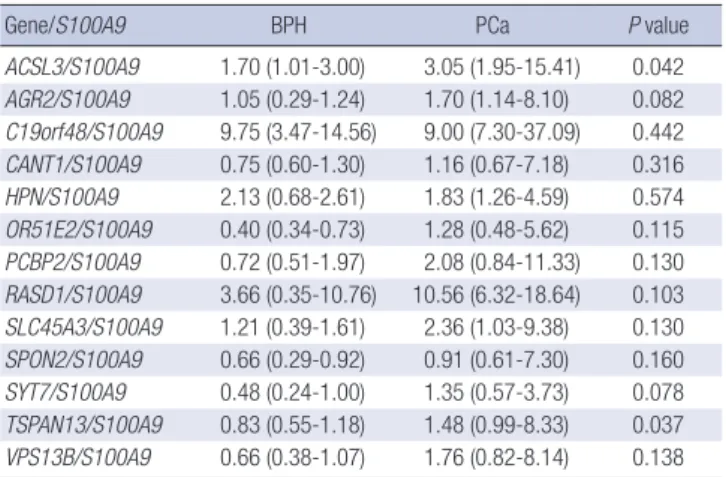
in PCa and BPH patients, respectively. The analysis showed that the TSPAN13-to-S100A9 urinary nucleic acid expression ratio was significantly different between PCa and BPH samples, with a P value of 0.037 (Table 3). Therefore, the TSPAN13-to-S100A9 ratio was selected for the validation study.
TSPAN13 and S100A9 tissue mRNA expression levels in PCa cases and BPH controls
Table 3 lists the baseline characteristics of the 129 PCa cases and 105 BPH controls. The mean age of PCa patients was 69.5 yr (range, 48-87 yr), and that of the BPH controls was 69.3 yr (range, 46-85 yr). The serum PSA level was higher in PCa pa- tients than in BPH controls (100.47 ± 239.65 ng/mL vs. 4.17 ± 7.79 ng/mL; P < 0.001). Of the 129 PCa cases, 66 (51.2%) under- went radical prostatectomy using open or laparoscopic proce- dures. The expression of TSPAN13 was significantly higher in
Table 2. Baseline characteristics of patients
Characteristics
Two-gene expression ratio
selection study P value
Tissue mRNA validation
cohort P value
Urinary nucleic acid
validation cohort P value
BPH PCa BPH PCa BPH PCa
No. 5 12 105 129 31 37
Age (yr; range) 69.60 (66-77) 70.83 (64-81) 0.791 69.30 (46-85) 69.50 (48-87) 0.838 69.26 (50-81) 67.30 (58-75) 0.189 PSA ± SD (ng/mL) 2.89 ± 1.81 47.52 ± 81.98 0.027 4.17 ± 7.79 100.47 ± 239.65 < 0.001 6.52 ± 13.87 20.64 ± 25.73 < 0.001 No. gleason score (%)
6 0 (0) 6 (4.7) 1 (2.7)
7 6 (50.0) 46 (35.7) 10 (27.0)
8 4 (33.3) 36 (27.9) 18 (48.6)
9 1 (8.3) 36 (27.9) 7 (18.9)
10 1 (8.3) 5 (3.9) 1 (2.7)
No. stage (%)
T1 0 (0) 8 (6.2) 1 (2.7)
T2 7 (58.3) 48 (37.2) 28 (75.7)
T3 4 (33.3) 34 (26.4) 8 (21.6)
T4 1 (8.3) 37 (28.7) 0 (0)
Unknown 0 (0) 2 (1.6) 0 (0)
P values were obtained from the Mann-Whitney U-test. BPH, benign prostatic hyperplasia; PCa, prostate cancer; PSA, prostate specific antigen.
Table 3. Comparison of urinary nucleic acid levels in the two-gene expression ratio selection study
Gene/S100A9 BPH PCa P value
ACSL3/S100A9 1.70 (1.01-3.00) 3.05 (1.95-15.41) 0.042 AGR2/S100A9 1.05 (0.29-1.24) 1.70 (1.14-8.10) 0.082 C19orf48/S100A9 9.75 (3.47-14.56) 9.00 (7.30-37.09) 0.442 CANT1/S100A9 0.75 (0.60-1.30) 1.16 (0.67-7.18) 0.316 HPN/S100A9 2.13 (0.68-2.61) 1.83 (1.26-4.59) 0.574 OR51E2/S100A9 0.40 (0.34-0.73) 1.28 (0.48-5.62) 0.115 PCBP2/S100A9 0.72 (0.51-1.97) 2.08 (0.84-11.33) 0.130 RASD1/S100A9 3.66 (0.35-10.76) 10.56 (6.32-18.64) 0.103 SLC45A3/S100A9 1.21 (0.39-1.61) 2.36 (1.03-9.38) 0.130 SPON2/S100A9 0.66 (0.29-0.92) 0.91 (0.61-7.30) 0.160 SYT7/S100A9 0.48 (0.24-1.00) 1.35 (0.57-3.73) 0.078 TSPAN13/S100A9 0.83 (0.55-1.18) 1.48 (0.99-8.33) 0.037 VPS13B/S100A9 0.66 (0.38-1.07) 1.76 (0.82-8.14) 0.138 P values were obtained from the Mann-Whitney U-test.
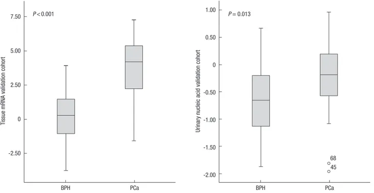
PCa than in BPH, whereas the expression of S100A9 was signifi- cantly lower in PCa than in BPH (each P < 0.001) (Table 4). The TSPAN13-to-S100A9 mRNA expression ratio, which was ana- lyzed with the same methods used in urine study, was signifi- cantly higher in PCa than in BPH (P < 0.001) (Table 4 and Fig.
2). Receiver operating characteristics (ROC) analysis was per- formed to assess the predictive value for PCa, which showed that the area under the curve (AUC) was 0.898 for the TSPAN13- to-S100A9 expression ratio (Fig. 3). No differences in theTSPAN13 and S100A9 mRNA expression levels and ratio were observed Fig. 2. Box plot of TSPAN13-to-S100A9 expression ratios in the tissue mRNA cohort and urinary nucleic acid cohort.
Tissue mRNA validation cohort
7.50
5.00
2.50
0
-2.50
BPH PCa
P < 0.001
Urinary nucleic acid validation cohort
1.00
0.50
0
-0.50
-1.00
-1.50
-2.00
BPH PCa
68 45 P = 0.013
Fig. 3. ROC curve analysis of TSPAN13-to-S100A9 ratios in the tissue mRNA cohort and urinary nucleic acid cohort.
Sensitivity
1.0
0.8
0.6
0.4
0.2
0
1-Specificity
0 0.2 0.4 0.6
Variable AUC P value
TSPAN13-to-S100A9 0.898 < 0.001
0.8 1.0
Tissue mRNA validation cohort
Sensitivity
1.0
0.8
0.6
0.4
0.2
0
1-Specificity
0 0.2 0.4 0.6
Variable AUC P value
TSPAN13-to-S100A9 0.676 0.013
0.8 1.0
Urinary nucleic acid validation cohort
according to clinicopathological variables such as age, PSA, grade, and stage (data not shown).
The ratio of urinary TSPAN13-to-S100A9 nucleic acid levels in the urinary nucleic acid validation cohort
Table 4 shows the clinicopathological characteristics of 37 PCa cases and 31 BPH controls included in the urinary nucleic acid validation cohort. The TSPAN13-to-S100A9 ratio was signifi- cantly higher in PCa cases than in BPH controls (P = 0.013) (Ta- ble 4 and Fig. 2). ROC analysis showed that the AUC value was 0.676 for the urinary nucleic acid TSPAN13-to-S100A9 ratio (Fig.
3). These results were consistent with those of the tissue mRNA expression study and demonstrated the diagnostic value of the urinary TSPAN13-to-S100A9 nucleic acid ratio for PCa. In the analysis of TSPAN13-to-S100A9 ratio according to clinical char- acteristics, there were no differences based on clinicopatholog- ical variables such as age, PSA, grade, and stage (data not shown).
DISCUSSION
The results of the present study indicated that the urinary TSPAN13- to-S100A9 nucleic acid ratio has diagnostic value as a biomarker for PCa. The TSPAN13-to-S100A9 ratio of urinary nucleic acids was significantly higher in PCa cases than in BPH controls (P = 0.013), and ROC curve analysis showed an AUC value of 0.676, which was consistent with the AUC value in the tissue mRNA expression study (AUC = 0.898, P < 0.001). These results dem- onstrate the possibility of finding diagnostic markers for PCa, since this outcome was obtained using urine supernatants and a new method, namely, the two-gene expression ratio.
Increasing research efforts have focused on identifying new non-invasive diagnostic markers for solid tumors. Significant advances have been made in certain malignancies, such as co- lon cancer (16). In urological cancer, numerous articles have reported results on genetic and epigenetic modifications in urine sediment as biomarkers for early diagnosis (17-19); how- ever, few studies have investigated markers in the urine super- natant (20,21). In the present study, we extracted CNAs from the urine supernatant rather than using the urine sediment as a source of material. The urine supernatant is more suitable for
identifying cancer biomarkers than the urine sediment because the sediment contains normal DNA derived from non-cancer- ous cells which might hamper the analysis. This was supported by Szarvas et al. (22), who suggested that the detection rate of genetic changes is higher in CNAs isolated from the urine su- pernatant than in those from the urine sediment. However, the origin of CNAs in the urine supernatant remains poorly under- stood. It is possible that circulating DNA or RNA is released from either necrotic or apoptotic cells (9). Zancan et al. (23) re- ported the usefulness of cell-free DNA measurements from urine supernatants to differentiate between patients with blad- der cancer and control cases. In addition, two studies showed reliable discrimination between cancer and non-cancer pa- tients using urine supernatant (21,24). These studies support the use of urine supernatants for the analysis of nucleic acids for PCa detection.
Although the CNAs extracted from the urine supernatant are valuable as cancer biomarker, the numeric values obtained by real-time PCR from urinary nucleic acids are difficult to nor- malize, whereas in tissue mRNA real-time PCR studies, house- keeping genes such as GAPDH can be used as reference mate- rials. Therefore, in the present study, we used a plasmid stan- dard and the two-gene expression ratio. Plasmids were used as reference materials in urinary nucleic acid real-time PCR to generate standard curves. The PCR fragments of target genes were cloned into suitable plasmid vectors and experiments were performed using the same dilutions of each plasmid stan- dard. The use of the same copy number of the plasmid standard allowed the adjustment of data obtained from real-time PCR.
Furthermore, plasmid standards have additional advantages.
One advantage of using plasmid standards is that once they are constructed by cloning the target PCR fragment into a suitable plasmid vector, they can be easily prepared in large amounts. In addition, plasmids can be stored for long periods and aliquoted at -20°C without significant degradation. Therefore, plasmid standards can be used as reference material to minimize inter- assay variation, and may offer a solution to the normalization problem associated with the use of urinary nucleic acids.
In the present study, the two-gene expression ratio was used.
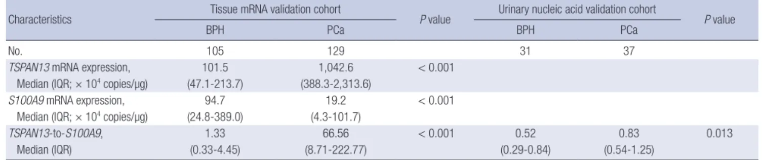
In a tissue mRNA study, Ma et al. (11) identified the two-gene Table 4. TSPAN13-to-S100A9 expression ratios in the tissue mRNA validation cohort and urinary nucleic acid validation cohort
Characteristics Tissue mRNA validation cohort
P value Urinary nucleic acid validation cohort
P value
BPH PCa BPH PCa
No. 105 129 31 37
TSPAN13 mRNA expression, 101.5 1,042.6 < 0.001
Median (IQR; × 104 copies/µg) (47.1-213.7) (388.3-2,313.6)
S100A9 mRNA expression, 94.7 19.2 < 0.001
Median (IQR; × 104 copies/µg) (24.8-389.0) (4.3-101.7)
TSPAN13-to-S100A9, 1.33 66.56 < 0.001 0.52 0.83 0.013
Median (IQR) (0.33-4.45) (8.71-222.77) (0.29-0.84) (0.54-1.25)
P values were obtained from the Mann-Whitney U-test. IQR, interquartile range.
expression ratio, HOXB13-to-IL17BR, as a predictor of breast cancer outcome during the early period. The HOXB13 gene was up-regulated and the IL17BR gene was down-regulated in breast cancer patients with disease recurrence. Reid et al. (25) failed to validate this two-gene ratio on frozen samples from 58 patients; however, a large scale (1,252 primary breast tumor specimens) study confirmed the reliability of the two-gene ratio as a predictor of breast cancer outcome (26). The two-gene ex- pression ratio in individual patients was also identified as a bio- marker signature for lung cancer diagnosis and prognosis (27,28). Although these studies were based on tissue mRNA ex- pression analyses, they highlight the value of the two-gene ex- pression ratio as a reliable biomarker, and suggest that it could be adapted to the analysis of CNAs. The objective of the present study was to identify a two-gene expression ratio in urinary nu- cleic acids consisting of one up-regulated and one down-regu- lated gene as a diagnostic marker for PCa. The TSPAN13-to- S100A9 ratio showed the same pattern in tissue mRNA and in urinary nucleic acids. These results demonstrate the reliability of urinary nucleic acid TSPAN13-to-S100A9 ratio as a biomarker.
S100A9, the down-regulated gene in the two-gene expression ratio in our study, has been investigated extensively. S100A9 is a member of the S100 family of proteins containing two EF hand calcium-binding motifs (29,30). S100A9 forms a complex with S100A8, another member of the S100 family, and the S100A9/
S100A8 heterodimer is released in virtually all inflammatory disorders (31). Tissue mRNA expression analyses showed ele- vated levels of S100A9 in many malignancies such as breast, lung, gastric, colorectal, and pancreatic cancer (32). However, in PCa, the expression of S100A9 was reported to be up-regulat- ed, whereas our previous study showed the opposite result (10,33). Our previous study analyzed a greater number of sam- ples than that of the study reporting the up-regulation of S100A9 in PCa, which increases the accuracy of our results. Moreover, our study demonstrated the down-regulation of S100A9 in PCa using both tissue mRNA expression and urinary nucleic acid analyses. Therefore, in the present study, we used S100A9 as the down-regulated gene in the two-gene expression ratio to exam- ine the reliability of urinary nucleic acids as biomarkers of PCa.
The present study had several limitations. First, the number of samples used in the validation study was relatively small; fur- ther investigation with a greater number of cases is needed to confirm the diagnostic value of the TSPAN13-to-S100A9 ratio.
In addition, the micro-array data used for the selection of can- didate genes in this study was derived from Western patients, and differences between Western and Korean patients were not taken into consideration. Future studies should include the se- lection of candidate genes from a Korean cohort to improve the accuracy of our findings. Finally, additional studies are neces- sary to confirm the relationship between S100A9 and TSPAN13.
Although S100A9 is a well studied gene, TSPAN13 as a member
of the transmembrane-4 superfamily has not been investigated extensively, although it is known to be involved in signal trans- duction events that play a role in the regulation of cell develop- ment, activation, growth and motility.
In patients with urothelial carcinoma, urine is a particularly desirable source of diagnostic marker, since its collection is more convenient and less invasive than that of blood. In the present study, analysis of urinary nucleic acids using the two- gene expression ratio yielded positive results regarding the identification of urinary diagnostic markers for PCa. In addi- tion, the procedure of detecting the two-gene expression ratio in nucleic acids isolated from the urine supernatant is simple and applicable to the clinical setting. However, the AUC value in the urinary nucleic acid cohort did not show sufficient detec- tion power, despite the fact that the TSPAN13-to-S100A9 ratio pattern was the same in urinary nucleic acids and in the tissue mRNA validation cohort. The identification of additional two- gene expression ratios in urinary nucleic acids is necessary to develop an accurate diagnostic marker for PCa using the meth- ods described in the present study.
In conclusion, the urinary nucleic acid TSPAN13-to-S100A9 ratio shows potential as a diagnostic marker for PCa. The results of the present study indicate that analysis of the urine superna- tant is a simple diagnostic method for PCa that could be adapt- ed to the clinical setting in the future.
ACKNOWLEDGMENTS
The biospecimens for this study were provided by the Chung- buk National University Hospital, a member of the National Biobank of Korea, which is supported by the Ministry of Health, Welfare, and Family Affairs. All samples derived from the Na- tional Biobank of Korea were obtained with informed consent under institutional review board-approved protocols. The au- thors wish to thank Ms. Eun-Ju Shim from the National Biobank of Korea at Chungbuk National University Hospital for the sam- ple preparations and her excellent technical assistance.
DISCLOSURE
The authors have no potential conflicts of interest to disclose.
AUTHOR CONTRIBUTION
Conception and design of the study: Yan C, Kim YH, Yun SJ. Ac- quisition of data: Kim YH, Seo SP, Jeong P, Lee IS. Statistical analysis: Seo SP, Kim D, Kim JM. First draft of manuscript: Yan C, Kim YH, Kang HW. Revision and critical review of the manu- script: Choi YH, Moon SK, Yun SJ, Kim WJ. Manuscript approv- al: all authors.
ORCID
Chunri Yan http://orcid.org/0000-0002-3043-6687 Ye-Hwan Kim http://orcid.org/0000-0002-8676-7119 Ho Won Kang http://orcid.org/0000-0002-8164-4427 Yung Hyun Choi http://orcid.org/0000-0002-1454-3124 Sung-Kwon Moon http://orcid.org/0000-0002-4514-3457 Seok-Joong Yun http://orcid.org/0000-0001-7737-4746 Wun-Jae Kim http://orcid.org/0000-0002-8060-8926
REFERENCES
1. Siegel R, Naishadham D, Jemal A. Cancer statistics, 2012. CA Cancer J Clin 2012; 62: 10-29.
2. Jung KW, Park S, Kong HJ, Won YJ, Boo YK, Shin HR, Park EC, Lee JS.
Cancer statistics in Korea: incidence, mortality and survival in 2006- 2007. J Korean Med Sci 2010; 25: 1113-21.
3. Jung KW, Yim SH, Kong HJ, Hwang SY, Won YJ, Lee JK, Shin HR. Cancer survival in Korea 1993-2002: a population-based study. J Korean Med Sci 2007; 22: S5-S10.
4. Roobol MJ, Zappa M, Määttänen L, Ciatto S. The value of different screening tests in predicting prostate biopsy outcome in screening for prostate cancer data from a multicenter study (ERSPC). Prostate 2007;
67: 439-46.
5. Ralla B, Stephan C, Meller S, Dietrich D, Kristiansen G, Jung K. Nucleic acid-based biomarkers in body fluids of patients with urologic malig- nancies. Crit Rev Clin Lab Sci 2014; 51: 200-31.
6. Gahan PB, Swaminathan R. Circulating nucleic acids in plasma and se- rum. Recent developments. Ann N Y Acad Sci 2008; 1137: 1-6.
7. Casadio V, Calistri D, Salvi S, Gunelli R, Carretta E, Amadori D, Silves- trini R, Zoli W. Urine cell-free DNA integrity as a marker for early pros- tate cancer diagnosis: a pilot study. Biomed Res Int 2013; 2013: 270457.
8. De Maio G, Rengucci C, Zoli W, Calistri D. Circulating and stool nucleic acid analysis for colorectal cancer diagnosis. World J Gastroenterol 2014;
20: 957-67.
9. Jahr S, Hentze H, Englisch S, Hardt D, Fackelmayer FO, Hesch RD, Knippers R. DNA fragments in the blood plasma of cancer patients:
quantitations and evidence for their origin from apoptotic and necrotic cells. Cancer Res 2001; 61: 1659-65.
10. Yun SJ, Yan C, Jeong P, Kang HW, Kim YH, Kim EA, Lee OJ, Kim WT, Moon SK, Kim IY, et al. Comparison of mRNA, protein, and urinary nu- cleic acid levels of S100A8 and S100A9 between prostate cancer and BPH. Ann Surg Oncol 2015; 22: 2439-45.
11. Ma XJ, Wang Z, Ryan PD, Isakoff SJ, Barmettler A, Fuller A, Muir B, Mo- hapatra G, Salunga R, Tuggle JT, et al. A two-gene expression ratio pre- dicts clinical outcome in breast cancer patients treated with tamoxifen.
Cancer Cell 2004; 5: 607-16.
12. Tomlins SA, Mehra R, Rhodes DR, Shah RB, Rubin MA, Bruening E, Makarov V, Chinnaiyan AM. Whole transcriptome amplification for gene expression profiling and development of molecular archives. Neo- plasia 2006; 8: 153-62.
13. Tomlins SA, Mehra R, Rhodes DR, Cao X, Wang L, Dhanasekaran SM, Kalyana-Sundaram S, Wei JT, Rubin MA, Pienta KJ, et al. Integrative molecular concept modeling of prostate cancer progression. Nat Genet
2007; 39: 41-51.
14. Chandran UR, Ma C, Dhir R, Bisceglia M, Lyons-Weiler M, Liang W, Michalopoulos G, Becich M, Monzon FA. Gene expression profiles of prostate cancer reveal involvement of multiple molecular pathways in the metastatic process. BMC Cancer 2007; 7: 64.
15. Kim YJ, Yoon HY, Kim SK, Kim YW, Kim EJ, Kim IY, Kim WJ. EFEMP1 as a novel DNA methylation marker for prostate cancer: array-based DNA methylation and expression profiling. Clin Cancer Res 2011; 17: 4523-30.
16. Mead R, Duku M, Bhandari P, Cree IA. Circulating tumour markers can define patients with normal colons, benign polyps, and cancers. Br J Cancer 2011; 105: 239-45.
17. Yu J, Zhu T, Wang Z, Zhang H, Qian Z, Xu H, Gao B, Wang W, Gu L, Meng J, et al. A novel set of DNA methylation markers in urine sediments for sensitive/specific detection of bladder cancer. Clin Cancer Res 2007;
13: 7296-304.
18. Miyake M, Sugano K, Sugino H, Imai K, Matsumoto E, Maeda K, Fuku- zono S, Ichikawa H, Kawashima K, Hirabayashi K, et al. Fibroblast growth factor receptor 3 mutation in voided urine is a useful diagnostic marker and significant indicator of tumor recurrence in non-muscle in- vasive bladder cancer. Cancer Sci 2010; 101: 250-8.
19. Vinci S, Giannarini G, Selli C, Kuncova J, Villari D, Valent F, Orlando C.
Quantitative methylation analysis of BCL2, hTERT, and DAPK promot- ers in urine sediment for the detection of non-muscle-invasive urothelial carcinoma of the bladder: a prospective, two-center validation study.
Urol Oncol 2011; 29: 150-6.
20. Zancan M, Galdi F, Di Tonno F, Mazzariol C, Orlando C, Malentacchi F, Agostini M, Maran M, Del Bianco P, Fabricio AS, et al. Evaluation of cell-free DNA in urine as a marker for bladder cancer diagnosis. Int J Biol Markers 2009; 24: 147-55.
21. Chang HW, Tsui KH, Shen LC, Huang HW, Wang SN, Chang PL. Uri- nary cell-free DNA as a potential tumor marker for bladder cancer. Int J Biol Markers 2007; 22: 287-94.
22. Szarvas T, Kovalszky I, Bedi K, Szendroi A, Majoros A, Riesz P, Füle T, László V, Kiss A, Romics I. Deletion analysis of tumor and urinary DNA to detect bladder cancer: urine supernatant versus urine sediment. On- col Rep 2007; 18: 405-9.
23. Zancan M, Franceschini R, Mimmo C, Vianello M, Di Tonno F, Maz- zariol C, Malossini G, Gion M. Free DNA in urine: a new marker for bladder cancer? Preliminary data. Int J Biol Markers 2005; 20: 134-6.
24. Casadio V, Calistri D, Tebaldi M, Bravaccini S, Gunelli R, Martorana G, Bertaccini A, Serra L, Scarpi E, Amadori D, et al. Urine cell-free DNA in- tegrity as a marker for early bladder cancer diagnosis: preliminary data.
Urol Oncol 2013; 31: 1744-50.
25. Reid JF, Lusa L, De Cecco L, Coradini D, Veneroni S, Daidone MG, Gariboldi M, Pierotti MA. Limits of predictive models using microarray data for breast cancer clinical treatment outcome. J Natl Cancer Inst 2005; 97: 927-30.
26. Jansen MP, Sieuwerts AM, Look MP, Ritstier K, Meijer-van Gelder ME, van Staveren IL, Klijn JG, Foekens JA, Berns EM. HOXB13-to-IL17BR expression ratio is related with tumor aggressiveness and response to tamoxifen of recurrent breast cancer: a retrospective study. J Clin Oncol 2007; 25: 662-8.
27. Gordon GJ, Jensen RV, Hsiao LL, Gullans SR, Blumenstock JE, Ramas- wamy S, Richards WG, Sugarbaker DJ, Bueno R. Translation of micro- array data into clinically relevant cancer diagnostic tests using gene ex-
pression ratios in lung cancer and mesothelioma. Cancer Res 2002; 62:
4963-7.
28. De Rienzo A, Dong L, Yeap BY, Jensen RV, Richards WG, Gordon GJ, Sugarbaker DJ, Bueno R. Fine-needle aspiration biopsies for gene expres- sion ratio-based diagnostic and prognostic tests in malignant pleural mesothelioma. Clin Cancer Res 2011; 17: 310-6.
29. Zimmer DB, Cornwall EH, Landar A, Song W. The S100 protein family:
history, function, and expression. Brain Res Bull 1995; 37: 417-29.
30. Donato R. S100: a multigenic family of calcium-modulated proteins of the EF-hand type with intracellular and extracellular functional roles.
Int J Biochem Cell Biol 2001; 33: 637-68.
31. Ehrchen JM, Sunderkötter C, Foell D, Vogl T, Roth J. The endogenous Toll-like receptor 4 agonist S100A8/S100A9 (calprotectin) as innate am- plifier of infection, autoimmunity, and cancer. J Leukoc Biol 2009; 86:
557-66.
32. Gebhardt C, Németh J, Angel P, Hess J. S100A8 and S100A9 in inflam- mation and cancer. Biochem Pharmacol 2006; 72: 1622-31.
33. Hermani A, Hess J, De Servi B, Medunjanin S, Grobholz R, Trojan L, Angel P, Mayer D. Calcium-binding proteins S100A8 and S100A9 as novel diagnostic markers in human prostate cancer. Clin Cancer Res 2005; 11: 5146-52.