Transplantation of Neural Differentiated Human Mesenchymal Stem Cells into the Cochlea of an Auditory-neuropathy Guinea Pig Model
The aim of this study was to determine the effects of transplanted neural differentiated human mesenchymal stem cells (hMSCs) in a guinea pig model of auditory neuropathy. In this study, hMSCs were pretreated with a neural-induction protocol and transplanted into the scala tympani of the guinea pig cochlea 7 days after ouabain injury. A control model was made by injection of Hanks balanced salt solution alone into the scala tympani of the guinea pig cochlea 7 days after ouabain injury. We established the auditory neuropathy guinea pig model using 1 mM ouabain application to the round window niche. After application of ouabain to the round window niche, degeneration of most spiral ganglion neurons (SGNs) without the loss of hair cells within the organ of Corti and increasing the auditory brain responses (ABR) threshold were found. After transplantation of neural differentiated hMSCs, the number of SGNs was increased, and some of the SGNs expressed immunoreactivity with human nuclear antibody under confocal laser scanning microscopy.
ABR results showed mild hearing recovery after transplantation. Based on an auditory neuropathy animal model, these findings suggest that it may be possible to replace degenerated SGNs by grafting stem cells into the scala tympani.
Key Words: Mesenchymal Stem Cells; Auditory neuropathy; Hearing Loss; Spiral ganglion neuron
Yong-Bum Cho1,4, Hyong-Ho Cho1,4, Sujeong Jang2,3,4, Han-Seong Jeong2,3,4, and Jong-Seong Park2,3,4
Departments of 1Otolaryngology and 2Physiology, Chonnam National University Medical School;
3Brain Korea 21 Project, Center for Biomedical Human Resources at Chonnam National University;
4Research Institute of Medical Sciences, Chonnam National University, Gwangju, Korea
Received: 23 November 2010 Accepted: 8 February 2011 Address for Correspondence:
Hyong-Ho Cho, MD
Department of Otolaryngology and Head and Neck Surgery, Chonnam National University Medical School, 671 Jebong-ro, Dong-gu, Gwangju 501-757, Korea
Tel: +82.62-220-6772, Fax: +82.62-228-7743 E-mail: victocho@hanmail.net
This study was supported by the Korea Research Foundation Grant funded by the Korean Government (Basic Promotion Fund: KRF-2008-E00148) and a grant (CRI09062-1) of Chonnam National University Hospital Research Institute of Clinical Medicine.
DOI: 10.3346/jkms.2011.26.4.492 • J Korean Med Sci 2011; 26: 492-498
INTRODUCTION
The loss of auditory afferent neurons, the spiral ganglion neu- rons (SGNs), can lead to sensorineural hearing loss (SNHL).
This can occur from primary neuronal loss involving degenera- tion of neurons in the absence of hair cell degeneration (1) or secondary to the loss of hair cells that normally supply trophic support to SGNs (2). Auditory neuropathy (AN) results when degeneration of SGNs occurs in the presence of functional hair cells (3). A recent study demonstrated that application of oua- bain, a well-known cardiac glycoside, to the round window re- sulted in a loss of SGNs, which serves as a model of AN (4, 5).
Currently, the use of a cochlear implant is the only method to treat the deaf resulting from the loss of hair cells or the loss of SGNs. However, survival of SGNs is a significant issue for ob- taining the hearing benefits provided by cochlear implants.
Many researchers seek methods for protection of SGNs (6-8) because SGNs do not regenerate to any clinically significant ex- tent after neural degeneration.
Stem cell replacement therapy raises the hope for hearing loss (9, 12). Among the various types of stem cells, bone mar- row-derived mesenchymal stem cells (MSCs) are one of the most promising stem cell sources for cell replacement therapy because they can differentiate into neurons (13) and can be used by the patient’s own bone marrow (14). Moreover, bone marrow transplantation has been shown to be a safe procedure for decades without any incidents of tumor formation in host tissues after transplantation. Primary MSCs have been proven to survive in the inner ear and to replace the lost SGNs (15, 16).
However, it remains unclear whether neural differentiated MSCs can serve as better sources for cell replacement therapy in in- jured cochleae. Previously, it has been reported that neuronal differentiation of MSCs could be induced by application of ba- sic fibroblast growth factor (bFGF), forskolin, and ciliary neuro- trophic factor (14, 17, 18). In this study we have shown that neu- ral differentiated human MSCs have the potential not only to replace the lost SGNs, but also to promote hearing recovery in the AN guinea pig model.
MATERIALS AND METHODS
Preparation of human MSCs and in vitro neural differentiation
Previously, we have reported neural differentiation of human MSCs (19, 20). Briefly, human bone marrow was obtained from the mastoid process during mastoidectomy for ear surgery. In- formed consent was obtained from all donors according to the guidelines of the Ethics Committee of the Chonnam National University Hospital. Human MSCs were isolated from the bone marrow and grown as adherent cultures in Dulbecco’s Modi- fied Eagle’s Medium (DMEM; Hyclone, Logan, UT, USA) sup- plemented with 10% fetal bovine serum (FBS; Hyclone) and 1%
penicillin-streptomycin (Hyclone). After 4 weeks, MSCs were grown to approximately 80% confluence in a 37°C humidified incubator with a 5% CO2 and 95% air environment. This stage was termed passage 0 (P0). The human MSCs used in the pres- ent study were obtained from passages 3-6.
To induce neural differentiation, MSCs were grown in DMEM containing 1% FBS and supplementary 100 ng/mL of bFGF (In- vitrogen Corporation, Carlsbad, CA, USA) for 7 days. The cells were then incubated in the presence of 10 μM forskolin (Sigma Chemical Co., St. Louis, MO, USA). Over the next 7 days, the cells were characterized and used for transplantation into the AN guinea pig model.
AN animal model and human MSC transplantation Twenty-four female guinea pigs, weighing 250-350 g, were used in the present study. Animal care complied with institutional guidelines. All surgical procedures were performed under asep- tic conditions. The guinea pigs were anesthetized with pentobar- bital sodium (40 mg/kg) by intraperitoneal injection. The mi- crosurgical techniques were performed under an operating mi- croscope (Zeiss OPMI® pico; Carl Zeiss, Goettingen, Germany).
To make an AN model, the right post-auricular region of the guinea pigs was shaved and sterilized with potadine and 70%
ethanol following anesthesia (see above). The animal was then placed on a heating pad (37°C), and the right bulla was opened
widely and the round window niche was exposed. A piece of gel- atin sponge soaked with 1 mM ouabain (Sigma Chemical Co.) was placed on the round window membrane. Then, the inci- sion was closed with sutures. The animals were randomly di- vided into 2 groups (n = 8 per group), as follows: control group, Hanks balanced salt solution (HBSS; Hyclone) without neural- induced human mesenchymal stem cell (nhMSC) transplanta- tion into the cochlea; and stem cell group, HBSS with nhMSC transplantation into the cochlea.
Neural-induced human MSC transplantation
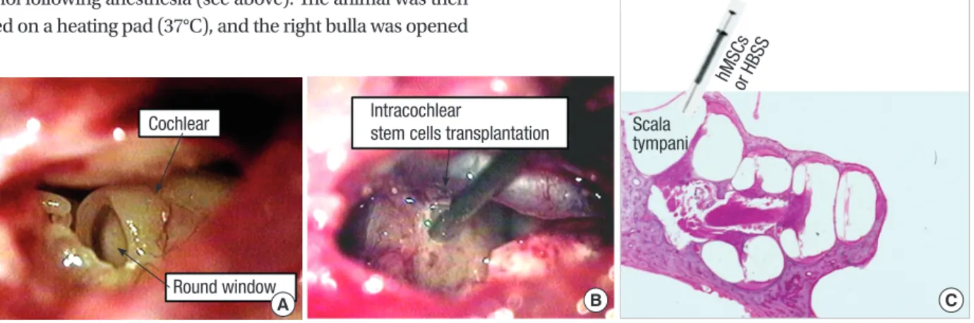
Transplantation of nhMSCs was performed 7 days after ouabain application. Procedures for animal preparation and anesthesia were similar to those described above. The right bulla was sur- gically exposed and opened to visualize the basal cochlea using a post-auricular incision. A small hole was made into the scala tympani at the basal cochlear turn using a 0.8-mm diamond drilling burr. HBSS with nhMSCs (1 × 10⁵ cells in 5 μL) or HBSS alone was then injected into the right scala tympani through the small hole using a Hamilton syringe and microinfusion pump at a speed of 2 μL/min (Fig. 1). After injection, the hole was plugged with small connective tissue and a fibrin-based adhe- sive agents (Greenplast®; Green-Cross, Seoul, Korea), and the incision was approximated with sutures.
Outcome measurement by histologic analysis
Histologic evaluation was performed 6 weeks after the transplan- tation. The animals were sacrificed by administration of a high dose of pentobarbital sodium. The cochleae were carefully re- moved, trimmed, fixed in 5% paraformaldehyde, then decalci- fied for 2 weeks in 10% EDTA. The fixed, decalcified cochleae were embedded in paraffin wax for paraffin section. The paraf- fin-embedded cochlea was sliced info 5-μm-thick serial paramo- diolar sections. The sections were mounted on glass slides and the morphology was checked via hematoxylin and eosin stain- ing. We obtained spiral ganglion images with a magnification of
Fig. 1. Photomicrographs of stem cell transplantation method. (A) Visualization of the round window after opening the bulla. (B, C) Note the tip of a needle of a Hamilton syringe is inserted into the most basal turn of the scala tympani, where neural-induced human mesenchymal stem cells (nhMSCs) or Hanks balanced salt solution (HBSS) were injected (C, H&E staining, × 40).
C Scala
tympani hMSCs
or HBSS
B Intracochlear
stem cells transplantation
A Cochlear
Round window
× 400 using a Nikon camera (Tokyo, Japan). These images were then printed on 10 × 7.5 cm photographic paper for neuron counting. Quantitative histology values were used to calculate the neuron density (neurons per field divided by the partial spi- ral ganglion cross-sectional area in each turn).
After blocking in 1% normal goat serum, the sections were incubated overnight at 4°C with a primary antibody diluted in phosphate-buffered saline (pH 7.4). The primary antibodies used in this study were anti-human nuclei (1:200), Math1 (1:200), and NF-H (1:300). Then, the sections were incubated with FITC- and rhodamine-conjugated secondary antibodies to visualize primary antibodies. 4’6-diamidino-2-phenylindole (DAPI) was used to label nuclei. All primary and secondary antibodies were purchased from Chemicon, except for Math1 (Ambion, Austin, TX, USA). Fluorescent images were obtained using a Zeiss LSM510 confocal microscope (Carl Zeiss Inc., Jena, Germany).
The captured images were processed using Zeiss LSM Image Browser version 4, 2, 0, 121 (Carl Zeiss Inc.) and Adobe photo- shop CS.
Auditory Brain Responses (ABR) measurement and analysis
ABR recordings were carried out using commercial auditory- evoked potential equipment (Navigator SE evoked potential system; Bio-logic Systems Corp., Mundelein, IL, USA) at vari- ous intervals (1, 3, and 5 weeks) after the transplantation. The animals were placed in a prone position after anesthesia (see above). ABRs were elicited using 11.1/sec alternating polarity clicks (150-3,000 Hz, stimulation rate 11/sec [reflecting lower frequency responses]) and 8 kHz (reflecting middle frequencies) alternating polarity tone bursts, a rise and fall time of 5 msec, and a plateau of 5 msec shaped with a Blackman window. The stimuli were delivered to the ear using an insert earphone. Re-
sponse signals were recorded by three silver needle electrodes.
The electrodes were placed in the vertex for an active electrode and in the bilateral retroauricular region for ground and refer- ence electrodes. The responses were digitally band-passed fil- tered (100-3,000 Hz), amplified, and time-averaged (n = 512 to 1,024) for a post-stimulus time window of 9.98 msec. Stimulus intensity from a 90-decibel (dB) sound pressure level (SPL) was used, down to threshold in 10 dB, then 5 dB steps around the threshold level. The thresholds of ABRs were measured by de- tecting the presence of wave V. At the threshold intensity, at least two sequences of recordings were made to confirm response reproducibility.
Statistical analysis
All results were reported as the mean ± standard error of the mean. Statistical analysis was performed with an independent sample t-test for comparison of two experimental groups using SPSS (version 17.0; SPSS, Inc., Chicago, IL, USA). A value of P <
0.05 was considered to be significant.
Ethics statement
This study protocol and procedures were reviewed and approved by the institutional review board (I-2009-03-016) and the insti- tutional animal care and use committee of the Chonnam Na- tional University Hospital.
RESULTS
Effect of ouabain on SGNs
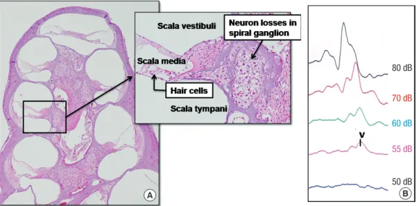
All experimental animals exhibited normal hearing with click- evoked ABR thresholds of -10 ± 10 dB prior to application of ouabain. Fig. 2 shows the effect of ouabain on SGNs. After 7 days of ouabain application, a severe decrease in SGNs was general-
Fig. 2. Ouabain-induced spiral ganglion neuropathy. (A) There is a severe loss of spiral ganglion neurons (SGNs) without a loss of hair cells after 7 days of ouabain application (H
&E staining, original magnification × 40 and × 200). (B) Also, an increase of the hearing threshold is noted (dB, decibel). V, wave v.
80 dB
70 dB 60 dB 55 dB 50 dB
A B
ly representative for all turns in the right ears. Despite the appar- ent loss of afferent innervation, hair cells were preserved in all turns. Also, the hearing threshold was increased in ABR mea- surements.
Transplanted neural differentiated hMSCs survive in the spiral ganglion
We explored the localization of nhMSCs after transplantation into the AN guinea pig cochlea 6 weeks after nhMSC transplan- tation. In the control group (Fig. 3A), severe loss of SGNs and sensory hair cells from the basal to the apical turns of the cochlea was noted. In contrast, in the stem cell group (Fig. 3B) the num- ber of SGNs and sensory hair cells was increased in all turns of the cochlea 6 weeks after stem cell grafting.
Even without the use of immunosuppressants, there was no evidence of acute rejection, which usually occurs within the first 6 months following transplantation. No significant inflamma- tion response was observed in the stem cell group.
We used human-specific anti-nuclei antibodies to specifical- ly identify human cells in the transplanted guinea pig inner ear cochlea. Transplanted human nuclei-positive cells located in
the spiral ganglion expressed NF-H, a neural cell marker, sug- gesting differentiation into neuronal cells (Fig. 4).
Based on quantitative histologic analysis, the average num- ber of neurons in each turn of the spiral ganglion was signifi- cantly greater in the stem cell group than in the control group (Fig. 5). In the control group, the average number of neurons in the basal, 2nd, 3rd, and 4th turns was 9.00 ± 1.27/1,000 μm2, 9.50 Fig. 3. Histologic findings of cochlea after stem cell tran- splantation. (A) Severe loss of SGNs and sensory hair cells from the basal to the apical turn of the cochlea is noted 6 weeks after HBSS injection (control group). (B) Numerous SGNs and sensory hair cells are observed in all turns of the cochlea 6 weeks after stem cell grafting (stem cell group). (H&E staining,
× 40 and × 200).
A B
Fig. 4. Confocal microscope image after immu- nostaining for anti-human nuclei and NF-H. (A) Numerous neurons are observed in the spiral ganglion (black arrow) after 6 weeks of stem cell transplantation (H&E staining, × 40). (B) the expression of anti-human nuclei (red) and NF-H (green) co-localizes with that of DAPI (blue)-positive cells in the spiral ganglion. Grafted stem cells (white arrow) are located in the spiral ganglion (× 200).
Average counts of neurons
Basal turn 2nd turn 3rd turn 4th turn 30
20
10
0
* * * *
Control Stem cell
Fig. 5. Average neuron counts in each turn of the spiral ganglion 6 weeks after trans- plantation. Average number of neurons in each turn of the spiral ganglion is significantly greater in the stem cell group than in the control group (*P < 0.05).
A B
ABR threshold (dB)ABR threshold (dB)
Click evoked ABR
ABRs for tone bursts of 8 kHz
Normal ABR
HBSS control
Ouabain apply
Stem cells transpl.
1 week 3 week 5 week
1 week 3 week 5 week 80
60 40 20
90 80 70 60 50 40
* *
*
50 dB
90 dB
80 dB
90 dB 30 dB
80 dB
70 dB
70 dB 10 dB
70 dB
60 dB
50 dB 0 dB
-10 dB
65 dB
55 dB
35 dB -20 dB
60 dB
50 dB
40 dB
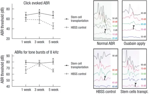
Fig. 6. Auditory brainstem response (ABR) results after nhMSC transplan tation. Click-evoked ABR waves are recorded up to -10 ± 10 dB in normal guinea pigs. Seven days after application of 1 mM ouabain to the round win- dow, an increase of ABR threshold is noted. After recei- ving a nhMSC transplantation, the auditory neuropathy guinea pigs show a significant improvement in hearing threshold compared to the control group (*P < 0.05).
Stem cell transplantation
Stem cell transplantation HBSS control
HBSS control
± 2.63/1,000 μm2, 7.63 ± 1.36/1,000 μm2, and 11.88 ± 3.10/1,000 μm2, respectively. In the stem cell group, the average number of neurons in the basal, 2nd, 3rd, and 4th turns was 15.30 ± 1.51/
1,000 μm2, 26.64 ± 1.46/1,000 μm2, 22.00 ± 1.99/1,000 μm2, and 21.44 ± 0.77/1,000 μm2, respectively.
Hearing recovery results after nhMSC transplantation Although evidence exists that transplanted nhMSCs differenti- ate to NF-H-positive neuronal cells in the spiral ganglion to re- place the lost cells in the AN guinea pig model, it is not clear if transplanted stem cells can enhance the hearing threshold. To evaluate the effect of nhMSC transplantation on hearing recov- ery in an AN guinea pig, we measured the ABR thresholds be- fore, and 1, 3, and 5 weeks after the transplantation. The ABR was evoked by two types of stimuli (alternative click sound and an 8 kHz pure tone burst sound). All experimental animals ex- hibited normal hearing with click-evoked ABR thresholds of 0 ± 10 dB prior to AN modeling. Seven days after the application of 1 mM ouabain on the round window, an increase of hearing threshold (63.00 ± 4.90 dB at click-evoked stimuli and 70.00 ± 5.70 dB at 8 kHz tone burst stimuli) was noted. After receiving a transplantation of nhMSCs, the guinea pigs showed a signifi- cant improvement in hearing threshold compared to the con- trol group. Five weeks after the transplantation, average ABR thresholds for the click and 8 kHz tone burst stimuli were de- creased to 46.00 ± 6.00 dB and 66.00 ± 2.45 dB, respectively (Fig.
6). The considerable improvement in hearing function observed in the ABR test is consistent with the restoration of SGNs in the stem cell group.
DISCUSSION
Recent advance in studies on stem cells have suggested the pos- sibility of clinical use of stem cell transplantation for ischemic
brain disease and neurodegenerative disorders (21, 22). Also, restoration of SGNs is one of the targets for stem cell therapy in the treatment of hearing loss (23). The present study provides the scientific basis for a requirement of neural differentiation of MSCs for better treatment and presents the novel demonstra- tion of a therapeutic approach leading to substantial recovery of hearing in AN patients’ ears. Some researchers have reported that transplantation of undifferentiated MSCs into the inner ear repairs injured cochlear fibrocytes in an animal SNHL model and increases survival in the spiral ganglion in an AN model (15, 16).
Various ototoxic treatments (5, 24) and the aging process lead to loss of SGNs (25). The application of ouabain to the round window membrane is a well-known method which induces the death of most SGNs, and thus provides an excellent model for AN (4, 5, 16). In this study, we placed a piece of gelatin sponge soaked with 1 mM ouabain on the round window membrane.
One week after ouabain application, there was a selective and permanent destruction of SGNs without loss of hair cells and an increase of the ABR threshold.
In the present study, the number of SGNs was increased at all turns of the cochlea after nhMSC transplantation, and the trans- planted nhMSCs were positively stained for NF-H, a mature neu- ronal marker, at the damaged spiral ganglion. This result sug- gests that the nhMSCs differentiate into neurons and replace the lost or damaged neural cells in the spiral ganglion. This find- ing suggests that the migration and differentiation of grafted stem cells are influenced by the cues or signals in the microen- vironment of the damaged tissue. It is known that damaged cells and tissues of the host brain are capable of releasing molecules that stimulate the production of neurotrophic factors in trans- planted MSCs (26).
The ultimate goal of stem cell therapy in AN patients is not only restoration of missing SGNs, but also to increase hearing
function. In this study, the decreased average ABR thresholds for click and 8 kHz tone burst stimuli indicated that nhMSC transplantation is an effective treatment method for AN. Be- cause the presence of some SGNs is known to enhance the func- tion of cochlear implants, the regeneration or protection of SGNs via nhMSC transplantation would also improve the therapeutic effect of cochlear implantation (27, 28). Thus, our results sug- gest that the transplantation of nhMSCs at the time of cochlear implant surgery could improve hearing recovery.
In this study, it is possible that immunologic rejection of trans- planted cells occurred in this experiment because we used the xenotransplantation procedure. Xenotransplantation was per- formed in this study because we sought the effectiveness of hu- man MSCs on cochlear repair. Although immunologic rejection was expected in this study, there was no evidence of acute rejec- tion in any of the animals used in our study. Escape from im- mune surveillance is partly due to the fact that MSCs lack MHC class II or inhibit T-cell proliferation (29, 30). Escape from im- mune surveillance was also caused by some degree of immu- noprotection of the cochlea provided by the blood-labyrinth barrier (16). However, further studies will be required to eluci- date the long-term immunologic reactions.
In conclusion, neural differentiated MSCs are able to restore damaged SGNs and decrease hearing thresholds in an AN mod- el. Furthermore, transplantation into the scala tympani is a suit- able method because numerous grafted nhMSCs were found in the spiral ganglion. These findings suggest that it may be possi- ble to replace degenerated SGNs by grafting stem cells into the scala tympani.
REFERENCES
1. Suzuka Y, Schuknecht HF. Retrograde cochlear neuronal degeneration in human subjects. Acta Otolaryngol Suppl 1988; 450: 1-20.
2. Keithley EM, Croskrey KL. Spiral ganglion cell endings in the cochlear nucleus of young and old rats. Hear Res 1990; 49: 169-77.
3. Starr A, Sininger YS, Pratt H. The varieties of auditory neuropathy. J Basic Clin Physiol Pharmacol 2000; 11: 215-30.
4. Lang H, Schulte BA, Goddard JC, Hedrick M, Schulte JB, Wei L, Schmiedt RA. Transplantation of mouse embryonic stem cells into the cochlea of an auditory-neuropathy animal model: effects of timing after injury. J Assoc Res Otolaryngol 2008; 9: 225-40.
5. Schmiedt RA, Okamura HO, Lang H, Schulte BA. Ouabain application to the round window of the gerbil cochlea: a model of auditory neuropa- thy and apoptosis. J Assoc Res Otolaryngol 2002; 3: 223-33.
6. Iguchi F, Nakagawa T, Tateya I, Kim TS, Endo T, Taniguchi Z, Naito Y, Ito J. Trophic support of mouse inner ear by neural stem cell transplantation.
Neuroreport 2003; 14: 77-80.
7. Shinohara T, Bredberg G, Ulfendahl M, Pyykkö I, Olivius NP, Kaksonen R, Lindström B, Altschuler R, Miller JM. Neurotrophic factor interven- tion restores auditory function in deafened animals. Proc Natl Acad Sci U S A 2002; 99: 1657-60.
8. Yagi M, Kanzaki S, Kawamoto K, Shin B, Shah PP, Magal E, Sheng J, Ra- phael Y. Spiral ganglion neurons are protected from degeneration by GDNF gene therapy. J Assoc Res Otolaryngol 2000; 1: 315-25.
9. Li H, Corrales CE, Edge A, Heller S. Stem cells as therapy for hearing loss.
Trends Mol Med 2004; 10: 309-15.
10. Li H, Roblin G, Liu H, Heller S. Generation of hair cells by stepwise dif- ferentiation of embryonic stem cells. Proc Natl Acad Sci U S A 2003; 100:
13495-500.
11. Oshima K, Grimm CM, Corrales CE, Senn P, Martinez Monedero R, Géléoc GS, Edge A, Holt JR, Heller S. Differential distribution of stem cells in the auditory and vestibular organs of the inner ear. J Assoc Res Otolaryngol 2007; 8: 18-31.
12. Parker MA, Cotanche DA. The potential use of stem cells for cochlear re- pair. Audiol Neurootol 2004; 9: 72-80.
13. Deng W, Obrocka M, Fischer I, Prockop DJ. In vitro differentiation of human marrow stromal cells into early progenitors of neural cells by con- ditions that increase intracellular cyclic AMP. Biochem Biophys Res Com- mun 2001; 282: 148-52.
14. Dezawa M, Kanno H, Hoshino M, Cho H, Matsumoto N, Itokazu Y, Ta- jima N, Yamada H, Sawada H, Ishikawa H, Mimura T, Kitada M, Suzuki Y, Ide C. Specific induction of neuronal cells from bone marrow stromal cells and application for autologous transplantation. J Clin Invest 2004;
113: 1701-10.
15. Kamiya K, Fujinami Y, Hoya N, Okamoto Y, Kouike H, Komatsuzaki R, Kusano R, Nakagawa S, Satoh H, Fujii M, Matsunaga T. Mesenchymal stem cell transplantation accelerates hearing recovery through the repair of injured cochlear fibrocytes. Am J Pathol 2007; 171: 214-26.
16. Matsuoka AJ, Kondo T, Miyamoto RT, Hashino E. Enhanced survival of bone-marrow-derived pluripotent stem cells in an animal model of au- ditory neuropathy. Laryngoscope 2007; 117: 1629-35.
17. Kim SS, Yoo SW, Park TS, Ahn SC, Jeong HS, Kim JW, Chang DY, Cho KG, Kim SU, Huh Y, Lee JE, Lee SY, Lee YD, Suh-Kim H. Neural induc- tion with neurogenin 1 increases the therapeutic effects of mesenchymal stem cells in the ischemic brain. Stem Cells 2008; 26: 2217-28.
18. Nagai A, Kim WK, Lee HJ, Jeong HS, Kim KS, Hong SH, Park IH, Kim SU. Multilineage potential of stable human mesenchymal stem cell line derived from fetal marrow. PLoS One 2007; 2: e1272.
19. Cho HH, Jeong HS, Jang SJ, Park JS, Cho HC, Jang CH, Cho YB. Neural differentiation of mesenchymal stem cells from bone marrow of human mastoid process. Korean J Otorhinolaryngol-Head Neck Surg 2008; 51:
422-8.
20. Cho HH, Jang S, Lee SC, Jeong HS, Park JS, Han JY, Lee KH, Cho YB. Ef- fect of neural-induced mesenchymal stem cells and platelet-rich plasma on facial nerve regeneration in an acute nerve injury model. Laryngo- scope 2010; 120: 907-13.
21. Lindvall O, Kokaia Z. Stem cells in human neurodegenerative disorders- -time for clinical translation? J Clin Invest 2010; 120: 29-40.
22. Nakayama D, Matsuyama T, Ishibashi-Ueda H, Nakagomi T, Kasahara Y, Hirose H, Kikuchi-Taura A, Stern DM, Mori H, Taguchi A. Injury-induced neural stem/progenitor cells in post-stroke human cerebral cortex. Eur J Neurosci 2010; 31: 90-8.
23. Tamura T, Nakagawa T, Iguchi F, Tateya I, Endo T, Kim TS, Dong Y, Kita T, Kojima K, Naito Y, Omori K, Ito J. Transplantation of neural stem cells into the modiolus of mouse cochleae injured by cisplatin. Acta Otolaryn- gol Suppl 2004; 551: 65-8.
24. Lee JE, Nakagawa T, Kim TS, Iguchi F, Endo T, Dong Y, Yuki K, Naito Y, Lee SH, Ito J. A novel model for rapid induction of apoptosis in spiral ganglions of mice. Laryngoscope 2003; 113: 994-9.
25. Shiga A, Nakagawa T, Nakayama M, Endo T, Iguchi F, Kim TS, Naito Y, Ito J. Aging effects on vestibulo-ocular responses in C57BL/6 mice: com- parison with alteration in auditory function. Audiol Neurootol 2005; 10:
97-104.
26. Chen X, Li Y, Wang L, Katakowski M, Zhang L, Chen J, Xu Y, Gautam SC, Chopp M. Ischemic rat brain extracts induce human marrow stromal cell growth factor production. Neuropathology 2002; 22: 275-9.
27. Coleman B, de Silva MG, Shepherd RK. Concise review: the potential of stem cells for auditory neuron generation and replacement. Stem Cells
2007; 25: 2685-94.
28. Nadol JB Jr, Shiao JY, Burgess BJ, Ketten DR, Eddington DK, Gantz BJ, Kos I, Montandon P, Coker NJ, Roland JT Jr, Shallop JK. Histopathology of cochlear implants in humans. Ann Otol Rhinol Laryngol 2001; 110:
883-91.
29. Krampera M, Glennie S, Dyson J, Scott D, Laylor R, Simpson E, Dazzi F.
Bone marrow mesenchymal stem cells inhibit the response of naive and memory antigen-specific T cells to their cognate peptide. Blood 2003; 101:
3722-9.
30. Tse WT, Pendleton JD, Beyer WM, Egalka MC, Guinan EC. Suppression of allogeneic T-cell proliferation by human marrow stromal cells: impli- cations in transplantation. Transplantation 2003; 75: 389-97.
AUTHOR SUMMARY
Transplantation of Neural Differentiated Human Mesenchymal Stem Cells into the Cochlea of an Auditory-neuropathy Guinea Pig Model
Yong-Bum Cho, Hyong-Ho Cho, Sujeong Jang, Han-Seong Jeong, and Jong-Seong Park
We investigated the effects of human mesenchymal stem cells (hMSCs) differentiated into neural cells on auditory neuropathy. The auditory neuropathy was induced in guinea pigs using 1mM ouabain applied to the round window niche. The differentiated hMSCs were transplanted into the scala tympani of the guinea pig cochlea 7 days after the ouabain-induced injury. The application of ouabain induced attenuated auditory brainstem response (ABR) and degeneration of spiral ganglion neurons (SGNs) without loss of hair cells in the organ of Corti. After transplantation of neural differentiated hMSCs, the number of SGNs was increased and the ABR results showed mild hearing recovery. It might be possible to replace degenerated SGNs by grafting stem cells into the scala tympani.