• 검색 결과가 없습니다.

Poly(Dimethylaminoethyl Methacrylate)-Based pH-Responsive Hydrogels Regulate Doxorubicin Release at Acidic Condition

N/A
N/A
Protected

Academic year: 2021

Share "Poly(Dimethylaminoethyl Methacrylate)-Based pH-Responsive Hydrogels Regulate Doxorubicin Release at Acidic Condition"

Copied!
13
0
0

로드 중.... (전체 텍스트 보기)

전체 글

(1)

http://dx.doi.org/10.12925/jkocs.2015.32.2.202

Poly(Dimethylaminoethyl Methacrylate)-Based pH-Responsive Hydrogels Regulate Doxorubicin Release at Acidic Condition

Seung-Hun LeeㆍJin-Oh You

Department of Engineering Chemistry, Chungbuk National University, Cheongju, 362-763, Republic of Korea

(Received May 2, 2015; Revised May 20, 2015; Accepted May 20, 2015)

Abstract : Stimuli-responsive biomaterials that alter their function through sensing local molecular cues may enable technological advances in the fields of drug delivery, gene delivery, actuators, biosensors, and tissue engineering. In this research, pH-responsive hydrogel which is comprised of dimethylaminoethyl methacylate (DMAEMA) and 2-hydroxyethyl methacrylate (HEMA) was synthesized for the effective delivery of doxorubicin (Dox) to breast cancer cells.

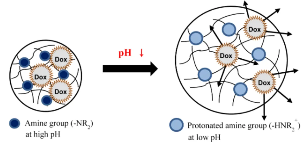
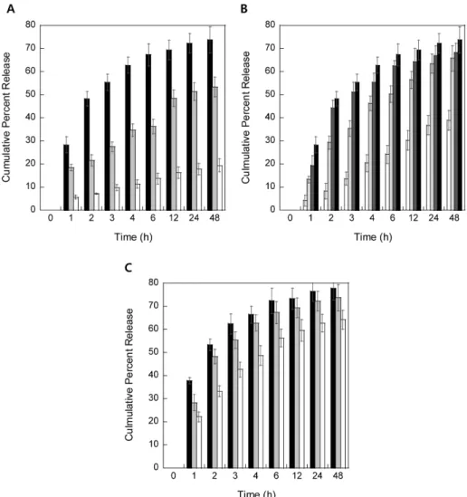
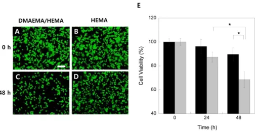
Cancer and tumor tissues show a lower extracellular pH than normal tissues. DMAEMA/HEMA hydrogels showed significant sensitivity by small pH changes and each formulation of hydrogels was examined by scanning electron microscopy, mechanical test, equilibrium mass swelling, controlled Dox release, and cytotoxicity. High swelling ratios and Dox release were obtained at low pH buffer condition, low cross-linker concentration, and high content of DMAEMA. Dox release was accelerated to 67.3% at pH 5.5 for 6-h incubation at 37

o

C, while it was limited to 13.8% at pH7.4 at the same time and temperature. Cell toxicity results to breast cancer cells indicate that pH-responsive DMAEMA/HEMA hydrogels may be used as an efficient matrix for anti-cancer drug delivery with various transporting manners. Also, pH-responsive DMAEMA/

HEMA hydrogels may be useful in therapeutic treatment which is required a triggered release at low pH range such as gene delivery, ischemia, and diabetic ketoacidosis.

Keywords : pH-responsive hydrogel, DMAEMA, drug delivery, doxorubicin, cancer therapy

1. Introduction

Polymeric materials that respond to a stimulus such as temperature,[1, 2] pH,[3, 4]

light,[5] electric field,[6] and ionic strength[7]

have been used to fabricate various types of hydrogels for biomedical applications. Among



Corresponding author (E-mail: [email protected])

the stimuli-sensitive polymers, pH-responsive polymers have garnered much attention in the fields of oral drug delivery,[8] gene delivery,[9]

insulin delivery,[10] actuators and sensors[11]

in the form of hydrogels due to their feasible swelling or deswelling properties.

Although natural and synthetic pH-sensitive polymers such as alginic acid,[12] chitosan,[13]

poly(acrylic acid),[14] and poly(methacrylic

acid)[15] have been used in systemic drug

delivery systems so far, their applications have

(2)

been limited that they have swelling behavior at high pH[16] due to ionizable functional groups on the polymer backbone or side chain.[17] Therefore, those polymers are not effective for drug delivery to the acidic environments like tumor[18] as well as intracellular delivery.[19] Recently, new strategies which have a reversed swelling behavior from the common pH-sensitive polymers have introduced to overcome the bottlenecks from conventional delivery manners.[20] In our previous reports, pH- sensitive hydrogels using dimethylaminoethyl methacrylate (DMAEMA) were prepared by photopolymerization using UV light source for drug and gene deliveries.[21] However, there was a severe problem that cells or biomolecules encapsulated in polymeric materials were directly exposed under UV light during the synthesis and there was highly possible to damage by long UV exposure time.[22] In this research, a series of pH- responsive hydrogels with DMAEMA and HEMA is synthesized by safe and simple polymerization technique and characterized for effective anti-cancer drug delivery to breast cancer cells, SK-BR-3.

DMAEMA is a pH-responsive monomer that has tertiary amine functional groups on its polymer backbone with a p K a of 7.5.[23]

DMAEMA has been used for drug delivery applications with other monomers because its excellent pH sensitivity with small pH changes.[24] Also, HEMA is a good biocompatible monomer and it uses for various biomedical applications such as drug delivery,[25] gene delivery,[26] biosensors,[27]

and scaffolds for tissue engineering.[28]

Doxorubicin (Dox) as a model drug was loaded in pH-responsive hydrogel. Dox is an effective chemotherapeutic agent and widely used for breast cancer therapy due to its high toxicity.[29]

In the present study, DMAEMA-based pH-responsive hydrogels were simply and safely synthesized compared to the hydrogels

by photopolymerization technique which is previously reported

21

and evaluated for effective Dox release in acidic condition, as shown in the schematic illustration in Figure1.

We demonstrated that pH sensitivity of DMAEMA/HEMA hydrogels was dominated by the content of pH-responsive monomer DMAEMA and the concentration of cross-linker TEGDMA. And, Dox-loaded DMAEMA/HEMA hydrogels with high Dox loading efficiency were induced to facilitate the cytotoxicity to breast cancer cells, SK-BR-3, compared to non-pH-responsive HEMA hydrogels. Our results show that pH- responsive DMAEMA/HEMA hydrogels were synthesized by simple polymerization and have excellent swelling property in acidic buffered medium even with a small pH drop. We also demonstrated the cytotoxicity of SK-BR-3 cells using Dox-loaded DMAEMA/HEMA hydrogels placed in the low pH medium which is a similar condition to the vicinity of a tumor.[30] These pH-responsive DMAEMA/

HEMA hydrogels are potentially useful for various biomedical applications such as drug delivery, gene delivery, diabetic ketoacidosis, biosensor, actuator, and tissue engineering.

2. Experimental

2.1. Materials

DMAEMA, a pH-responsive monomer, and HEMA, a non-pH-responsive co-monomer, were purchased from Acros (Morris Plains, NJ, USA) and used without further purification.

Tetraethylene glycol dimethacrylate (TEGDMA) as a cross-linking agent, ammonium persulfate, sodium metabisulfite, ethylene glycol were purchased from Sigma (St. Louis, MO, USA).

To prepare pH buffered media, sodium

phosphate monobasic and dibasic were

obtained from Sigma. Doxorubicin

hydrochloride as a model drug was purchased

from Sigma. All cell culture media and

reagents, unless otherwise mentioned, were

(3)

provided from Corning Cellgro (Manassas, VA, USA). Deionized water (18.2 MΩ) used in all experiments was from Milli-Q purification system (Millipore Corp., Billerica, MA, USA).

2.2. Fabrication of pH-responsive hydrogels

Two monomers, DMAEMA and HEMA, were mixed with predetermined ratios (10/90, 20/80, or 30/70, mol/mol%) to synthesize pH-responsive hydrogels. Then, TEGDMA as a cross-linker was added in mixed monomer solution with different concentrations (0.5, 1.0, or 1.5 wt%). Ammonium persulfate and sodium metabisulfite were added to initiate polymerization and finally equimolar ratio of distilled water and ethylene glycol was poured in mixed monomer solution. The molar ratio between the monomers and the solvent was 1.

The solution was gently swirled for 20 seconds to avoid air bubbles and poured into glass petri dish. Hydrogels synthesized from the different molar ratios of two monomers were investigated for pH responsibility at different pH conditions. Also, the different concentrations of cross-linker, TEGDMA, were also tested to obtain different elasticity of hydrogels which is directly relevant to swelling properties. The polymerization took about 15 minutes at room temperature. After polymerization, hydrogel in a glass petri dish was cut into 22.5 mm disk in diameter using a cork borer (U.5322, Usbeck, Germany) and washed with deionized water for 1 hour in order to remove unreacted monomers. To synthesize Dox-loaded hydrogels, 20 mg of Dox was added in monomer solution before hydrogel synthesis which showed a good cytotoxicity in another literature.[31] Also, non-pH-responsive HEMA hydrogels were synthesized by the same procedure for the control experiments.

2.3. Preparation of pH buffer solution Sodium phosphate monobasic, NaH

2

PO

4,

and dibasic, Na

2

HPO

4,

solutions (each 0.2 M

concentration) was used to make pH buffered media. Two solutions of NaH

2

PO

4

and Na

2

HPO

4

were mixed with predetermined ratios and put into glass jar placed on the shaker. The pH of the buffered medium was adjusted using 0.1 N of HCl and NaOH to obtain pH 5.5, 6.5, and 7.4 solutions and the final pHs were measured by pH meter (PHI 255 pH meter, Beckman Coulter, Brea, CA, USA).

2.4. Equilibrium swelling studies

Equilibrium mass swelling tests of hydrogels were performed in phosphate buffered media of known pH and composition at 37

o

C.

Hydrogel disks with 22.5 mm in diameter were placed into a glass jar containing 30 mL of buffered medium on the shaker (100 rpm) setup in incubator maintained at 37

o

C. The mass swelling ratio of the hydrogels as a function of pH was measured the mass of the hydrogels every hour until 4 hours and then every two hours until 12 hours and calculated as follows:

Mass swelling ratio = M

s

/ M

i

where M

s

is the mass of the swollen hydrogel in phosphate buffered medium and M

i

is the mass of the initial hydrogel before swelling.

4

2.5. Mechanical test

The tensile test of each hydrogel (13 × 35

× 20 mm, 10/90, 20/80, and 30/70 DMAEMA/HEMA, mol/mol%) cross-linked with 0.5, 1.0, and 1.5 wt% TEGDMA were measured by Instron Bioplus 5543 (Instron, Norwood, MA, USA) using a 500 N loading cells. Hydrogels were strained to failure at a rate of 2 mm/min. The Young’s modulus was calculated from the initial 40% strain. Seven hydrogel samples of each formulation were tested.

2.6. Scanning electron microscopy

Hydrogel with 30/70 DMAEMA/HEMA

(mol/mol%) was synthesized and dried for 48

hours at room temperature in a fume hood.

(4)

Then, hydrogel was coated with Pt/Pd for 90 sec with 40 mA with a sputter coater (208HR, Cressington Scientific Instruments, England) and imaged with SEM (FESEM Ultra55, Zeiss, Thornwood, NY, USA) at a beam voltage of 5 kV. HEMA hydrogel (100%) under the equal condition was also synthesized and visualized by SEM for comparison.

2.7. Drug Release

Controlled Dox release from Dox-loaded DMAEMA/HEMA hydrogels was performed in a phosphate buffered medium of known pH, composition, and temperature. Each hydrogel disk was put into the glass jar with 20 mL of different pH buffered medium and placed on a shaker with a shaking rate of 100 ± 1 rpm in an incubator maintained at 37°C. Samples were collected once every hour for 6 hours and then every 6 hours until 48 hours (Data were only shown at 12, 24, and 48 hours after 6 hour incubation). The sample volume was 500 µL and new fresh buffered medium was added after sampling to maintain a uniform concentration of Dox. The concentration of released Dox was determined by a Gemini XPS microplate spectrophotometer (Molecular Devices Corp, Sunnyvale, CA, USA) at an excitation wavelength 485 nm and emission wavelength 590 nm based on a standard fluorescence concentration calibration curve. To determine the loading efficiency of Dox, hydrogel was put into the pH 2 phosphate buffered medium and vigorously stirred for 24 h and then the final concentration of Dox was calculated from the standard curve.

2.8. pH change measurements

To demonstrate the pH-responsive swelling of hydrogels under cell culture system, pH-responsive DMAEMA/HEMA (30/70, mol/mol%) and non-pH-responsive HEMA hydrogels were placed in Transwell

®

plate (12-well plate, Costar, Corning Inc., Corning,

NY, USA) with SK-BR-3 cells at a seeding density of 1.0 × 10

6

cells/well, as shown in Figure 5A-B. The pH change of growth medium in Transwell

®

plate was measured at different times (0, 24, and 48 h) with cell density using a pH electrode (PHI 255 pH meter).

2.9. Cytotoxicity test

The cellular cytotoxicity from Dox-loaded pH-responsive DMAEMA/HEMA hydrogels and Dox-loaded non-pH-responsive HEMA hydrogels with SK-BR-3 breast cancer cells was evaluated by calcein AM staining and resazurin-based cell toxicology assay kit (Sigma). Cells were seeded in 12-well glass-bottom cell culture plates (MatTek Corp., Ashland, MA, USA) at a seeding density of 1.0 × 10

6

cells/well and grown for 24 hours. Dox-loaded DMAEMA/HEMA and HEMA hydrogels were put into each well in culture dishes and incubated at 37

o

C. After 24- and 48-h incubations, cell viability was examined by using calcein AM (1 µM). Cells showing green-colored fluorescence which are considering live cells were taken images using an inverted fluorescent microscope (IX53, Olympus, Japan). To perform the resazurin- based cell toxicology assay, cells were seeded at a seeding density of 1.0 × 10

6

cells/well and grown for 24 h in the 12-well Transwell

®

plate with 3 µm pore size filter membrane. Dox-loaded hydrogels were transferred to the top of Transwell

®

insert as shown in the schematic diagram in Figure 5A-B and incubated at 37

o

C for 24 and 48 hours. To quantify the cell damage by Dox, resazurin dye solution (100 µL) was added to each well (1000 µL of growth medium) in Transwell

®

plate and the plate was incubated in a humidified chamber at 37

o

C with 5%

CO

2

for 4 hours. The sample (100 µL) was

taken from each well and transferred to

96-well UV plate (Corning Inc., Acton, MA,

USA). The number of viable cells in each

well was determined using a Gemini XPS

(5)

Fig. 1. Schematic diagram of pH-triggered Dox release from DMAEMA/HEMA hydrogel in acidic condition.

microplate spectrophotometer at an excitation of 560 nm and an emission of 590 nm by comparison to a standard curve. To investigate the cytotoxicity of Dox-free DMAEMA/HEMA hydrogels for comparison, resazurin-based cell toxicology assay was also performed with SK-BR-3 cells for 24- and 48-h incubation (data not shown). All cell viability assays were performed three times.

2.10. Statistical analysis

Unless otherwise mentioned, triplicate data were obtained and presented as mean ± standard deviation. Statistical difference was analyzed using analysis of variance with Student’s t -test on the significance level of p <

0.05.

3. Results and Discussion

3.1. Synthesis of pH-responsive DMAEMA/HEMA hydrogels

DMAEMA/HEMA hydrogels which have pH responsibility were synthesized by a simple polymerization technique using TEGDMA as a cross-linker and ammonium persulfate/sodium metabisulfite as initiators. The three different molar ratios of DMAEMA and HEMA (10/90,

20/80, and 30/70, mol/mol%) were used to investigate pH sensitivity at different pH conditions. Also, the three different concentrations of TEGDMA (0.5, 1.0, and 1.5 wt%) were examined to obtain different hydrogel elasticity. Figure 2A shows the SEM images of pH-responsive DMAEMA/HEMA (30/70, mol/mol%) and non-pH-responsive HEMA (100%) hydrogels cross-linked with 1.0 wt% TEGDMA. It was visually confirmed by SEM images that both hydrogels had the smooth surface and didn’t show cracks or pores during the synthesis which are directly affected to hydrogel swelling and Young’s modulus. It was examined at least in five electron microscopic fields. Although the different molar contents of DMAEMA from 10 to 30 mol% and TEGDMA concentration as a cross-linker from 0.5 to 1.5 wt% were used for synthesis, hydrogels didn’t show any significant difference regarding their morphology or color.

Also, as shown in Figure 2B,

DMAEMA/HEMA (10/90 and 30/70,

mol/mol%) and HEMA hydrogels were

synthesized and put into different pH buffer

solutions (pH 7.4 or 5.5) to observe

morphology change such as crack or damage

for 6 hours before the mass swelling

(6)

Fig. 2. SEM images of (A) pH-responsive DMAEMA/HEMA (30/70, mol/mol%) and non- pH-responsive HEMA hydrogels cross-linked with 1.0 wt% TEGDMA. Scale bar = 100 µm. (B) is the photos of HEMA (100%) and DMAEMA/HEMA (10/90 and 30/70, mol/mol%) hydrogels taken by a digital camera in pH 7.5 and 5.5 buffered media for 6-h swelling. Scale bar =10 mm. (C) Mass swelling ratios of 30/70 DMAEMA/HEMA (mol/mol%) hydrogels cross-linked with 1.0 wt% TEGDMA at pH 5.5 (●), 6.5 (■), and 7.4 (▲), respectively. (D) Mass swelling ratios of 30/70 (●), 20/80 (■), and 10/90 (▲) DMAEMA/HEMA (mol/mol%) hydrogels cross-linked with 1.0 wt% TEGDMA in pH 5.5 buffered medium. (E) Mass swelling ratios of 30/70 DMAEMA/HEMA (mol/mol%) hydrogels cross-linked with 0.5 (●), 1.0 (■), and 1.5 (▲) wt% TEGDMA in pH 5.5 buffered medium. The error is the standard deviation of the mean, where n = 5.

experiments. It didn’t show any hydrogel damage visually during the swelling process, although there was significant size change with different ratios of DMAEMA/HEMA and pH values. While the size of HEMA hydrogel was not changed for 6-h swelling,

DMAEMA/HEMA hydrogels showed

significant size change especially at low pH condition (pH 5.5) and size change was also

accelerated when DMAEMA content was

increased from 10 to 30% in hydrogel. These

trends were confirmed by mass swelling studies

with different molar ratios of DMAEMA and

HEMA, cross-linker TEGDMA concentrations,

and different swelling buffered medium

conditions.

(7)

3.2. Mass swelling ratio

DMAEMA/HEMA hydrogels were successfully synthesized and equilibrium swelling studies from different formulations were performed in phosphate buffered media which are adjusted to predetermined pH. We measured the mass swelling ratio of the hydrogels with different molar ratio of

DMAEMA and HEMA, TEGDMA

concentration, and pH of the swelling medium, as shown in Figure 2C-E. First, the mass swelling ratio of hydrogels was examined at the different pH buffered medium conditions in Figure 2C. To observe the extent of hydrogel swelling, 30/70 (mol/mol%) DMAEMA/HEMA hydrogels cross-linked with 1.0 wt% TEGDMA were examined in pH 5.5, 6.5, and 7.4 phosphate buffered media for 24 hours. Hydrogel at pH 7.4 phosphate buffer showed a 1.14 ± 0.08% increased mass after 2 hours, whereas hydrogel at pH 5.5 was increased up to 1.74 ± 0.09%. At 12-h incubation, the mass swelling of hydrogels at pH 5.5, 6.5, and 7.4 were 3.03 ± 0.14, 2.36

± 0.12 and 1.16 ± 0.10, respectively.

Equilibrium swelling was obtained after 12-h swelling time. Swelling is directly related to the function of water sorption and electrostatic interactions due to the protonated DMAEMA groups and the ions in solution. Although we also measured the swelling ratio of 30/70 (mol/mol%) DMAEMA/HEMA hydrogels cross-linked with 1.0 wt% TEGDMA at pH 8.5 phosphate buffered medium for 24 h, we didn’t find any significant swelling behavior (data not shown). In Figure 2D, the swelling ratios of 10/90, 20/80, and 30/70 (mol/mol%) DMAEMA/HEMA hydrogels cross-linked with 1.0 wt% TEGDMA were 1.12 ± 0.07, 1.38

± 0.08, and 1.45 ± 0.09 after 1 h swelling time at pH 5.5, respectively. Then, the swelling ratio of 30/70 (mol/mol) DMAEMA/HEMA hydrogels (2.91 ± 0.13) was significantly increased for 6 hours at pH 5.5 compared to 10/90 (mol/mol%) DMAEMA/HEMA hydrogels (1.39 ± 0.13).

Due to the increase of protonated amine groups in DMAEMA polymer backbone, higher DMAEMA content hydrogels had significantly higher mass swelling ratio.

The 30/70 (mol/mol%) DMAEMA/HEMA hydrogels were also synthesized with 0.5, 1.0, and 1.5 wt% TEGDMA to determine the mass swelling ratio as a function of cross-linker concentration. Swelling tests were performed in a pH 5.5 phosphate buffered medium to obtain maximal swelling effect. As shown in Figure 2E, the hydrogels cross-linked with 1.5 wt% TEGDMA had lower mass swelling ratio relative to 0.5 wt% TEGDMA hydrogels. For example, the swelling ratio of 30/70 (mol/mol%) DMAEMA/HEMA hydrogels cross-linked with 1.5 wt% TEGDMA was 2.16 ± 0.10 after 4 h at pH 5.5, while the swelling ratio of hydrogels cross-linked with 0.5 wt% TEGDMA was 2.57 ± 0.13 under the same swelling conditions. Increasing the TEGDMA concentration raised the cross-linking density of the hydrogel network, which the rigidity of matrix was also increased and thus swelling property was decreased.

3.3. Mechanical properties

To determine the elasticity of pH-responsive

DMAEMA/HEMA hydrogels, the Young’s

modulus was examined by the Instron BioPuls

5543 using a tensile mode. Three molar ratios

of DMAEMA/HEMA (10/90, 20/80, and

30/70, mol/mol%) and three concentrations of

TEGDMA (0.5, 1.0, and 1.5 wt%) were

tested. As shown in Figure 3A, the Young’s

moduli of pH-responsive DMAEMA/HEMA

hydrogels were increased with decreasing

DMAEMA content. For example, the Young’s

moduli of 10/90, 20/80, and 30/70

(mol/mol%) DMAEMA/HEMA hydrogels

cross-linked with 1.0 wt% TEGDMA were

0.69 ± 0.06, 0.64 ± 0.06, and 0.54 ± 0.07,

respectively. The Young’s modulus was also

increased with the increasing of cross-linking

agent concentration. The Young’s modulus of

hydrogel cross-linked with 0.5 wt% TEGDMA

(8)

Fig. 3. Young’s moduli of (A) 30/70, 20/80, and 10/90 DMAEMA/HEMA (mol/mol%) hydrogels cross-linked with 1.0 wt% TEGDMA were measured. Also, Young’s moduli of (B) 30/70 DMAEMA/HEMA (mol/mol%) hydrogels cross-linked with 0.5, 1.0, and 1.5 wt% TEGDMA were measured. The error is the standard deviation from the mean ( n = 5, * is p < 0.05 and ** is p < 0.01).

was 0.32 ± 0.01, while the Young’s modulus of hydrogel cross-linked with 1.5 wt%

TEGDMA was 0.73 ± 0.09. Thus, the mechanical properties of DMAEMA/HEMA hydrogels can be tuned in the range of 0.32 to 0.73 MPa by the molar ratio of DMAEMA to HEMA and cross-linking density.

3.4. Controlled Dox release

The controlled release of Dox from pH-responsive DMAEMA/HEMA hydrogels showed a dependence on the content of

pH-sensitive monomer DMAEMA,

concentration of cross-linker TEGDMA, and pH buffer condition, as shown in Figure

4A-C. The 30/70 DMAEMA/HEMA

hydrogels cross-linked with 1.0 wt%

TEGDMA were selected to investigate the controlled release of Dox at different pH conditions. Controlled release of Dox was examined by the released Dox amount from hydrogels in different pH buffered media. To determine the loading efficiency of Dox in

hydrogels, Dox-loaded DMAEMA/HEMA hydrogel was put into pH 2 phosphate buffered medium for 24 hours. The Dox encapsulation efficiency was 72 ± 5% and drug loss was occurred during the washing process of hydrogel in distilled water twice.

As shown in Figure 4, the trends of Dox release were apparently observed by different initial slopes at different DMAEMA content, TEGDMA concentration, and pH buffered medium until 12 hours. This is exactly coincident with the slope of mass swelling ratio in Figure 2C-E. It means that hydrogel swelling is directly related to the Dox release until 12 h. For examples, the swelling ratios of 30/70 DMAEMA/HEMA (mol/mol%) hydrogels were obtained 3.04 ± 0.14, 2.36 ± 0.12, and 1.16 ± 0.10 at pH 5.5, 6.5, and 7.5 and accumulated Dox release was observed 69.3 ± 4.26, 48.5 ± 3.52 and 16.3 ± 2.46%

at the same pH conditions, respectively.

Figure 4B shows that Dox release is also

dependent on the DMAEMA content. When

(9)

the DMAEMA content was increased from 10 to 30%, Dox release was also raised up to 12.9% (from 56.4 to 69.3%) due to the increase of pH-sensitive swelling. It revealed that cross-linker concentration was also one of main factors to control the Dox release, as shown in Figure 4C. When the cross-linker concentration was 0.5 wt%, Dox release was released quickly compared to 1.0 and 1.5 wt%

cross-linker concentrations. For example, Dox release was 73.3 ± 4.62% at 0.5 wt%

TEGDMA concentrations for 12-h release, whereas it was 59.6 ± 4.63% at 1.5 wt%

TEGDMA concentration at the same time.

Increasing of cross-linker concentration brings to the higher crosslinking density which makes hydrogel matrix network dense.

3.5. Cytotoxicity

Before the investigation of cytotoxicity to breast cancer cells with pH-responsive hydrogels, we measured the pH change of growth media under cell culture system using the Transwell

®

plate to make the similar acidic environmental condition like tumor or cancer tissues, as shown in schematic diagrams in Figure 5A-B. According to previous report regarding cancer and tumor tissues, those tissues had the acidic pH values (approximately pH 6.8 to 7.2) compared to the normal tissues. pH-responsive DMAEMA/

HEMA (30/70, mol/mol%) hydrogel cross- linked with 1 wt% TEGDMA and non-pH- responsive HEMA hydrogels cross-linked with the same concentration of TEGDMA were placed in Transwell

®

plate with culturing of SK-BR-3 cells at the seeding density 10

6

cells/well. And, the pH changes of growth media were measured at 0, 24, and 48 hours.

In Figure 5C, the pH of growth media with cells was significantly dropped with the increasing of cell density and pH-responsive hydrogel size was apparently larger due to increased cellular metabolic waste and environmental hypoxic condition in growth media. For example, the pH drop was

observed 7.41 ± 0.03, 7.08 ± 0.09, and 6.64

± 0.13 at 0, 24, and 48 hours and the cell densities were increased to 1.00 × 10

6

, 1.56

× 10

6

, and 2.78 × 10

6

cells/well at the same time, respectively. The increase of cell density led to decrease of pH as a function of time.

It means that pH-responsive hydrogel is able to swell drastically in acidic growth media and Dox can be released out quickly compared to non-pH-sensitive hydrogel.

To investigate the therapeutic efficacy of

pH-responsive DMAEMA/HEMA (30/70,

mol/mol%) hydrogels cross-linked with 1.0

wt% TEGDMA through pH-triggered Dox

release, we treated SK-BR-3 cells with

hydrogels for 0, 24, 48 hours observing with

pH changes in growth media using the

Transwell

®

plate, as shownin Figure 6. As a

control, non-pH-responsive HEMA hydrogel

cross-linked 1.0 wt% TEGDMA was also

tested as described above. Cell viability was

measured quantitatively by a fluorescent

microplate reader and qualitatively by a

fluorescent microscopy. The cell viability of

SK-BR-3 cells treated with non-pH-

responsive HEMA hydrogel exhibited 100.0 ±

3.3, 96.3 ± 5.7, and 89.6 ± 5.5 at 0, 24,

and 48 hours, respectively. However,

SK-BR-3 cell viability treated with

pH-responsive DMAEMA/HEMA hydrogel

was significantly decreased to 100.0 ± 2.9,

87.3 ± 4.4 and 68.5 ± 6.6 at 0, 24, and 48

hours, respectively. The cell viability treated

with pH-responsive hydrogels (Figure 6A and

C) showed an enhanced tumoricidal effect

compared to non-pH-responsive hydrogels

(Figure 6B and D) by calcein AM staining

method. At 48-hour treatment, tumoricidal

effect was significantly accelerated in

pH-responsive hydrogels. Recently, there are

many reports with PLGA (poly lactic-co-g

lycolic acid) as drug delivery matrixes for

enhanced anti-cancer drug delivery. However,

PLGA encapsulated with anti-cancer drugs

showed slow release due to slow PLGA

degradation.[32] Dox release with pH-

(10)

Fig. 4. Controlled Dox release from pH-responsive DMAEMA/HEMA hydrogels. (A) Dox- loaded 30/70 DMAEMA/HEMA (mol/mol%) hydrogels cross-linked with 1.0 wt%

TEGDMA were monitored for pH-triggered Dox release in phosphate buffered media

at pH 7.4 (white), 6.5 (gray), and 5.5 (black), respectively. (B) Controlled Dox release

was measured from 10/90 (light gray), 20/80 (dark gray), and 30/70 (black)

DMAEMA/HEMA (mol/mol%) hydrogels cross-linked with 1 wt% TEGDMA in pH

5.5 phosphate buffered medium. As a control, non-pH-responsive HEMA hydrogel

loaded with Dox was also tested with the same method in pH 5.5 (white) buffered

medium. (C) Controlled Dox release was also measured from 30/70

DMAEMA/HEMA (mol/mol%) hydrogels cross-linked with 0.5 (black), 1.0 (gray), and

1.5 (white) wt% TEGDMA at pH 5.5 buffered medium. The error is the standard

deviation of the mean, where n = 5.

(11)

Fig. 5. Schematic illustration of Transwell

®

system which was used for efficient Dox release to SK-BR-3 cells and swelling effects of (A) non-pH-responsive HEMA (100%) and (B) pH-responsive DMAEMA/HEMA (30/70, mol/mol%) hydrogels in cell growth media.

(C) pH measurement of the growth medium for 30/70 DMAEMA/HEMA (mol/mol%) hydrogel seeded with SK-BR-3 cells at an initial density of 10

6

cells/well. The error is the standard deviation of the mean (where n = 5, * is p < 0.05 and ** is p < 0.01).

responsive DMAEMA/HEMA hydrogel would be quite fast and can be easily tuned with other factors such as DMAEMA content, cross-linker concentration, and pH values.

Therefore, pH-triggered Dox delivery with DMAEMA/HEMA hydrogel could be beneficial for chemotherapy. The polymers for conventional pH-triggered release (alginate, chitosan, polymethacrylate, etc.) showed high swelling property at high pH area and they are not useful for the treatment of cancer.

However, DMAEMA/HEMA hydrogel which is proposed in this research shows an excellent swelling effect at low pH even in the small pH change and can control the release of anti-cancer drugs to avoid side effects.

Additionally, our hydrogels can be synthesized by simple polymerization technique and easily scale-up for various biomedical applications such as ischemia, diabetic ketoacidosis,

morphine overdoses, and tumorigenic cancers.

4. Conclusions

pH-responsive DMAEMA/HEMA hydrogels were synthesized and characterized for the controlled Dox release and enhanced cytotoxicity to breast cancer cells, SK-BR-3.

The hydrogels showed a high mass swelling

ratio at low pH, low cross-linking density,

and high DMAEMA content. Controlled Dox

release was directly related to the pH

condition of buffered medium, cross-linker

concentration, and ratio of DMAEMA in

hydrogel. From the cell toxicity test with

SK-BR-3 cells, it was found that

pH-responsive hydrogels showed rapid swelling

in acidic culture medium and Dox release was

accelerated. Thus, enhanced cytotoxicity with

(12)

Fig. 6. Fluorescent microscopy images of SK-BR-3 cells at (A-B) 0 and (C-D) 48 hours treated with (A-C) pH-responsive Dox-loaded 30/70 DMAEMA/HEMA (mol/mol%) hydrogel cross-linked with 1.0 wt% TEGDMA and (B-D) non-pH-responsive Dox-loaded HEMA hydrogel cross-linked with 1.0 wt% TEGDMA in Transwell

®

at0and48hours. Scale bar = 100 µm. (E) Cell viability of SK-BR-3 cells were determined by a resazurin-based toxicology assay. Cells were treated with pH-responsive Dox-loaded 30/70 DMAEMA/HEMA (mol/mol%) hydrogel cross-linked with 1.0 wt% TEGDMA and non-pH-responsive Dox-loaded HEMA hydrogel cross-linked with 1.0 wt% TEGDMA in Transwell

®

for 0, 24, and 48 hours, respectively. The error is the standard deviation from the mean (where n = 3, * is p <

0.05).

pH-responsive DMAEMA/HEMA hydrogels was observed compared to non-pH-responsive HEMA hydrogels. These hydrogels are relatively simple to fabricate and may be useful to deliver the anti-cancer drugs to the acidic environments with high drug loading efficiency as well as tissue engineering application.

Acknowledgements

This work was supported by the research grant of the Chungbuk National University in 2013.

References

1. S. Cui, Y. Hu, Z. Huang, C. Ma, L. Yu, X. Hu, Int.J. Therm. Sci. 79 (2014) 276-282.

2. D.P. Huynh, G.J. Im, S.Y. Chae, K.C. Lee, D.S. Lee, J. Control. Release 137 (2009) 20-24.

3. N. Zalachas, S. Cai, Z. Suo, Y. Lapusta, Int. J. Solids. Struct. 50 (2013) 920-927.

4. Y. Hu, J.O. You, D.T. Auguste, Z. Suo, J.J. Vlassak, J. Mater. Res. 27 (2012) 152-160.

5. J. Liu, J. Nie, Y. Zhao, Y. He, J. Photoch.

Photobio. A 211 (2010) 20-25.

6. H. Li, Z. Yuan, K.Y. Lam, H.P. Lee, J.

Chen, J. Hanes, J. Fu, Biosens. Bioelectron.

19 (2004) 1097-1107.

(13)

7. X.-W. Liu, S. Zhu, S.-R. Wu, P. Wang, G.-Z. Han, Colloid Surface A 417 (2013) 140-145.

8. A.M. Lowman, M. Morishita, M. Kajita, T. Nagai, N.A. Peppas, J. Pharm. Sci. 88 (1999) 933-937.

9. J.O. You, D.T. Auguste, Nano Lett. 9 (2009) 4467-4473.

10. X. Gao, Y. Cao, X. Song, Z. Zhang, X.

Zhuang, C. He, X. Chen, Macromol.

Biosci. 14 (2014) 565-575.

11. N.I. Georgiev, A.M. Asiri, A.H. Qusti, K.A. Alamry, V.B. Bojinov, Sensor. Actuat.

B-Chem. 190 (2014) 185-198.

12. S.W. Jang, J.W. Lee, D.S. Ryu, M. Son, M.J. Kang, Int. J. Biol. Macromol. 70C (2014) 174-178.

13. N. Solomko, O. Budishevska, S. Voronov, K. Landfester, A. Musyanovych, Macromol.

Biosci. 14 (2014) 1076-1083.

14. S.S. Halacheva, D.J. Adlam, E.K. Hendow, T.J. Freemont, J. Hoyland, B.R. Saunders, Biomacromolecules 15 (2014) 1814-1827.

15. P. Yang, D. Li, S. Jin, J. Ding, J. Guo, W. Shi, C. Wang, Biomaterials 35 (2014) 2079-2088.

16. J.O. You, D.T. Auguste, Biomaterials 31 (2010) 6859-6866.

17. Y. Tian, Chinese Chem. Lett. 19 (2008) 1111-1114.

18. S. Peppicelli, F. Bianchini, L. Calorini, Cancer metastasis Rev. 33 (2014) 823-832.

19. S. Quader, H. Cabral, Y. Mochida, T.

Ishii, X. Liu, K. Toh, H. Kinoh, Y. Miura, N. Nishiyama, K. Kataoka, J. Control.

Release 188 (2014) 67-77.

20. A.L. Daniel-da-Silva, L. Ferreira, A.M.

Gil, T. Trindade, J. Colloid Interface Sci.

355 (2011) 512-517.

21. J.O. You, D.T. Auguste, Biomaterials 29 (2008) 1950-1957.

22. F.R. Gruijil, H.J. Kranen, L.H.F.

Mullenders, J. Photoch. Photobio. B 63 (2001) 19-27.

23 X. Zhang, K. Matyjaszewski, Macromolecules 32 (1999) 1763-1766.

24. S. Brahim, D. Narinesingh, A. Guiseppi- Elie, Biomacromolecules 4 (2003) 1224- 1231.

25. Y. Li, K. Xiao, W. Zhu, W. Deng, K.S.

Lam, Adv. Drug Deliv. Rev. 66 (2014) 58-73.

26. R.P. Johnson, S. Uthaman, J.V. John, M.S.

Heo, I.K. Park, H. Suh, I. Kim, Macromol.

Bio. 14 (2014) 1239-1248.

27. G. Slaughter, J. Sunday, IEEE Sens. J. 14 (2014) 1573-1576.

28. N. Ramalingam, T.S. Natarajan, S. Rajiv, J. Biomed. Mater. Res. A 103 (2014) 16-24.

29. A. Gabizon, H. Shmeeda, T. Grenader, Eur. J. Pharm. Sci. 45 (2012) 388-398.

30. J.O. You, P. Guo, D.T. Auguste, Angew.

Chem. Int. Edit. 52 (2013) 4141-4146.

31. G.D. Kang, S.H. Cheon, S.C. Song, Int. J.

Pharm. 319 (2006) 29-36.

32. L. Mu, S.S. Feng, J. Contorl. Release 86

(2003) 33-48.

수치

Fig.  1.    Schematic  diagram  of  pH-triggered  Dox  release  from  DMAEMA/HEMA  hydrogel  in  acidic  condition.
Fig.  2.  SEM  images  of  (A)  pH-responsive  DMAEMA/HEMA  (30/70,  mol/mol%)  and  non-  pH-responsive  HEMA  hydrogels  cross-linked  with  1.0  wt%  TEGDMA
Fig.  3.  Young’s  moduli  of  (A)  30/70,  20/80,  and  10/90  DMAEMA/HEMA  (mol/mol%)  hydrogels  cross-linked  with  1.0  wt%  TEGDMA  were  measured
Fig.  4.  Controlled  Dox  release  from  pH-responsive  DMAEMA/HEMA  hydrogels.    (A)  Dox-  loaded  30/70  DMAEMA/HEMA  (mol/mol%)  hydrogels  cross-linked  with  1.0  wt%
+3

참조

관련 문서