Antioxidant Activities and Hepato-protective Effects of Stauntonia hexaphylla Fruit Extract Against H
2O
2-induced Oxidative Stress and Acetaminophen- induced Toxicity
Gyuok Lee, Jaeyong Kim, Huwan Kang, Donghyuck Bae and Chul-yung Choi*
Jeonnam Institute of Natural Resources Research, Jangheung-gun, Jeollanamdo 59338, Korea Received April 27, 2018 /Revised May 22, 2018 /Accepted May 24, 2018
The antioxidant activity and protective effects of a hot water extract from the Stauntonia hexaphylla fruit (WESHF) were investigated in vitro and in vivo. The total polyphenol and flavonoid contents of WESHF were 16.13±0.27 mg gallic acid equivalent/g and 4.7±0.80 mg catechin equivalent/g, re- spectively. In addition, the DPPH radical-scavenging activity (SC
50) and the Oxygen Radical Absorb- ance capacity of WESHF were 63.62±4.10 μg/ml and 90.63±5.29 μM trolox equivalent/g, respectively.
The hepatoprotective effect of WESHF against hydrogen peroxide-induced oxidative damage was investigated. H
2O
2-induced liver damage on HepG2 cells was prevented by 200 μg/ml of WESHF.
Furthermore, to investigate the protection mechanism of WESHF on hydrogen peroxide-induced cyto- toxicity in HepG2 cells, pre-treatment with 200 μg/ml of WESHF significantly attenuated a decrease in the activities of CAT, SOD, GR, and GPx. The hepatoprotective activity of WESHF was evaluated in an experimental model of hepatic damage induced by acetaminophen (APAP). The levels of alanine aminotransferase (ALT) and aspartate aminotransferase (AST) were significantly decreased in the liv- ers of mice treated with 200 mg/kg of WESHF compared to the APAP-treated group. The lipid perox- idation level, which increased after APAP administration, was significantly reduced in the WESHF group. In addition, histological examinations of the liver showed the same protective effect of WESHF treatment. Based on these findings, it is suggested that WESHF has potent hepatoprotective effects, and the mechanism that causes this type of protection could be related to antioxidant pathways.
Key words : Acetaminophen, antioxidant, hepatoprotective effect, oxidative stress, Stauntonia hexaphylla
*Corresponding author
*Tel : +82-61-860-2620, Fax : +82-61-864-7105
*E-mail : [email protected]
This is an Open-Access article distributed under the terms of the Creative Commons Attribution Non-Commercial License (http://creativecommons.org/licenses/by-nc/3.0) which permits unrestricted non-commercial use, distribution, and reproduction in any medium, provided the original work is properly cited.
Journal of Life Science 2018 Vol. 28. No. 6. 708~717 DOI : https://doi.org/10.5352/JLS.2018.28.6.708
서 론
최근 생활수준의 향상과 건강에 대한 관심이 증가하면서 노화와 질병을 일으키는 원인물질에 대한 연구가 활발하게 진행되고 있다. 생체 내 대사산물인 과산화수소(H
2O
2), 과산화 물 음이온(O
2-), 하이드록실 라디칼(OH)과 같은 활성산소종 (reactive oxygen species, ROS)은 흡연, 음주, 자외선, 그리고 과도한 스트레스와 같은 외부 요인에 의해서도 생성될 수 있으 며[20], 생성된 과량의 활성산소는 심혈관계질환, 당뇨병, 암, 파킨슨과 같은 각종 질병의 원인으로 알려져 있다[44]. 과량 생산된 ROS로 인한 산화적 손상을 막기 위해 다양한 항산화 물질 및 항산화효소가 작용하여 체내 항상성을 유지한다. 항 산화 효소에는 superoxide dismutase (SOD), catalase (CAT), glutathione peroxidase (GPx), 및 glutathione생성 효소 등이
있으며[31, 36] 이와 같은 항산화 효소는 세포막의 지질 과산화 손상, sulfhydryl-함유 효소의 불활성화, 및 구성 단백질의 교 차결합 등을 일으키는 ROS를 불활성화 시키거나 제거함으로 써 항산화 작용을 한다[3, 23].
식물계에 널리 분포하는 폴리페놀 화합물 및 플라보노이드 화합물은 식물의 2차 대사산물로서 다양한 구조와 분자량을 가지며, 이러한 화합물이 활성산소종을 억제하여 항산화, 항 암, 항염 효과를 보이는 것으로 알려져 있다[17, 34, 46].
간은 에너지대사와 내인성, 외인성 물질에 대한 해독 작용 을 수행하는 기관으로 물질대사 과정에서 많은 활성산소를 생성하고 과도하게 발생된 활성산소는 산화적 스트레스를 유 발하여 다양한 급 만성 질환을 유발시킬 수 있다[8]. 아세트아미 노펜(acetaminophen, APAP)은 진통제 및 해열제로 사용되는 의약품으로 과용량 복용시 간 내 cytochrome P450효소에 의해 대사된 독성 중간산물이 증가하면서 무독화반응의 균형이 깨져 잔여 중간대사체가 세포칼슘기능 이상, 지질과산화유발, 미토 콘드리아 기능 저하 등을 일으켜 간 독성을 유발한다[4, 14].
멀꿀(Stauntonia hexaphylla)은 남부해안지역에서 자생하는
으름덩굴과 상록덩굴식물로[1], 4월~5월 중순에 흰색 꽃이 피
고, 검붉은 색의 열매가 9월~11월에 열린다[42]. 한국의 남부
해안지역을 비롯하여 일본, 중국 등 따뜻한 저지대 지역과 산
Table 1. Levels of phenolic compounds, flavonoids, radical scavenging activities of hot water extract from Stauntonia hexaphylla fruit
Extract Phenolic compound
(mg GAE2)/g)
Flavonoid content (mg CE3)/g )
ORAC4) (μM TE/g5))
DPPH SC506)
(μg/ml)
WESHF 1) 16.13±0.27 4.7±0.80 90.63±5.29 63.62±4.10
1)WESHF : hot water extract from Stauntonia hexaphylla fruit. 2)GAE : gallic acid equivalent. 3)CE : catechin hydrate equivalent.
4)ORAC : oxygen radical absorbance capacity. 5)TE : trolox equivalent. 6)SC50: concentration required for 50% scavenging of radicals.
기슭에서 자라고, 중국에서는 전통적으로 진통, 진정 및 이뇨 제로 사용했다[51]. 멀꿀에 관한 연구들은 항HIV-1 [52], 항염 증[13], 세포독성효과[56]가 있는 것으로 알려져 있으며, 멀꿀 잎의 생리활성성분으로는 triterpenoids, glucosides, flavonoids, phenylpropanoids, phenolic glucosides, 그리고 chlorogenic acid analogues 등 있으며, 이러한 성분에 대한 알도즈 환원효 소 억제효능과 최종당화산물생성저해활성을 확인하였다[19].
또한 멀꿀 열매에 대한 연구는 LPS로 유도된 랫 족부종을 억 제하는 항염증 효능[24]에 대한 보고가 있을 뿐 간세포 보호효 과에 관한 연구 자료는 거의 없는 실정이다. 따라서 본 연구에 서는 멀꿀 열매 추출물을 대상으로 과산화수소로 유발된 산화 적 스트레스에 대한 간세포 보호 효과 및 아세트아미노펜 유 도 간 독성 생쥐모델에서 간 기능 보호 효과를 검토하고자 하였다.
재료 및 방법
재료 및 추출물의 제조
본 연구에서 사용한 멀꿀 열매는 전라남도 고흥군에서 채취 한 열매를 세척 후 2.1 kg에 증류수 40 l를 가하여 100℃에서 3시간 동안 가열, 추출하였다. 추출된 용액은 400메쉬 여과포 로 여과한 다음 감압회전농축기(R-114, Buchi Labortechnik, Flawil, Switzerland)로 농축하였고, 여과 후 남은 잔사에 다시 동량의 증류수를 사용하여 동일 과정을 2번 더 추출, 여과 및 감압 농축하였다. 농축된 열수추출물을 동결건조기(FDS8508, Ilshin Co., Seoul, Korea)에서 동결 건조하여 멀꿀 열매 추출물 (WESHF)을 얻었으며, 이를 -70℃에 냉동 보관하면서 사용하 였다.
총 페놀성화합물 및 총플라보노이드 함량
총 폴리페놀 함량은 Foiln-Ciocalteu's 방법을 이용하여 측 정하였다[16]. 표준물질로는 gallic acid (Sigma-Aldrich Co., St. Louis, MO, USA)를 사용하여 추출물 g당 mg gallic acid equivalent (GAE/g)로 환산하여 나타내었다. 총 플라보노이 드 함량은 Moreno 방법[38]을 응용하여 측정하였다. 표준물질 로는 catechin (Sigma-Aldrich Co., St. Louis, MO, USA)를 사 용하여 추출물 g당 mg catechin equivalent (CE/g)로 환산하 여 나타내었다.
항산화 활성 측정
DPPH 라디칼 소거활성은 1~1,000 μg/ml의 농도범위에서 0.2 mM의 DPPH 용액 150 μl와 멀꿀 열매 추출물을 각각 100 μl씩 혼합하여 실온에서 30분간 반응 시켰다. 반응 후 micro- titer plate spectrophotometer (Molecular devices, CA, USA) 로 517 nm에서 흡광도를 측정하였다[12]. Oxygen radical ab- sorbance capacity (ORAC) 시험법은 fluorescent 표준액 150 μl, trolox 25 μl, 멀꿀 열매 추출물 25 μl을 각각 혼합하여 37℃
에서 15분 반응시킨 후 과산화 라디칼 유발물질인 2,2'-azobis (2-methylpropionamidine) dihydrochloride (AAPH) 25 μl를 첨가하여 반응시킨 후, spectrofluorometer (SpectraMax 190, Silicon Valley, CA, USA)를 사용하여 excitation 485 nm, emission 538 nm에서 2시간 동안 5분마다 형광을 측정하여 Trolox equivalent (TE/g) 값으로 나타내었다[22].
세포배양
HepG2 세포주는 American Type Culture Collection (Manassas, VA, USA)사로부터 분양 받아, 20% fetal bovine serum (FBS, gibco, Carlsbad, CA, USA)과 0.5%(V/V)의 streptomycin (50 g/ml)과 penicillin (50 IU/ml)을 첨가한 Minimum Essential Medium Eagle (MEM, Gibo) 배지를 사용 하여 37℃, CO
2, 95% humid air로 조절된 배양기(HERAcell 150, Thermo Electron Corp)에서 배양하였다.
세포독성
세포독성은 HepG2 세포를 5×10
4cell/well로 24 well plate 에 배양하고 20시간 후 3% FBS를 함유한 배지로 교환한 후, WESHF를 처리하고 1시간 후에 300 μM 과산화수소를 처리하 여 24시간 배양하였다. 세포생존율은 MTT방법[6]으로 측정하 였다. 간세포의 세포보호 효과를 확인하기 위해 세포막 손상 에 따라 배지로 유출된 젖산 수소제거효소(lactate dehydroge- nase, LDH)량을 측정하는 LDH kit (Lactate Dehydrogenase activity colorimetric assay kit, BioVision, CA, USA)를 이용하 여 측정하였다.
항산화 효소 활성 측정
SOD 활성은 xanthine과 xanthine oxidase의 반응에서 형성
된 superoxide anion radical이 tetrazolium blue와 formazan
을 형성하는 원리를 이용한 방법[35], CAT 활성은 20 mM 과 산화수소를 기질로 균질액 내의 CAT에 의해 감소하는 과산화 수소량을 측정하는 방법[18], GPx는 Paglia와 Valentine[40]의 방법을 GR은 Carlberg와 Mannervik [5]의 방법을 사용하여 측정하였다.
뇌의 지질과산화 측정
항산화제에 의한 활성 산소종으로 유도된 산화적 스트레스 의 저해 효과를 알아보기 위해 생성된 지질과산화량을 측정하 였다. 정상적인 rat의 뇌를 이용하여 trichloroacetic acid와 thi- obarbituric acid (TBA)를 반응시켜 반응산물을 532 nm에서 흡광도를 측정하였다[50].
실험동물
실험에 사용한 동물은 샘타코에서 5주령의 ICR계 수컷 생 쥐를 구입하여 사용하였다. 실험동물은 일정한 조건(기온 20±
2℃, 습도 50%, 명암주기 12시간)에서 1주일 적응시킨 후 체중 이 25~30 g의 생쥐를 선별하여 5마리를 한 군으로 난괴법에 의해 총 5군으로 분류하였으며, 고형사료와 물은 자유로이 공 급하였다. 정상군(control)과 아세트아미노펜(APAP)군은 증 류수 0.1 ml/day를 투여하고, 실험군은 멀꿀 열매 추출물(50, 100, 200 mg/kg)를 3일 동안 경구 투여하였다. 최종 경구 투여 3시간 후에 APAP (400 mg/kg, i.p)를 복강 투여하고 18시간 절식시킨 후 희생하여, 생체시료를 획득하여 본 연구에 사용 하였다. 본 동물실험은 천연자원연구센터 동물실험윤리위원 회의 승인을 받아 진행하였다(JINR1413).
혈청 중 ALT 및 AST 활성 측정
Alanine aminotransferase (ALT)와 aspartate aminotrans- ferase (AST) 활성도는 각 기질과 효소반응을 이용한 비색법 [29, 47]에 의해 제조된 assay kit (Asan Pharmaceutical, Ko- rea)로 측정하였다.
간 조직의 MDA측정
Malondialdehyde (MDA)의 양은 조직을 0.38% TBA와 15% TCA로 구성된 TBA reagent에 넣어 30분간 끓인 후, 원심 분리(I-15PK, Sartorius, Göttingen, Germany)하여 상층액을 획득하여 microtiter plate spectrophotometer (Molecular de- vices, CA, USA)로 535 nm에서 측정한 후 MDA 표준곡선에 따라 nmol/mg로 표기하였다.
조직의 병리학적 검사
간 조직을 10% Neutral buffered formalin (Sigma, Saint louis, USA) 용액에 48시간 고정시킨 후 흐르는 물에 5시간 수세하고 70%, 80%, 90%, 100% ethanol에서 순차적으로 1시 간씩 탈수시켰다. 탈수시킨 조직을 xylene으로 처리 한 후 파
라핀으로 포매하고 5 μm로 박절한 후 유리 슬라이드에 부착하 고 Hematoxylin-Eosin (H&E)으로 염색한 다음 광학현미경으 로 관찰하였다.
통계처리
실험결과는 Graphpad Prism 5.01 software (GraphPad software, CA, USA)을 사용하여 통계 처리하였다. 결과는 시 료의 평균 및 표준편차로 나타내었고 각 실험군간 비교는 일 원배치분산분석(one way ANOVA)으로 분석한 후 던네트의 다중검정(Dunnnett’s multiple range tests)으로 p<0.05수준에 서 시료간 유의성 여부를 검증하였다.
결과 및 고찰
총 페놀, 총 플라보노이드, 항산화 활성 측정(DPPH 라디 컬 소거능, ORAC)
항산화 효능을 확인하기 위해서 먼저 멀꿀 열매 추출물의 총 페놀성 화합물, 총 플라보노이드 함량, DPPH 라디컬 소거 능을 확인하였다. 페놀 화합물과 플라보노이드는 식물에 많이 포함되어 있으며, 유리 라디컬의 제거를 통한 항산화 효과를 나타낸다[45, 49]. 멀꿀 열매 추출물의 총 페놀성 화합물 및 총 플라보노이드 함량을 측정한 결과는 각각 16.13±0.27 mg GAE/g, 4.70±0.80 mg CE/g이다. 멀꿀 부위별 메탄올 추출물 의 총 페놀화합물 함량이 과피, 종자, 과육에서 각각 9.6 mg/l, 8.1 mg/l, 7.3 mg/l, 총 플라보노이드 함량은 과피, 과육, 종자 에서 각각 6.7 mg/l, 6.3 mg/l, 5.1 mg/l이라는 연구보고[43]에 비해 낮은 수준으로 측정되었다. 이러한 차이는 추출용매, 시 료부위, 추출온도 등에서 기인한 것으로 사료된다. 한편, 대표 적인 항산화 물질인 녹차에 함유된 페놀성 화합물과 총 플라 보노이드의 함량을 각각 85.62 mg GAE/g과 242.74 mg CE/g 으로 보고되었으며[21], 서양민들레 지하부 열수 추출물의 물 분획물에서의 총 페놀성 화합물 함량이 11.01±0.22 mg GAE/
g, 산국대 추출물의 총 플라보노이드함량이 4.75±0.03 mg CE/g로 멀꿀과 유사한 값을 나타내는 것으로 보고[28, 37]되 었다. 본 연구에서 멀꿀 열매 추출물은 총 폴리페놀함량이 총 플라보노이드 함량보다 높은 것으로 측정되었는데, 이는 폴리 페놀이 플라보노이드를 포함하는 큰 범주이기 때문에 해당 시료 안에 비플라보노이드계 폴리페놀함량이 높아 나타나는 결과로 사료된다.
DPPH 라디컬 소거능은 항산화물질의 전자공여능에 의하
여 수소 혹은 전자를 받아 짙은 자색에서 노란색으로 변하는
정도에 따라 항산화능을 평가하며, 페놀성 화합물, 방향족 화
합물 및 아민류 등이 환원되는 것을 지표로 하여 짧은 시간
내에 활성을 측정하는 방법이다[25]. 본 연구에서 멀꿀 열매
추출물의 DPPH 라디컬 소거능(SC
50)은 630.62±4.1 μg/ml으
로 측정되었다. 멀꿀 부위별 메탄올 추출물의 DPPH 라디컬
Contro
l 0 0.2 2 20 200
0 50 100 150
** ** ***
###
WESHF (μg/ml) H2O2(300μM)
Cell viability (% of control)
Con
trol 0.2 2 20 200 400
0 50 100 150
**
WESHF (μg/ml)
Cell viability (% of control)
Contr
ol 100 200
300 400 600
800 0
50 100 150
** ***
H2O2
Cell viability (% of control)
Con
trol 0 0.2 2 20 200
0 10 20 30
40 ###
* ***
WESHF (μg/ml) H2O2(300μM)
LDH Activity(mU/ml)
A B
C D
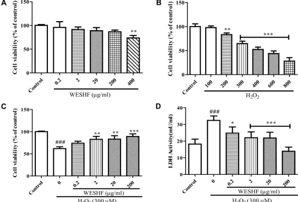
Fig. 1. Protective effects of hot water extract from Stauntonia hexaphylla fruit (WESHF) on hydrogen peroxide (H2O2)-induced cytotox- icity in HepG2 cells. (A) Cytotoxicity to cell viability after treated alone with WESHF. Cells were treated with DW or WESHF (0.2~400 μg/ml) for 24 hr, and cell viability was determined by MTT assay. (B) Cytotoxicity to cell viability after alone with hydrogen peroxide. Cells were treated with DW or H2O2 (100~800 μM) for 24 hr, and cell viability was determined by MTT assay. (C) Effects of WESHF treatment on cell viability by H2O2-induced cytotoxicity. (D) Effects of WESHF treatment on lactate dehydrogenase (LDH) release of cell damaged by hydrogen peroxide. Cells were treated with WESHF for 1 hr and incubated with H2O2 (300 μM) for 24 hr (C and D). Cell was measured by MTT assay and LDH leakage assay. ###
p<0.005, comparison between hydrogen peroxide and control. * p<0.05; ** p<0.01; ***p<0.005, comparison between hydrogen peroxide and sample group.
소거활성(IC
50, mg/l)은 과피, 과육, 종자가 각각 1513.0, 6109.7, 4378.0로 보고되어 있다[43]. 또한, 멀꿀 잎 70%에탄올 추출물 과 그 분획물의 DPPH 라디컬 소거능(SC
50, μg/ml)을 측정한 결과는 70% 에탄올추출물, 클로로포름 분획, 에틸아세테이트 분획, 부탄올 분획, 물 분획에서 각각 193.18±19.01, 420.28±
41.23, 63.00±6.20, 254.84±21.52, 1109.58±98.17로 보고되었다 [19]. ORAC assay는 2004년 항산화 작용의 표준화를 위한 세 계학술대회에서 선정된 방법으로 수소전자전달과 관련된 항 산화 물질의 유리 라디컬 소거 능력을 측정할 수 있으며, 식품 내 존재하는 hydrophobic성분과 hydrophilic 성분 모두에 반 응하는 특성으로 광범위한 항산화 활성 측정에 사용되고 있다 [26]. 멀꿀 열매 추출물의 항산화 활성을 알아보기 위해 멀꿀 열매 추출물을 각각 인산 완충액에 용해하여 AAPH에 의한 peroxy radical의 생성과 소멸에 따른 fluorescent의 감소율을 ORAC assay로 측정한 결과는 90.63±5.29 μM TE/g로 측정되 었다. Park 등의 연구결과[41]에 따르면 블루베리의 경우 57.59±1.01 μM TE/g으로 보고하였는데, 이와 비교할 때 멀꿀 열매가 1.58배 높은 활성을 나타내고 있음을 확인하였다.
세포독성 및 간세포 보호 효과
멀꿀 열매 추출물의 세포 독성을 MTT방법을 이용하여 측 정한 결과는 Fig. 1과 같다. 멀꿀 열매 추출물을 각각 0.2, 2, 200 및 400 μg/ml 농도로 처리하였을 때, 세포생존율은 각각 95.87±6.9, 91.53±3.08, 88.87±3.83, 73.63±3.08%로 나타났다(Fig.
1A). 따라서 멀꿀 열매 추출물을 HepG2 세포주에 처리하였을 때 200 μg/ml농도까지는 세포독성이 없음을 확인하였다.
과산화수소는 세포에서 산화적 손상을 유발하는 가장 중요
한 요인으로 세포 내에 과도한 산화적 스트레스가 유발되면
결과적으로 세포 사멸이 이루어져 세포생존율이 감소된다. 본
연구에서는 과산화수소가 산화적인 스트레스를 유발하는 적
정 농도를 결정하기 위해 HepG2 세포주에 여러 농도(100,
200, 300, 400, 600, 800)의 과산화수소를 24시간 동안 처리한
후 세포생존율을 구하였다. 연구 결과 300 μM 과산화수소에
서 64.6%의 세포 생존율를 확인하였으며, 이 농도를 산화적
스트레스 유도 농도로 결정하였다(Fig. 1B). 과산화수소로 유
도된 세포독성에 대한 멀꿀 열매 추출물의 간 세포 보호효과
를 측정하기 위해 과산화수소(300 μM) 처리 1시간 전 멀꿀
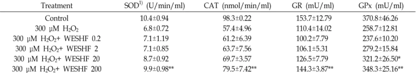
Table 2. Effects of hot water extract from Stauntonia hexaphylla fruit (WESHF) on antioxidant enzyme in HepG2 cells
Treatment SOD1) (U/min/ml) CAT (nmol/min/ml) GR (mU/ml) GPx (mU/ml)
Control 300 μM H2O2
300 μM H2O2+ WESHF 0.2 300 μM H2O2+ WESHF 2 300 μM H2O2+ WESHF 20 300 μM H2O2+ WESHF 200
10.4±0.94 6.8±0.72 7.1±1.19 7.1±0.85 8.7±0.92 9.9±0.98**
98.3±0.22 57.4±4.96 61.2±6.39 63.7±7.56 69.7±3.57 79.5±7.42**
153.7±12.79 110.4±14.02 100.2±7.79 106.1±5.31 126.5±7.79 144.3±3.87**
370.8±46.26 258.7±12.81 237.6±10.20 279.2±15.84 321.2±26.50*
348.3±25.16**
Cell were pre-treated with various concentrations of WESHF for 1 h and then were treated with H2O2 (300 μM) for 24 hr. Values are expressed as mean ± S.D. (n=3).
1)SOD : superoxide dismutase, CAT: catalase, GR: glutathione reductase, GPx: glutathione peroxidase.
** p<0.01, comparison between 300 μM H2O2 and sample group.
열매 추출물을 HepG2 세포주에 처리한 후 세포 생존율을 측 정하였다. Fig. 1C 와 같이 과산화수소 처리군의 세포생존율은 61.67±4.2%로 가장 낮았으며, 멀꿀 열매 추출물 0.2, 2, 20, 200 μg/ml로 처리 후 세포생존율이 각각 73.9±4.58, 82.86±6.40, 83.13±7.66, 89.09±5.98%로 농도의존적으로 증가하였다. 이는 이미 보고된 HepG2 세포에 커큐민을 처리하였을 때 500 μM 과산화수소 처리에 의한 세포사멸 정도가 감소한 연구 결과[7]
및 미더덕 껍질을 2 mM 과산화수소 처리하여 세포사를 유도 한 연구 결과[30]와 유사하다. LDH (lactate dehydrogenase)는 nicotinamide adenine dinucleotide (NAD)의 존재 하에 혐기 적 해당 작용의 최종산물인 L-lactate와 pyruvate 간의 가역적 산화환원반응을 촉매 하는 효소로서 간, 심장, 근육 내에 다량 존재한다[33]. 본 연구에서 LDH활성을 측정한 결과 대조군과 과산화수소 처리군이 각각 18.2±2.97 mU/ml, 32±2.67 mU/
ml로 과산화수소 처리로 LDH활성이 증가함을 확인하였다 (Fig. 1D). 이와 같은 증가는 멀꿀 열매 추출물 0.2, 2, 20, 200 μg/ml로 처리하였을 때 LDH활성이 각각 24.78±3.75 mU/ml, 22.06±3.50 mU/ml, 21.09±3.41 mU/ml, 13.94±2.45 mU/ml로 농도의존적으로 감소하였다. 따라서 HepG2 세포주에 과산화 수소를 처리하여 산화적 스트레스를 준 대조군과 멀꿀 열매 추출물 처리군을 비교한 결과, 과산화수소 처리로 HepG2 세 포주의 세포생존율은 감소하고, LDH 활성이 증가하였으나 멀 꿀 열매 추출물 처리로 세포독성과 LDH 활성이 감소하여 멀 꿀 열매 추출물의 간 세포 보호활성을 확인할 수 있었다.
간 세포의 항산화 효소 활성
Table 2는 멀꿀 열매 추출물이 과산화수소로 산화적 손상이 유도된 HepG2 세포주의 SOD, CAT, GPx, GR 활성에 미치는 영향을 나타난 결과이다. SOD활성도는 대조군, 과산화수소 처리군이 각각 10.38±0.94, 6.78±0.72 U/min/ml으로 과산화 수소 처리로 감소를 확인하였으며, 이러한 감소는 멀꿀 열매 추출물 200 μg/ml처리로 증가하였다(9.9±0.98 U/min/ml,
p<0.01). CAT활성도는 대조군, 과산화수소 처리군이 각각98.28±0.23, 57.36±4.96 nmol/min/ml으로 과산화수소 처리로
CAT활성의 감소를 확인하였으며, 멀꿀 열매 추출물 200 μg/
ml처리한 후 그 값이 증가함을 확인하였다(79.52±7.42 nmol/
min/ml, p<0.01). GPx활성도를 측정한 결과 대조군, 과산화수 소 처리군이 각각 370.8±46.26, 258.7±12.81 mU/ml으로 대조 군에 비해 과산화수소 처리군의 값이 감소함을 확인하였으며, 멀꿀 열매 추출물 20, 200 μg/ml처리로 각각 321.2±26.50(p<
0.05), 348.3±25.16 mU(p<0.01)로 감소된 값이 증가한 것을 확 인하였다. 또한 GR활성도는 대조군, 과산화수소 처리군이 각 각 153.7±12.79, 110.4±14.02 mU/ml으로 과산화수소 처리로 인한 감소를 확인하였으며, 이러한 GR활성도 감소는 멀꿀 열 매 추출물 200 μg/ml 처리로 그 값이 증가함을 확인하였다 (144.3±3.87 mU/ml, p<0.01). Yu 등[55]는 ursolic acid가 과산 화수소로 손상된 청각세포에서 CAT, GPx활성을 증가시켰다 고 보고하였으며, Alia 등[2]은 HepG2 세포에 tert-butyl hy- droperoxide로 산화스트레스를 유도한 후 말채나무를 처리한 결과 GSH-Px, CAT, SOD, GR과 같은 항산화 효소의 활성이 유의적으로 증가하였다고 보고하였다. 위의 결과로 볼 때 멀 꿀 열매 추출물은 HepG2 세포 내에서 항산화 효소를 활성화 시켜 과산화수소로 유도된 산화적 손상으로부터 세포를 보호 하는 효과를 갖는 것을 확인할 수 있었다.
뇌 조직의 지질과산화
활성산소종으로 유도된 산화적 스트레스에 의해 생성된 지 질과산화물에 대한 항산화제의 저해 효과를 알아보기 위해 쥐 뇌 조직에 FeCl
2를 처리한 후 멀꿀 열매 추출물에 의한 지질 과산화물 함량 변화를 측정한 결과는 Fig. 2와 같다. FeCl
2처 리로 증가한 지질과산화물 함량(0.61±0.02)은 멀꿀열매 50, 100, 200 μg/ml처리로 각각 0.30±0.02, 0.33±0.03, 0.33±0.04로 감소하였다. 이는 Kim 등[27], Eu 등[10]이 보고한 결과와 동일 함을 확인하였다.
혈청 중 ALT와 AST 활성
혈청 aminotransferase 활성은 간질환의 진단에 널리 사용
되는 효소로서 간 손상이 간세포의 수송기능 및 막 투과성에
Con
trol 0 50 100
200 0.0
0.2 0.4 0.6 0.8
***
WESHF (μg/ml) FeCl
2OD
Fig. 2. Protective effects of FeCl2-induced malondialdehyde (MDA) formation in brain tissue on hot water extract from Stauntonia hexaphylla fruit (WESHF). *** p<0.005, comparison between FeCl2 and sample group.
Table 3. Effects of hot water extract from Stauntonia hexaphylla fruit (WESHF) on serum transaminases activity in the acetaminophen (APAP)-induced liver injury in mice
Group ALT (mU/l) AST (mU/l)
Control APAP APAP+ WESHF 50 APAP+ WESHF 100 APAP+ WESHF 200
44.80±6.86 97.40±49.06 48.99±13.22*
65.12±13.19 51.08±5.86*
31.66±8.62 99.74±54.72 53.56±29.92 54.78±15.46 47.66±10.69*
The mice were pretreated with WESHF (50, 100 or 200 mg/kg, i.g.) once daily for three consecutive days. The control mice were given saline. Three hours after the final treatment, the mice were treated with APAP (400 mg/kg, i.p.). Hapatotoxicity was determined 18 h after by quantifying the serum activities of alanine aminotransferase (ALT) and aspartate amino- transferase (AST). Values are expressed as mean ± S.D. for five mice. * p<0.05, comparison between APAP and sample group.
Control 0 50 100
200 0
5 10 15 20
**
***
WESHF (mg/kg) APAP (400 mg/kg)
Malondialdehyde(nmol/mg)
Fig. 3. Inhibitory effects of hot water extract from Stauntonia hexaphylla fruit (WESHF) on acetaminophen (APAP)-in- duced lipid peroxidation. The mice were pretreated with WESHF (50, 100 or 200 mg/kg, i.g.) once daily for three consecutive days. The control mice were given saline.
Three hours after the final treatment, the mice were treated with APAP (400 mg/kg, i.p.). Mice were sacri- ficed 18 h after APAP administration. Values are ex- pressed as mean ± S.D. for five mice. **p<0.01, ***
p<0.005, comparison between APAP and sample group.
변화를 초래하여 이들 효소가 혈중으로 유리되어 혈장 내 활 성이 증가함으로 간 손상의 지표로 이용된다[9, 32]. 본 연구에 서는 아세트아미노펜으로 간 독성을 유발한 생쥐 모델에서 WESHF의 보호효과를 관찰하기 위해 간 기능의 혈액학적 지 표인 ALT 및 AST 활성을 측정하였다. 아세트아미노펜 단독투 여는 혈청 ALT, AST의 활성도를 정상군에 비해 각각 약 2.1배, 2.2배 이상 증가시켰다(Table 3). 이는 Eu 등[10], Gum 등[15]
이 아세트아미노펜을 복강투여 후 관찰한 결과와 동일하고, APAP에 의한 간 손상이 정상적으로 유도되었음을 보여주는 결과이다. 아세트아미노펜에 의해 증가된 혈청 속의 ALT, AST 효소는 멀꿀 열매 추출물 처리로 아세트아미노펜 단독 처리군에 비하여 이들 효소의 활성이 농도 의존적으로 감소하 는 것을 확인할 수 있었다(Table 3). 본 연구 결과 멀꿀 열매
추출물 200 mg/kg로 처리한 생쥐에서 가장 강한 간 보호 효과 를 확인하였다.
간 조직 중 지질과산화물 함량
간 조직의 지질과산화도를 측정하기 위해 멀꿀 열매 추출물 을 농도별로 전 처리한 후 APAP를 복강 내로 주사하여 간 손상을 유도한 후 생쥐 모델에서 간 조직 내 지질과산화물의 함량을 측정 결과는 Fig. 3이다. 아세트아미노펜으로 간 손상 을 유도한 결과, 정상군은 지질과산화물량이 1.31±0.69 nmol/
mg인데 비하여 아세트아미노펜처리군은 14.72±3.89 nmol/
mg로 증가한 것을 확인할 수 있었다. 이러한 증가한 수치는
3일간 멀꿀 열매 추출물 50, 100, 200 mg/kg 투여한 결과 각각
11.35±1.77, 7.41±3.68, 5.42±1.79 nmol/mg로 유의성 있는 감
소를 보였다(Fig. 3). 아세트아미노펜은 급성 산화적 간 손상
유발에 널리 사용되는 약물[39]로써 해열·진통제의 주성분으
로 적정 농도를 복용할 경우 생체 내 항산화 방어 기전에 의해
무독화되어 배설됨으로써 안전하다고 알려져 있으나, 과 용량
복용 시 활성대사체인 N-acetyl-p-benzoquinoneimine (NAPQI)
을 무독화시키는 glutathione이 오히려 고갈됨으로써 세포막
의 산화적 손상 및 세포 내 거대분자가 파괴되어 간세포가
손상되는 것으로 알려져 있다[53]. 아세트아미노펜에 의한 급
성 간 손상 과정에는 glutathione 함량 감소와 대사 활성체에
의한 지질과산화 반응이 일어난다[11]. 지질과산화 반응은 간
기능의 부전을 야기하는 기본적 기전 중의 하나로 중요시되어
A B
C D E
Fig. 4. Effects of hot water extract from Stauntonia hexaphylla fruit (WESHF) pretreatment on the acetaminophen (APAP)-induced liver damage in mice. Histological examination of liver tissues was performed 18 hr after the APAP administration. Liver tissues were fixed, sectioned at 5 μm thickness, and stained with hematoxylin and eosin (H&E; magnification, ×10). (A) Liver from a mouse treated with saline; (B) liver from a mouse treated with APAP (400 mg/kg, i.p.); (C) liver from a mouse treated with WESHF (50 mg/kg, i.g.) and APAP (400 mg/kg, i.p.); (D) liver from a mouse treated with WESHF (100 mg/kg, i.g.) and APAP (400 mg/kg, i.p.); (E) liver from a mouse treated with WESHF (200 mg/kg, i.g.) and APAP (400 mg/kg, i.p.).
왔으며 이러한 지질과산화 반응에 의한 간 손상 예방과 관련 한 기전은 glutathione의 대사와 밀접하게 관련되어 있을 뿐만 아니라[54], 간 조직 내에서 superoxide를 제거하는 SOD, 그리 고 과산화수소를 제거하는 CAT, glutathione peroxidase과 같 은 항산화 효소들도 중요한 역할을 담당하는데 이러한 효소들 은 대사 활성체에 의한 지질과산화 반응의 연쇄적 진행을 차 단한다[48]. 본 연구에서는 동물실험에서 CYPE효소활성, glu- tathione함량의 변화를 측정하지 않았으나 세포수준에서 멀꿀 열매 추출물의 항산화 활성 향상을 확인한 바 아세트아미노펜 에 의한 간 손상 회복 기전에 항산화 효소가 관련되었을 것으 로 사료되며 앞으로의 연구에서 간 독성 작용기전에 대한 간 약물대사효소의 변화와 GSH availability 변화에 대한 연구가 진행되어야 할 것으로 사료된다.
조직병리학적 변화
간 조직을 관찰한 결과 정상군은 간세포들이 중심정맥을 중심으로 정상적인 소엽구조를 나타내었으며, 기타의 조직학 적인 구조도 정상으로 배열되어 있었으나(Fig. 4A), 아세트아 미노펜으로 간 독성을 유발한 군의 간 조직(Fig. 4B)은 간 소엽 의 구조가 명확하지 않으며, 중심정맥 주위의 울혈과 간세포 괴사가 관찰되었다. 이에 비해 멀꿀 열매 추출물 처리군(Fig.
4C~Fig. 4E)은 울혈과 세포 괴사가 완화되어 정상세포의 소엽 구조로 회복되는 것을 확인할 수 있었다.
최근 나고야의정서 발효로 자생식물에 대한 관심이 증가되 어 국내 토종 식물인 멀꿀에 대한 경제적 가치가 증대되고
있다. 따라서 본 논문은 멀꿀 열매에 대한 총페놀함량과 플라 보노이드 함량을 측정하였고, 항산화 활성을 평가하였으며,
in vitro 및 in vivo계에서 손상된 간세포에 대한 보호 효과를확인하였다. 기존에 멀꿀에 대한 다양한 연구가 진행되지 않 았으므로 본 연구는 멀꿀을 기능성 소재로 개발 시 기초자료 로 활용될 수 있을 것으로 판단된다
감사의 글
본 연구는 산업통상자원부 창의산업거점기관지원사업 바 이오상용기술고도화 플랫폼 구축사업의 지원을 받아 수행하 였다.
References
1. Akira, I. and Hideji, I. 1989. The triterpenes from Stauntonia hexaphylla call tissues and their biosynthetic significance. J.
Nat. Prod. 52, 623-628.
2. Alia, M., Ramis, S., Mateos, R., Granado-Serrano, A. B., Bravo, L. and Goya, L. 2013. Antioxidant activities of hot water extract from cornus walteri wanger against oxidative stress induced by tert-butyl hydroperoxide in HepG2 cells.
J. Kor. Soc. Food Sci. Nutr. 42, 1525-1532.
3. Bradford, M. M. 1976. A rapid and sensitive method for the quantitation of microgram quantities of protein utilizing the principle of protein-dye binding. Anal. Biochem. 72, 248-254.
4. Bray, B. J. and Rosengren, R. J. 2001. Retinol potentiates acet- aminopheninduced hepatotoxicity in the mouse: Mechanis- tic studies. Toxicol. Appl. Pharm. 173, 129-136.
5. Carlberg, I. and Mannervik, B. 1975. Purification and charac- terization of the flavoenzyme glutathione reductase from rat liver. J. Biol. Chem. 250, 5475-5480.
6. Carmichael, J., DeGraff, W. G., Gazdar, A. F., Minna, J. D.
and Mitchell, J. B. 1987. Evaluation of a tetrazolium-based semiautomated colorimetric assay: assessment of chemo- sensitivity testing. Cancer Res. 47, 936-942.
7. Chen, X., Zhong, Z., Xu, Z., Chen, L. and Wang, Y. 2011.
No protective effect of curcumin on hydrogen peroxide in- duced cytotoxicity in HepG2 cells. Pharmacol. Rep. 63, 724- 732.
8. Cichoz-Lach, H. and Michalak, A. 2014. Oxidative stress as a crucial factor in liver diseases. World J. Gastroenterol. 20, 8082-8091.
9. Clissold, S. P. 1986. Pracetamol and phenacetin. Drugs 4, 46-59.
10. Eu, J. B., Kim, S. O., Seoung, T. J., Choi, S. G., Cho, S. H.
and Choi, C. Y. 2010. Protective effect of theanine on the acetaminophen-induced hepatotoxicity. J. Kor. Soc. Food Sci.
Nutr. 39, 350-355.
11. Fisher, L., Green, M. D. and Harman, A. W. 1982. Levels of acetaminophen and its metabolites in mouse tissues after a toxic does. J. Pharmacol. Exp. Ther. 221, 407-413.
12. Floege, l. A., Kim, D. O., Chung, S. J., Koo, S. I. and Chun, O. K. 2011. Comparison of ABTS/DPPH assays to measure antioxidant capacity in popular antioxidant-rich US foods.
J. Food Compos. Anal. 24, 1043-1048.
13. Gao, H., Zhao, F. and Chen, G. D. 2009. Bidesmoside tri- terpenoid glycosides from Stauntonia chinensis and relation- ship to antiinflammation. Phytochemistry 70, 795-806.
14. Guengerich, F. P., Kim, D. H. and Iwasaki, M. 1991. Role of human cytochrome P-450 IIEL in the oxidation of many low molecular weight cancer suspects. Chem. Res. Toxicol.
4, 168-179.
15. Gum, S. I., Lee, D. U. and Cho, M. K. 2007. Protective effects of water extracts composed of Adenophora triphylla var. japon- ica Hara on the acetaminophen-induced hepatotoxicity. Kor.
J. Food Sci. Technol. 39, 688-693.
16. Gutfinger, T. 1981. Polyphenols in olive oils. J. Am. Oil Chem.
Soc. 58, 966-968.
17. Heim, K. E., Tagliaferro, A. R. and Bobilya, D. J. 2002.
Flavonoid antioxidant: chemistry, metabolism, and struc- ture-activity relationships. J. Nutr. Biochem. 13, 572-584.
18. Hugo, A. 1984. Catalase in vitro. Methods Enzymol. 105, 121- 126.
19. Hwang, S. H., Kwon, S. H., Kim, S. B. and Lim, S. S. 2017.
Inhibitory activities of Stauntonia hexaphylla leaf constituents on rat lens aldose reductase and formation of advanced gly- cation end products and antioxidant. BioMed. Res. Int. 2017, 1-8.
20. Jeong, C. H., Choi, G. N., Kim, J. H., Kwak, J. H., Kim, D. O., Kim, Y. J. and Heo, H. J. 2010. Antioxidant activities from the aerial parts of platycodon grandiflorum. Food Chem.
118, 278-282.
21. Jeong, C. H., Jang, S. T., Joo, O. S., Lee, S. C., Shin, Y. H., Shim, K. H., Cho, S. H., Choi, S. G. and Heo, H. J. 2009.
Phenolic content, antioxidant effect and acetylcholinesterase inhibitory activity of Korean commercial green, puer, oo- long, and black teas. Kor. J. Food Preserv. 16, 230-237.
22. Karadag, A., Ozcelik, B. and Saner, S. 2009. Review of meth- ods to determine antioxidant capacities. Food Anal. Methods 2, 41-60.
23. Kim, A. K. and Kim, J. H. 2001. Alterations of antioxidant enzymes in response to oxidative stress and antioxidants.
Biomol. Ther. 9, 249-257.
24. Kim, J. Y., Kim, H. S., Choi, H. J., Jo, A, Kang, H. W., Yun, H. J., Choi, S. Y. and Im, S. J. 2018. Anti-inflammatory effects of a stauntonia hexaphylla fruit extract in lipopolysaccharide- activated raw-264.7 macrophages and rats by carra- geenan-induced hind paw swelling. Nutrients 10, 1-12.
25. Kim, O. S., Park, S. S. and Sung, J. M. 2012. Antioxidant activity and fermentation characteristics of traditional black rice wine. J. Kor. Soc. Food Sci. Nutr. 41, 1693-1700.
26. Kim, S. H. and Kim, Y. M. 2007. Determination of the anti- oxidant capacity of Korean ginseng using an ORAC Assay.
J. East Asian Soc. Dietary Life 17, 393-401.
27. Kim, S. J. and Han, D. S. 2005. Effect of plants extract on lipid peroxidation of rat brain tissue induced by reactive oxygen species. Kor. J. Food Sci. Technol. 17, 393-401.
28. Kim, Y. S., Hwang, J. W., Park, P. J. and Jeong, J. H. 2014.
Antioxidant activity and protective effects of extracts from Chrysanthemum boreale on t-BHP induced oxidative stress in Chang cells. J. Kor. Soc. Food Sci. Nutr. 43, 60-66.
29. Kind, P. R. N. and King, E. J. 1954. Estimation of plasma phosphatase by determination of hydrolysed phenol with amino antipyrine. J. Clin. Pathol. 7, 322-326.
30. Koh, E. K., Lee, Y. J., Kim, J. E., Kwak, M. H., Go, J., Son, H. J., Lee, H. S., Jung, Y. J. and Hwang, D. Y. 2014. Protective Effect of aqueous extracts of Styela Clava Tunic against apop- tosis of HepG2 Cells induced by hydrogen peroxide. J. Life Sci. 24, 595-602.
31. Krinsky, M. 1992. Mechanism of action of biological anti- oxidants. Proc. Soc. Exp. Biol. Med. 200, 248-234.
32. Lee, W. M. 2004. Acetaminophen and the US Acute liver failure study group: lowering the risks of hepatic failure.
Hepatology 40, 6-9.
33. Lim, T. S., Kim, J. S. and Kim, J. M. 1984. Electrophoretic patterns of GGT, ALP and LDH isoenzyme in various hep- atic diseases. Med. J. 25, 268-285.
34. Lu, Y. and Foo, L. Y. 2000. Antioxidant and radical scaveng- ing activities of polyphenols from apple pomace. Food Chem.
68, 81-85.
35. McCord, J. M. and Fridovich, I. 1969. Superoxide dismutase.
An enzymic function for erythrocuprein (hemocuprein). J.
Biol. Chem. 244, 6049-6055.
36. Michiels, C., Raes, M., Toussaint, O. and Remacle, J. 1994.
Importance of se-glutathione peroxidase, catalase, and Cu/Zn SOD for cell survival against oxidative stress. Free Radic. Biol. Med. 17, 235-248.
37. Min, K. C. and Jhoo, J. W. 2013. Antioxidant activity and inhibitory effect of Taraxacum officinale extracts on nitric ox- ide production. Kor. J. Food Sci. Technol. 45, 206-212.
38. Moreno, M. I. N., Isla, M. I., Sampietro, A. R. and Vattuone, M. A. 2000. Comparison of the free radical-scavenging activ- ity of propolis from several regions of Argentina. J. Ethno- pharmacol. 71, 109-114.
39. Oleg, M., Miriam, W. L., Kenneth, R., Laishun, C., Ghung, Y. and Masayori, L. 1999. Acetaminophen toxicity. J. Biol.
Chem. 274, 10348-10335.
40. Paglia, D. E. and Valentine, W. N. 1967. Studies on the quan- titative and qualitative characterization of erythrocyte gluta- thione peroxidase. J. Lab. Clin. Med. 70, 158-169.
41. Park, J. H., Park, H. M., Kang, S. J., Kang, E. J., Lee, D. H.
and Kim, D. I. 2012. Originals: Quality characteristics and granule manufacture of mulberry and blueberry fruit extracts. Kor. J. Food Cookery Sci. 28, 375-382.
42. Park, J. O., Jeong, B. J., Park, M. Y., Kang, S. Y., Kang, J.
G., Park, Y. J. and Heo, B. G. 2010. Effect of gamma ray irradiation on the seed germination, growth and variant in- duction of Hibiscus hamabo and stauntonia hexaphylla. J. Life Sci. Nat. Res. 32, 85-95.
43. Park, Y. J., Park, Y. S., Towantakavanit, K., Park, J. O., Kim, Y. M., Jung, K. J., Cho, J. Y., Lee, K. D. and Heo, B. G.
2009. Chemical components and biological activity of staun- tonia hexaphylla. Kor. J. Plant Res. 22, 403-411.
44. Pellegrini, M. and Baldari, C. T. 2009. Apoptosis and oxida- tive stress-related disease: the p66shc connection. Curr. Mol.
Med. 9, 392-398.
45. Pietta, P. G. 2000. Flavonoids as antioxidants. J. Nat. Prod.
63, 1035-1042.
46. Proestos, C., Sereli, D. and Komaitis, M. 2006. Determination of phenolic compounds in aromatic plants by RP-HPLC and GC-MS. Food Chem. 95, 44-52.
47. Reitman, S. and Frankel, S. A. 1957. Colorimetric method
for determination of serum glutamic oxalacetic and glutamic pyruvic transaminases. Am. J. Clin. Pathol. 28, 56-63.
48. Vendemiale, G., Altomare, E., Grattagliano, I. and Albano, O. 1989. Increased plasma levels of glutathione and ma- londialdehyde after acute ethanol ingestion in humans. J.
Hepatol. 9, 359-365.
49. Vinson, J. A., Su, X., Zubik, L. and Bose, P. 2001. Phenol anti- oxidant quantity and quality in foods: fruits. J. Agric. Food Chem. 49 ,5315-5321.
50. Wang, E. J., Li, Y., Lin, M., Chen, L., Stein, A. P., Reuhl, K. R. and Yang, C. S. 1996. Protective effects of garlic and related organosulfurcompounds on acetaminophen-induced hepatotoxicity in mice. Toxicol. Appl. Pharmacol. 136, 146-154.
51. Wang, H. B., Mayer, R., Rucker, G., Yang, J. J. and Matteson, D. S. 1998. A phenolic glycoside and triterpenoids from Stauntonia hexaphylla. Phytochemistry 47, 467-470.
52. Wei, Y., Ma, C., Chen, D. and Hattori, M. 2008. Anti-HIV-1 protease triterpenoids from Stauntonia obovatifoliola Hayata subsp. Phytochemistry 69, 1875-1879.
53. Yonamine, M., Aniya, Y., Yokomakura, T., Koyama, T., Nagamine, T. and Nakanishi, H. 1996. Acetaminophen-de- rived activation of liver microsomal glutathione S-transferse of rats. J. Pharmacol. 72, 175-181.
54. Yung, J. H., Chiu, L. C. and Ooi-Clin, V. 1994. Effect of poly- saccaride peptide on glutathione and protection against par- acetamol-induced hepatotoxity in the rat. Clin. Pharmacol.
16, 723-729.
55. Yu, Y. H., Seo, S. J., Hur, J. M., Park, R. K., So, H. S., Jeon, B. H. and You, Y. O. 2006. Protective effect of urolic acid from corni fructus on the hydrogen peroxide-induced dam- age of HEI-OC1 auditory cells. Kor. J. Oriental Physiol. Pathol.
20, 1524-1529.
56. Zhao, J., Yim, S. H. and Um, J. I. 2014. Cytotoxic component in an extract from the leaves and stems of Stauntonia hexaphylla. Nat. Prod. Sci. 20, 130-134.
초록:멀꿀 열매 추출물의 항산화 활성 및 H
2O
2로 유도된 산화적 스트레스와 아세트아미노펜 독성 모델 에서의 간 보호효과
이규옥․김재용․강후원․배동혁․최철웅*
(천연자원연구센터)