39(11), 1647~1653(2010) DOI: 10.3746/jkfn.2010.39.11.1647
군소( Aplysia kurodai )에 분포하는 글루코사미노글리칸의 추출과 기능특성
2. 글루코사미노글리칸의 구조 특성 윤보영1․최병대1․배동원2․최영준1†
1경상대학교 해양식품공학과/해양산업연구소
2경상대학교 공동실험실습관
Extraction of Glycosaminoglycan from Sea Hare, Aplysia kurodai , and Its Functional Properties 2. Structural Properties of Purified Glycosaminoglycan
Bo-Yeong Yoon1, Byeong Dai Choi1, Dong-Won Bae2, and Yeung Joon Choi1†
1Dept. of Seafood Science and Technology/Institute of Marine Industry, Gyeongsang National University, Gyeongnam 650-160, Korea
2Central Instrument Facility, Gyeongsang National University, Gyeongnam 660-701, Korea
Abstract
Glycosaminoglycan (GAG) was purified from polysaccharide extracted from sea hare muscle on DEAE- Sepharose column and investigated for the functional groups, distribution of sugars, composition of disaccharide and structure of GAG. Purified GAG was composed of disaccharide above 55% of total sugar. Purified GAG showed amide I peak in 1648/cm and C-O stretch peak as properties of carbohydrate, amino acid peak in 1457/cm, and peak in 866/cm as properties of monosaccharide by FT-IR. Fucose, N-acetylgalactosamine, N-acetylglu- cosamine, glucose, galactose, mannose and xylose were found in MALDI-TOF MS/MS spectra of hydrolysates by chondroitin sulfate ABC lyase and heparanase I. Purified GAG was expected to be heparan sulfate including N-acetylgalactosamine and N-acetylglucosamine above 70% of total sugar. The structure of GAG was supposed as GlyUA(2S)-GlcNS and GlyUA-GlcNS(6S) with O-linkage on protein core.
Key words: sea hare, Aplysia kurodai, glycosaminoglycan, purification, structure
†Corresponding author. E-mail: [email protected]
†Phone: 82-55-640-3115, Fax: 82-55-640-3111
서 론
Glycosaminoglycan(GAG)은 반복하는 이당류 단위를 가 진 음이온성 이형다당류(1)로, 산성당(uronic acid)과 아미노 당(N-acetylglucoamine, N-acetylgalactosamine)의 결합을 통해 사슬이 무수히 반복되는 구조를 가지고 있다. 당 잔기 에 황산기(SO3-)나 카르복실기(COO-)를 가져 음전하를 띠 며, 당 잔기들 간의 결합 형태와 sulfate의 수와 위치에 따라 그 특성이 분류된다(1,2). GAG는 황산기의 유무에 따라 황 산화 다당과 비황산화 다당으로 크게 나누어지며, 대표적인 GAG에는 hyaluronic acid, chondroitin sulfate, dermatan sulfate, heparin/heparan sulfate와 keratan sulfate 등이 있 다(3). Hyaluronic acid를 제외한 GAG는 당단백질과는 다른 프로테오글리칸으로 존재하며, 음이온기가 많은 chondroitin sulfate, keratan sulfate(또는 dermatan sulfate)의 콘드로이 틴군(chondroitin family)과 헤파린군(heparin family)은 결 합 부위에 중심단백질을 가지고 있다(4). 당과 단백질로 되어 있는 일군의 화학 물질을 당단백질이라 하며, 당 부분은 2~
6종류의 단당으로 되어 있고 단백질과 공유 결합하는 특성 을 가지고 있다(3). GAG류는 소량의 펩티드와 결합하고 있 으며, 당단백질과는 구별하여 프로테오글리칸이라 한다. 프 로테오글리칸은 2개의 당이 반복 구조를 형성하고 있지만, 당단백질의 경우는 이들 이당류의 반복 구조가 없고 일반적 으로 uronic acid와 황산기(-SO3H)가 없기 때문에 당단백질 과 프로테오글리칸은 서로 구분해야 할 필요성이 있다. 프로 테오글리칸으로 결합하는 당사슬의 종류에는 크게 poly- peptide chain의 Asn 잔기에 결합하는 N-linkage와 Ser/Thr 잔기에 결합하는 O-linkage가 있다(3). N-linkage는 N- acetylglucosamine의 환원성 말단이 peptide chain의 Asn 잔기와 결합하고 Man3-GlcNAc2를 기본으로 여러 구조로 이루어지며, O-linkage는 N-acetylgalactosamine의 환원성 말단이 peptide chain의 Ser/Thr 잔기와 결합하여, 공통적인 구조를 가지는 N-linkage와는 달리 더욱 다양한 구조를 가 진다.
Chondroitin sulfate는 포유동물의 연골뿐만 아니라, 각 조 직과 체액에 이르기까지 널리 분포하고 있다(5). 쥐의 늑골
이나 소의 태아(6) 및 사람의 위와 신장조직(7), 사람이나 토끼의 혈액과 뇨(8)에 분포하며, 이 밖에 닭의 태아, 각막 상피조직(9), 상어연골(10), 무척추동물(11), 소, 돼지, 오징 어 연골 등의 관절연골(12)에서 sulfate를 가진 GAG가 분리 되었으며, 해삼에서도 당단백질이 발견되어 포유동물 이외 에서도 고루 분포하고 있음이 확인되었다(13-16).
한편 헤파린군(heparin/heparan sulfate family)도 여러 동물조직에서 발견되고 있으며 황산기의 수와 위치에 따라 다양한 형태의 구조를 나타내고 지금까지 알려진 구조만 하 더라도 6가지에 이른다. 척추동물의 조직에서 분리한 hep- arin과 heparan sulfate은 antithrombin-III과 결합하는 특성 을 가져 항응고 활성을 보이며, post-translation 기작을 통 해 heparin은 유선 세포 내에서 양으로 하전한 단백질 분해 효소의 생성량을 조절한다(17). 이는 모든 포유동물의 매우 중요한 기능 중 하나로 임신 중 태아와 산모의 영양공급 통 로인 태반의 항응고 활성에 관련한 모든 혈액 응고 방지 기 능을 조절해, 임신 중 발생하는 임신중독증을 비롯한 여러 가지 질병을 예방한다(18). 그러나 수서 척추동물에서는 헤 파린군의 분포가 매우 제한적이라고 보고하였다(19).
군소(Aplysia kurodai)는 패각이 퇴화되어 몸속에 얇은 판 모양으로 들어 있고, 얇고 볼록한 렌즈모양으로 생겼으 며, 주로 해조류를 섭취하며 봄부터 여름까지 해안에서 다량 생산된다(20). 군소의 생리적인 기능으로서 항암(21-23)효 능, purple gland의 항균효능(24)과 군소의 신경 펩티드에 관한 연구(25,26)는 이루어져 있으나, 군소의 GAG와 관련하 여 Hovingh와 Linker(27)의 Aplysia californica와 Helix aspersa의 chondrotin sulfate와 heparan sulfate에 관한 연 구 외에는 거의 이루어져 있지 않다.
본 연구에서는 남해안 일대에서 여름철에 어획되어 부산 과 경남지역을 중심으로 소비되고 있는 군소에서 GAG를 정제하고 IR spectrum, 이당류 분석 및 질량분석을 통하여 GAG의 구조적 특성을 조사하였다.
재료 및 방법 재료 및 시약
끓는 물에서 5분 동안 자숙하여 색소와 냄새를 제거한 군 소를 통영시 잠수기조합에서 구입하여, 육질과 내장을 분리 한 다음 동결건조 하여 -20oC의 냉동고에 보관하면서 시료 로 사용하였다. 단백질 분해효소로서 Flavourzyme 500 MG 와 Neutrase는 Novozymes(Seoul, Korea) 에서 구입하였으 며, DEAE-Sepharose 수지와 Superdex HR-200 칼럼은 GE healthcare Korea(Seoul, Korea)에서 구입하였고, papain (P3375), 분자량 측정을 위한 표준단백질과 GAG 표준물질 및 그 외의 모든 시약은 Sigma-Aldrich사(St. Louis, MO, USA)에서 구입하였다.
동결 건조한 군소의 일반성분은 AOAC법(28)에 따라 수
분은 상압가열건조법, 조회분은 건식회화법, 조단백질은 micro Kjeldahl법으로 분석하였으며, 조지방은 Folch법(29) 으로 분석하고, 탄수화물은 위의 성분을 감하여 구하였다.
다당류의 추출
동결 건조시료 1 g에 10 배량의 50 mM sodium phosphate 완충액(pH 6.0)을 첨가하고, 기질에 대하여 단백질 농도비가 1/50이 되도록 Flavourzyme 500 MG을 첨가하여 60oC의 항 온 수조에서 흔들어 주면서 15시간 동안 가수분해하였다.
원심분리(3,000×g, 30 min)하여 얻은 가수분해물의 상등액 에 최종농도가 3%가 되도록 trichloroacetic acid를 첨가하여 실온에서 30분간 방치한 후, 원심분리(3,000×g, 30 min)하 여 침전한 단백질을 제거하였다. 회전진공증발기로 40oC 이 하에서 상등액의 Brix가 60이 될 때까지 감압농축하고, 농축 물에 5배량의 에탄올을 첨가하여 알코올 불용성 물질을 침전 시킨 다음, 원심분리(3,000×g, 30 min)한 침전물을 회수하 여 추출 다당류로 사용하였다.
DEAE-Sepharose에 의한 GAG의 분리 및 정제
최적 조건으로 추출한 다당류를 50 mM sodium phos- phate buffer(pH 6.0)에 50 mg/mL의 농도가 되도록 용해시 킨 후, 같은 완충액으로 평형화시킨 DEAE-Sepharose 칼럼 (1.6×15 cm)에 200 μL를 loading하여 칼럼 부피와 동량의 50 mM sodium phosphate buffer(pH 6.0)로 세척하고, 2 M NaCl을 함유한 같은 buffer로 유속 1 mL/min으로 선형균배 하였으며 용출액의 GAG는 254 nm에서 검출하였다. 용출액 은 0~70 min에서는 5 mL, 70~120 min에서는 2 mL씩 분획 하였다.
당 함량의 측정
각 획분의 총 탄수화물 함량은 phenol-sulfuric acid법(30), uronic acid 함량은 Bitter와 Muir(31)의 carbazole assay을 다소 변형한 방법으로, N-acetylhexosamines 함량은 MBTH (3-methyl-2-benzothiazolinone hydrazone hydrochloride) 법(32)으로 각각 측정하였다. 즉 phenol-sulfuric acid 법은 증류수에 녹인 시료 150 μL를 시험관에 취하고 5%(w/v) phenol 용액 150 μL와 혼합한 후, 신속히 진한 황산 750 μL를 첨가해 10분간 실온에서 반응시켰다. 한번 더 vortex를 이용 해 혼합하고 발색이 될 때까지 실온에서 30분간 반응시킨 다음 490 nm에서 흡광도를 측정하였으며, D-galactose로 작성한 검량곡선에 따라 함량을 계산하였다.
Uronic acid 함량은 증류수에 용해되어 있는 시료 200 μL 를 cap tube에 분취하고 4 M ammonium sulfate 20 μL를 가하여 혼합하였다. 진한 황산에 녹아있는 25 mM sodium tetraborate 1 mL를 첨가한 뒤 100oC에서 5분간 가열하고 실온에서 방냉시킨 다음, 0.1% cabazole 용액을 40 μL 가하 고 다시 100oC에서 15분간 반응시켰다. 실온까지 방냉한 후 520 nm에서 흡광도를 측정하였다. Uronic acid 함량은
0 0.1 0.2 0.3 0.4 0.5 0.6 0.7
0 5 10 15 20 25 30
Fraction Number
Absorbance .
Hexosamine 525 nm Uronic acid 520 nm Total sugar 490 nm
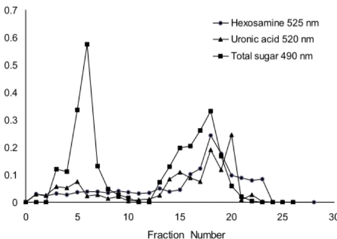
Fig. 1. Chromatogram of polysaccharide from sea hare mus- cle on DEAE-Sepharose fast flow. The constituents were elut- ed with linear gradient between 0~2 M NaCl containing 0.5 M sodium phosphate buffer (pH 6.0) at a flow rate of 1 mL/min.
Fraction was collected with 5 mL in number 1 to 12 and with 2 mL in from 13 to 26. Total carbohydrates, uronic acid, hexos- amines were assayed by the phenol-sulfuric method, MBTH and the carbazole reaction, respectively.
glucuronolactone으로 작성한 검량곡선으로 계산하였다.
MBTH 법에 의한 당 함량의 측정은 증류수에 녹아있는 시료 100 μL를 시험관에 분취하고, 1,9-dimethylmethylene blue(pH 3.0) 용액 2.5 mL를 가하여 혼합하고 525 nm에서 흡광도를 측정하였으며, N-acetyl-D-galactosamine으로 작성한 검량곡선으로 함량을 구하였다.
효소적 depolymerization에 의한 GAG의 확인
효소적 탈중합으로 GAG를 확인하기 위해 정제한 시료 (100 μg)를 chondroitin sulfate ABC lyase(C2905)와 he- parinase I(H2519)로 37oC에서 15시간 동안 가수분해 하였 다. 가수분해 후 100oC에서 2분 동안 가열하여 효소를 불활 성화 시키고, 0.45 μm 필터로 여과한 여액을 시험관에 50 μL를 분취하여 250 μL의 30 mM HCl을 첨가하여 탈중합시 켰다. 반응액은 분광광도계로 232 nm에서 흡광도를 측정하 였으며, chondroitin sulfate A(C9819)를 표준물질로 사용하 여 작성한 검량곡선에 따라 GAG의 함량을 측정하였다.
조성당과 기능기의 분석
정제한 GAG의 조성당 분석은 건조 시료를 10 mg/mL 농도로 용해하여 2 mL를 분취하고 최종농도가 2 M이 되도 록 trifluoroacetic acid를 첨가하여, 100oC에서 4시간 동안 산가수분해 하였다. 산가수분해물을 Speed-vac(DP23080A, Sunonwealth Elec Mach Ind Co. Ltd., Peking, China)으로 건조하여 소량의 탈이온수로 수회 세척한 후 탈이온수 100 μL에 용해하여 16 mM NaOH 용액으로 평형화된 CarboPac PA1 column(0.45×25 cm, Dionex Co., Bannockburn, IL, USA)에 loading하고, 동일 용액으로 유속 1.0 mL/min에서 용출하면서 HPAED-PAD(High-performance anion ex- change chromatography-pulsed amperometric detector, Dionex Co.)로 당을 검출하였다.
정제한 GAG의 기능기는 정제한 GAG에 소량의 KBr을 첨가하여 tablet를 형성시킨 후 적외선 분광광도계(Vertex 80v, Bruker Optics, Ettlingen, Germany)를 사용하여 스펙 트럼을 측정하였다.
SAX-HPLC에 의한 disaccharides 분석
정제한 GAG의 disaccharides 분석을 위해 건조된 시료 (50 μg)를 chondroitin sulfate ABC lyase와 heparinase I로 37oC에서 15시간 동안 가수분해하였다. 가수분해물 중의 효 소를 100oC에서 2분 동안 가열하여 불활성화 시키고, 0.45 μm 필터로 여과한 뒤 탈이온수로 평형화된 SAX column (Phenosphere 5u SAX 80A, 25×0.46 cm, Phenomenex Co., Torrance, CA, USA) 상에서 2 M NaCl(pH 3.5) 용액으로 선형균배하여 용출시키면서 232 nm에서 이당류를 검출하 였다. 표준품으로는 heparin/heparan sulfate disaccharide kit(Seikagaku Co., Tokyo, Japan)를 사용하였다.
질량 분석
정제 GAG의 질량은 electrospray ionization mass spec-
trometry(ESI-MS) 질량분석기인 Qtrap 3200 LC/MS/MS (Applied Biosystems, Forster, CA, USA)를 사용하여 pos- itive mode에서 ion voltage 5,500, curtain gas(CUR) 20으로 조건을 맞추어 syringe direction injection 방법으로 Q1 scan method로 실험을 수행하고 결과를 분석하였다.
결과 및 고찰 정제한 다당류의 특성
당은 2개의 peak 형태로 용출되었으며(Fig. 1), 초기의 peak는 중성 다당류 중에서 단당류가 칼럼에 흡착하지 않고 용출되며, 이후에 용출하는 주 peak는 GAG의 용출 peak인 것으로 추정하였다. GAG의 지표 물질인 uronic acid와 hexosamine 함량을 측정한 결과 앞서 얻은 용출 형태와 일 치하는 구간에서 uronic acid와 hexosamine이 용출되는 것 을 확인하였다. 대부분의 uronic acid는 GAG와 프로테오글 리칸에서 발견된다(33). GAG 지표물질을 사용하여 군소 육 질에서 추출한 다당류와 이들 정제물의 hexuronic acid와 hexosamine의 함량을 측정한 결과(Table 1), 아미노당의 유 도체로 작용하는 hexuronic acid 함량은 다당 추출물의 경우 1.0±0.0 g/100 g이었고 정제물의 경우에는 6.0±0.2 g/100 g이었다. GAG를 구성하는 성분으로 또 다른 지표물질인 hexosamine의 함량 또한 당 추출물에서 5.6±0.2 g/100 g의 함량을 나타내었으며 정제물의 경우에는 25.7±1.3 g/100 g 로서 상당히 높은 함량을 보였다.
효소특이적 탈중합으로 GAG를 확인한 결과(Table 1), 군 소 다당류 추출물에서 정제한 글리코사미노글리칸을 chon- droitin sulfate ABC lyase를 이용해 가수분해 후 측정한 결 과 이당류의 함량은 22.1±1.7 g/100 g으로 22% 이상을 함유
Table 1. Hexuronic acid, hexosamine and disaccharide con- tents in polysaccharide extract (ASE) and purified glyco-
saminoglycan (ASF) (unit: g/100 g)
Hexuronic acid Hexosamine Disaccharide ASEASF
ASF CS1) ASF HPI2)
1.0±0.0 6.0±0.2
--
5.6±0.2 25.7±1.3
--
-- 22.1±1.7 36.1±2.5
1)ASF CS was treated by chondroitin sulfate lyase ABC.
2)ASF HPI was treated by heparinase I.
2.0 4.0 6.0 8.0 10.0 12.0 14.0 16.0 18.0 20.8 Time (min)
30.2
20.0
10.0
0.0
-10.0
300
200
100
0 -50
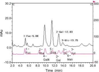
Fig. 3. Saccharides and amino saccharides constituents of purified glycosaminoglycan from sea hare muscle by Bio-LC (HPAEC-PAD system). The saccharides were eluted by 16 mM NaOH at a flow rate of 1 mL/min.
0 500 1000 1500 2000 2500 3000 3500 4000 4500 Wavelength number (cm-1)
0.5 0.4 0.3 0.2 0.1 0
A B
Fig. 2. FT-IR spectrum of heparan sulfate (A) and purified glycosaminoglycan (B) from sea hare muscle.
하고 있는 것으로 나타났고, heparinase I을 이용해 가수분해 후 측정한 결과에서는 이당류의 함량이 36.1±2.5 g/100 g으 로 36% 이상을 함유하고 있는 것으로 나타났다. 이 같은 결과는 GAG의 기본형태를 구성하는 이당류 단위가 전체 구성물 중 55% 이상인 것을 의미하는 것으로 정제물이 GAG로 이루어진 다당 복합체임을 제시한다.
정제한 GAG는 agarose 겔 전기영동 상으로 미루어 보아 단일물질이며, 표준물질과의 이동도를 비교할 때 주성분은 heparan sulfate인 것으로 추정하였고, 겔 크로마토그래피 상에서 분자량은 29.6 kDa으로 확인되었다(33).
FT-IR에 의한 기능기의 확인
Haparan 표준물의 FT-IR 스펙트럼은 3459.83, 1628.48, 1235.63, 1024.95, 994.43 cm-1에서 큰 peak를 보였으며, 1423.52, 1148.36, 819.50, 620.36 및 585.60 cm-1에서는 중간 크기의 peak를 나타낸 반면, 군소에서 정제한 GAG는 3455.91, 1648.03, 1083.52, 530.61 cm-1에서 큰 peak를, 984.04 cm-1에 서 중간 크기의 peak를 각각 나타내고 있었다(Fig. 2).
단백질 용액에서 1652 cm-1는 amide I, 1688과 1638 cm-1 는 대표적인 antiparallel β-sheet를 나타내며, 1644 cm-1는 무작위 구조를 반영한다(34). 1663~1630 cm-1의 영역은 물 의 HOH bending과 중복되는 amide I band의 특징이며, 1148 cm-1은 탄수화물, 1738 cm-1은 지질, 3100 cm-1(일차 아민)과 3300 cm-1(일차아민+이차아민)은 각각 amide-N 과 단백질-N의 지표로 사용될 수 있다. 그리고 850 cm-1부근 의 peak는 glucose, galactose, mannose의 C-O-C skeletal
을 반영한다(35). 본 실험 결과 정제한 GAG는 1648 cm-1에서 amide I의 특징적인 띠와 1457 cm-1에서 C-O stretch, 탄수 화물 및 아미노산의 특징, 866 cm-1에서 단당류의 특징을 보이는 것으로 나타났다.
정제 GAGs의 구성당의 분포
정제한 GAG는 fucose, N-acetylgalactosamine, N-acetyl- glucosamine, glucose, galactose, 미량의 mannose와 xylose 로 구성되어 있는 것으로 나타났고(Fig. 3), 이중에서 N- acetylgalactosamine, N-acetylglucosamine이 70% 이상을 차지하는 다당복합체임을 확인할 수 있었다. GAG는 glu- cose, galactose, N-acetyl-galactosamine, N-acetyl-glu- cosamine, mannose, xylose 등으로 연결되어 생물체내에서 proteoglycan의 형태로 존재하며(36), 구조를 해석하고 해당 분자들의 서열을 해석하기 위해 구성당 분석은 필수적인 항 목이다. 구성당 분석만으로 GAG의 구조적 해석은 불가능하 나 단당류, 산성 당, 아미노당의 존재 여부를 통해 GAG의 존재 여부를 판단할 수 있고, 이 결과를 토대로 구성 이당류 를 밝혀낼 수 있다면, 특정 GAG가 어떤 형태로 정제 대상 속에서 존재하고 있는지를 알아낼 수 있고, 이는 생물체내에 서 이루어지는 해당 GAG의 대사과정에 대한 이해로 이어져 그 기능성을 활용할 수 있는 방향을 제시해 줄 수 있게 된다 (37). 그러나 질산이나 TFA 등을 사용한 산 가수분해는 기 질 특이적이지 못하기 때문에 구성당의 서열을 분석할 수는 없고, 분석 시료의 단순한 구성 성분만을 확인할 수 있는데, 효소를 사용하게 되면 해당 시료의 당 서열을 알 수 있다(3).
이당류의 조성
정제된 GAG의 이당류를 분석한 결과(Fig. 4), heparinase I을 사용한 Fig. 4(A)는 heparin/heparan sulfate 이당류 kit 의 Δ4GlyUA→4GlcNS(6S)와 일치하는 35분대에서 peak가 용출되었으며, chondroitin sulfate ABC lyase를 사용한 Fig.
A
m/z, amu
m/z, amu
Fig. 5. MS spectra of chondroitin sulfate ABC lyase hydrolysate (A) and heparanase I hydrolysate (B) of purified glyco- saminoglycan from sea hare muscle.
30 32 34 36 38 40 42 44 46 48 50 Time (min)
25 20 15 10 5 0
Fig. 4. SAX-HPLC analysis of heparin/heparan sulfate dis- accharides produced by enzymatic depolymerization. The col- umn was eluted by deionized water with a linear gradient of 2 M NaCl at pH 3.5. The eluent was monitored at 232 nm. Capital letters indicate the purified glycosaminoglycan treated by hep- arinase I (A), by chondroitin sulfate ABC lyase (B); disaccharides kit: Δ4GlyUA→4GlcNS(6S) (C) and disaccharides kit: Δ4GlyUA (2s)→4GlcNS (D).
4(B)는 heparin/heparan sulfate disaccharide kit의 Δ4GlyUA (2s)→4GlcNS와 일치하는 36분대에서 peak가 용출되는 것 을 확인할 수 있었다. 이 같은 결과에 미루어 정제한 GAG는 heparan sulfate로 추정하였다. 그리고 2가지 형태의 heparan sulfate가 동정됨으로 인해 앞서 정제물을 효소 처리하여 전 기영동 한 결과(33)인 2개의 band와 일치하는 결과를 보였 다. 따라서 정제한 GAG가 heparan sulfate일 가능성을 제시 하였다.
질량분석기에 의한 당의 구조
Heparan ΔDi-OS, heparan ΔDi-NS, heparan ΔDi-6S, heparan ΔDi-di(U,N)S, heparan ΔDi-tri(U,6,N)S를 표준물 질로 사용하고, chondroitin sulfate ABC lyase와 he- parinase I를 이용하여 탈중합한 후 질량 분석한 결과(Fig.
5A), 정제한 GAG를 chondroitin sulfate ABC로 가수분해 질량 스펙트럼에서는 Thr(118.9), GlcA(176.90), GlcNAc
O H
-O
OH H
H
H
OSO3- H COO-
O
O H
OH H
H
H
NHSO3- H CH2OH
O-
GlyUA(2S)-GlcNS
O H
O
OH H
H
H
OH H COO-
O
O H
OH H
H
H
NHSO3- H CH2OSO3
O
GlyUA-GlcNS(6S)
Fig. 6. The supposed structure of disaccharide in purified glycosaminoglycan from sea hare muscle.
(220.9), glucuronic acid-2-N-acetyl-glucosamic acid(261.0), NeuAc(309.3), GlcNS(318.9)를 확인할 수 있었으며, hepa- ranase I로 가수분해한 질량 분석 스펙트럼(Fig. 5B)에서도 같은 질량의 물질을 확인할 수 있었다. 이 같은 결과로 미루 어 군소 GAG는 단백질 핵의 threonine 잔기에 O-연결된 GlyUA(2S)-GlcNS와 GlyUA-GlcNS(6S) 구조를 가지고 있음을 확인하였다(Fig. 6).
요 약
군소에서 추출한 다당류로부터 DEAE-Sepharose 상에 서 glycosaminoglycan(GAG)을 정제하여 기능기의 분포, 구성당의 분포, 이당류의 조성과 당 구조를 조사하였다. 정 제한 GAG는 기본 형태를 구성하는 이당류 단위가 전체 구성 물 중 55% 이상을 차지하고 있는 다당 복합체였다. 정제한 GAG는 1648 cm-1에서 amide I의 특징적인 띠와 1457 cm-1 에서 C-O stretch, 탄수화물 및 아미노산의 특징, 866 cm-1에 서 단당류의 특징을 보이는 것으로 나타났다. 정제한 GAG 는 fucose, N-acetylgalactosamine, N-acetylglucosamine, glucose, galactose, 미량의 mannose와 xylose로 구성되어 있는 것으로 나타났고, 이중에서 N-acetylgalactosamine, N-acetylglucosamine이 70% 이상을 차지하는 다당 복합체 인 heparan sulfate인 것으로 추정되었다. 군소 GAG는 단백 질핵의 threonine 잔기에 O-연결된 GlyUA(2S)-GlcNS와 GlyUA-GlcNS(6S) 구조를 가지고 있는 것으로 나타났다.
감사의 글
이 논문은 2008년도 정부(교육과학기술부)의 재원으로 한 국연구재단의 지원을 받아 수행된 기초연구사업임(No. KRF- 2008-313-F00089).
문 헌
1. Davies P, Roubin RH, Whitelock JM. 2008. Characterization and purification of glycosaminoglycans from crude bio- logical samples. J Agric Food Chem 56: 343-348.
2. Woods RJ. 1998. Computational carbohydrate chemistry:
what theoretical methods can tell us.Glycoconjugate J 15:
209-216.
3. Taylor ME, Drickamer K. 2006. Introduction to Glycobiol- ogy. 2nd ed. Oxford University Press, Oxford, UK. p 3-16.
4. Dinesh R, Garud VM, Mamoru K, Balagurunathan K. 2008.
Inhibition of heparan sulfate and chondroitin sulfate proteo- glycan biosynthesis. J Biol Chem 283: 28881-28887.
5. Mauro PAS, Dietrich CP. 1979. Chondroitin sulfates of the epiphysial cartila ages of different mammals. Comp Bio- chem Physiol Part B 62: 115-117.
6. Sampaio LO, Dietrich CP. 1981. Changes of sulfated muco- polysaccharides and mucopolysaccharides during fetal development. J Biol Chem 256: 9205-9210.
7. Sekino T, Murata K, Saito Y, Tsubura K. 1977. A study of acidic glycosaminoglycans in human gastric tissue.Dig- estion 16: 28-29.
8. Hata RI, Nagai Y. 1978. A low-sulfated chondroitin sulfate in human blood and urine.Biochem Biophys Acta543: 156- 9. Yonekura H, Oguri K, Nakazawa K, Shimizu S, Nakanish166.
Y, Okayma M. 1982. Isolation and partial characterization of sulfated glycoproteins synthesized by corneal epithelium.
J Biol Chem 257: 11166-11175.
10. Nadanaka S, Sugahara K. 1997. The unusual tetra- saccharide sequence GlcAβ1-3GalNAc(4-sulfate)β1-4GlcA (2-sulfate)β1-3GalNAc(6-sulfate) found in the hexasac- charides prepared by testicular hyaluronidase digestion of shark cartilage chondroitin sulfate D.Glycobiology7: 253- 11. Cassaro CMF, Dietrich CP. 1997. Distribution of sulfated263.
mucopolysaccharide in invertebrates.J Biol Sci252: 2254- 2261.
12. Kawai Y, Seno N, Anno K. 1966. Chondroitin polysulfate of squid cartilage. J Biochem 60: 317-321.
13. Moon JH, Ryu HS, You BJ, Moon SK. 1996. Physicochem- ical properties and dietary effect of glycoprotein from sea cucumber (Stichopus japonicus). J Korean Soc Food Sci Nutr 25: 240-248.
14. Ryu HS, Moon JH, Suh JS. 1997. Chemical compositions of glycoprotein and chondroitin sulfates from sea cucumber (Stichopus japonicus). J Korean Soc Food Sci Nutr 26:
72-80.
15. Moon JH, Ryu HS, Yang HS, Suh JS. 1998. Antimutagenic and anticancer effects of glycoprotein and chondroitin sul- fates from sea cucumber (Stichopus japonicus).J Korean Soc Food Sci Nutr 27: 350-358.
16. Vieira RP, Mulloy B, Mourao PAS. 1991. Structure of a fu- cose-branched chondroitin sulfate from sea cucumber. J Biol Chem 266: 13530-13536.
17. Humphries DE, Wong GW, Friend DS, Gurish MF. 1999.
Heparin is essential for the storage of specific granule pro- teases in mast cells. Nature 400: 769-772.
18. Tusar KG, Douglas MT. 2006. Placental dermatan sulfate:
isolation, anticoagulant activity, and association with hep- arin cofactor II. J Blood 107: 2753-2758.
19. Hovingh P, Piepkom M, Linker A. 1986. Biological im- plications of the structural, antithrombin affinity and anti- coagulant activity relationships among vertebrate heparins and heparin sulfates. J Biochem 237: 573-581.
20. Kwon OK, Min DK, Lee JR, Lee JS, Je JG, Choe BL. 2001.
Korean mollusks with color illustration. Hangul press, Busan, Korea. p 179
21. Yamada K, Ojika M, Ishigaki T, Yoshida Y. 1993. Aplyro- nine A, a potent antitumor substance, and the congeners aplyronines B and C isolated from the sea hare Aplysia kurodai. J Am Chem Soc115: 11020-11021.
22. Kigoshi H, Suenaga K, Mutou T, Ishigaki T, Atsumi T, Ishiwata H, Sakakura A, Ogawa T, Ojika M, Yamada K.
1996. Aplyronine A, a potent antitumor substance of marine origin, aplyronines B and C, and artificial analogues: Total synthesis and structure-cytotoxicity relationship. J Org Chem 61: 5326-5351.
23. Kuroda T, Suenaga K, Sakakura A, Handa T, Okamoto K, Kigoshi H. 2006. Study of the interaction between actin and antitumor substance aplyronine A with a novel fluorescent photoaffinity probe. Bioconjug Chem 17: 524-529.
24. Melo VMM, Duarte ABG, Carvalho AFFU, Siebra EA, Vasconcelos IM. 2000. Purification of a novel antibacterial and haemagglutinating protein from the purple gland of the sea hare, Aplysia dactylomela Rang, 1828. Toxicon 38:
1415-1427.
25. Vilim FS, Cropper EC, Rosen SC, Tenenbaum R, Kupfer- mann I, Weiss KR. 1994. Structure, localization, and action of buccalin B: A bioactive peptide from Aplysia. Peptides 15: 959-969.
26. Morishita F, Sasaki K, Kanemaru K, Nakanishi Y, Matsushima O, Furukawa Y. 2001. NdWFamide: A novel
excitatory peptide involved in cardiovascular regulation of Aplysia. Peptides 22: 183-189.
27. Hovingh P, Linker A. 1998. Glycosaminoglycans in two mollusks,Aplysia californicaandHelix aspersa, and in the leech,Nephelopsis obscura.Comp Biochem Physiol B119:
691-696.
28. AOAC. 1990.Official methods of analysis.15th ed. Associa- tion of Official Analytical Chemists, Washington, DC, USA.
p 868-931.
29. Folch J, Lees M, Sloane-Stanley GH. 1957. A simple meth- od for the isolation and purification of total lipids from ani- mal tissues. J Biol Chem 226: 497-509.
30. Mauro SGP, Rodolpho M, Cludio A, Silva AV. 1998. Highly sulfated dermatan sulfates from ascidians. J Biol Chem 264: 9972-9979.
31. Bitter T, Muir HM. 1962. A modified uronic acid carbazole reaction. Anal Biochem 4: 330-334.
32. Frederik MA, Roger B, Robert EK, David DM. 2000. Prep- aration and analysis of glycoconjugates (Chapter 17). In Current Protocols in Molecular Biology. John Wiley &
Sons, Inc., Hoboken, NJ, USA. p 17.9.
33. Yoon BY, Choi BD, Choi YJ. 2010. Extraction of glyco- saminoglycans from sea hare, Aplysia kurodai, and its functional properties 1. Optimum extraction of polysac- charide and purification of glycosaminoglycan. J Korean Soc Food Sci Nutr 39: 1640-1646.
34. Saguer E, Fort N, Alvarez PA, Sedman J, Ismail AA. 2008.
Structure-functionality relationships of porcine plasma proteins probed by FTIR spectroscopy and texture analysis.
Food Hydrocolloids 22: 459-467.
35. Thygesen LG, Lokke MM, Micklander E, Engelsen SB.
2003. Vibrational microspectroscopy of food. Raman vs.
FT-IR. Trends Food Sci Technol 14: 50-57.
36. Perez-Villar J, Hill RL. 1999. The structure and assembly of secreted mucins. J Biol Chem 274: 31751-31754.
37. Anderson RGV. 1998. The caveolae membrane system.
Annual Review Biochem 67: 199-225.
(2010년 9월 30일 접수; 2010년 11월 1일 채택)