• 검색 결과가 없습니다.

Current aspects and prospects of glass ionomer cements for clinical dentistry

N/A
N/A
Protected

Academic year: 2021

Share "Current aspects and prospects of glass ionomer cements for clinical dentistry"

Copied!
10
0
0

로드 중.... (전체 텍스트 보기)

전체 글

(1)

Introduction

Glass ionomer cement (GIC), an acid-base cement, is formed by the reaction of weak polymeric acids with inorganic glass powder [1]. GIC has multiple advantages: First, it adheres specifically to the teeth to prevent corrosion or leakage. Second, there is slow re- lease of fluoride ion over time to maintain dental health. Third, its color is very similar to that of human teeth [2,3]. Despite the ad- vantages of GIC, further improvement is required in terms of its mechanical characteristics. In order to improve the mechanical strength of GIC, the resin-modified glass ionomer (RMGI) was developed; it has an additional monomer compared to GIC and improved mechanical strength through photopolymerization and acid-base reaction [4,5]. RMGI obtained by resin curing has im- proved physical properties, but the amount of the released fluoride ion, which is important in preventing dental caries, is low [4].

Studies have reported on the manufacture of GIC using macro- monomer and viscosity dilution materials to exclude the effects of

Current aspects and prospects of glass ionomer cements for clinical dentistry

Eun Young Park

1

, Sohee Kang

2

1

Department of Dentistry, Yeungnam University College of Medicine, Daegu, Korea

2

Department of Dentistry, Yeungnam University Hospital, Daegu, Korea

Glass ionomer cement (GIC) is a tailor-made material that is used as a filling material in dentist- ry. GIC is cured by an acid-base reaction consisting of a glass filler and ionic polymers. When the glass filler and ionic polymers are mixed, ionic bonds of the material itself are formed. In addition, the extra polymer anion reacts with calcium in enamel or dentin to increase adhesion to the tooth tissue. GICs are widely used as adhesives for artificial crowns or orthodontic brackets, and are also used as tooth repair material, cavity liner, and filling materials. In this review, the current status of GIC research and development and its prospects for the future have been discussed in detail.

Keywords: Bioactive glass; Compomers; Glass ionomer cements; Hydroxyapatites; Resin-modified glass ionomer

Yeungnam Univ J Med 2020;37(3):169-178 https://doi.org/10.12701/yujm.2020.00374

Received: May 19, 2020 Revised: June 11, 2020 Accepted: June 15, 2020 Corresponding author:

Sohee Kang

Department of Dentistry, Yeungnam University Hospital,170 Hyeonchung-ro, Namgu, Daegu 42415, Korea

Tel: +82-53-620-3282 Fax: +82-53-629-1772 E-mail: [email protected]

water and the production of a material known as a compomer [6].

Clinically, GIC is applied close to the pulp. However, it is diffi- cult to use RMGI in deep underlined cavities. In dental clinics, ei- ther GIC or RMGI may be used, depending on the purpose.

There has been a recent focus on the study of “smart” materials that confer biocompatibility and cause remineralization, while maintaining the physical properties of materials [7]. Bioactive glass (BAG), composed of NaO, SiO, PO, and CaO, is known to be used for the loss of osseous tissue; therefore, a study was conduct- ed to increase the biocompatibility of GIC by adding BAG to GIC [3,8]. Studies have also reported an increase in biocompatibility with the addition of synthetic hydroxyapatite (HA) to the inorgan- ic components of GIC, since HA is highly analogous to the major components of tooth enamel or dentin in terms of structure [7].

In this review, we will describe the history of the development of GIC and determine the direction that GIC research should take in the future.

Copyright © 2020 Yeungnam University College of Medicine

This is an Open Access article distributed under the terms of the Creative Commons Attribution Non-Commercial License (http://creativecommons.org/licenses/by-nc/4.0/) which permits unrestricted non-commercial use, distribution, and reproduction in any medium, provided the original work is properly cited.

(2)

Glass ionomer cement

GIC is a combination of silicate and polycarboxylate that releases fluoride and attaches to dental tissue. It is used in a variety of appli- cations, including the filling material of dental cervical lesions; the restoration of children’s teeth; the core construction of tubular flu- id; and the adhesion of tooth fillings [9]. GIC was first introduced in 1972 by Wilson and Kent [10]. It consists of a water-soluble polyacrylic acid and fluoroaluminosilicate glass. When the silicate powder and polymeric liquid are mixed, an acid-base reaction takes place (Fig. 1). As metallic polymer salts begin to form, gela- tion begins, and continues until the cement hardens. Early GIC was considered an alternative to amalgam as tooth filling material.

However, the mechanical properties of early GIC were not as ad- vantageous as those of amalgam and required further improve- ment. Thus, the metal-reinforced GIC was first introduced in 1977. Williams et al. [11] described the addition of silver-amalgam alloy powder to GIC to increase the strength of the cement and provide radiopacity at the same time. However, both early GICs and metal-reinforced GICs had low viscosity, making them un- comfortable for clinical use. To overcome these issues, high viscosi- ty GICs called viscous or condensable GICs were developed [12,13]. These materials were used in atraumatic restorative treat- ment in the early 1990s [14]. The developed materials are com- posed of fine glass particles and high molecular weight anhydrous polyacrylic acids and possess a high powder/liquid mixing ratio, resulting in fast setting time and conferring high viscosity [13,14].

The setting reaction mechanism of high viscosity GICs is the same as that of conventional GICs based on the acid-base reaction.

GICs release biologically active ions, fluoride, sodium, phos- phate, and silicate that are biologically beneficial around the medi- um, therefore, these ions are naturally bioactive substances [15].

As more of these ions are released under acidic conditions when compared to neutral conditions, GIC can lower the pH of the sur- rounding medium under acidic conditions [15].

Resin-modified glass ionomer cement

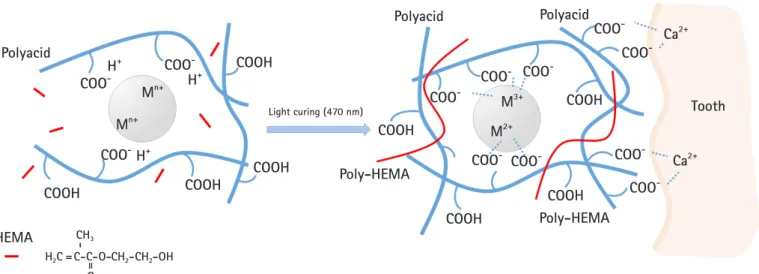
RMGI is composed of resin added to the GIC. Due to resin addi- tion, the binding strength, tensile strength, and compressive strength of the GICs are maintained and their solubility in aqueous environment is lowered, thereby improving the shortcomings of GIC [5,9]. Resin in RMGI is obtained by first putting the mono- mer into the liquid component of the GIC and then photo polym- erization. Ultraviolet irradiation results in monomer polymeriza- tion, followed by an acid-base reaction, which improves mechani- cal strength (Fig. 2). Owing to this improved mechanical strength,

RMGI is widely used as a dental filling material.

Earlier, Mathis and Ferracane [16] attempted to manufacture dental filling materials by mixing GIC and a composite prepared by mixing resin with commercial GIC. The resulting material did not exhibit clinically acceptable properties but it did demonstrate the possibility of combining acid-base and resin polymerization settings within a single material. RMGI, which is obtained by light curing, was developed in 1992 [5]. The basic acid-base reaction in these materials is mainly supplemented by the second resin created by light curing [5,17]. They are GICs containing a small number of monomers that can be polymerized in aqueous medium. An- other method has also been reported that alters the side chain of polyalkenoic acid, but the GIC is still prepared through mecha- nisms based on acid-base reactions [7]. The term ‘resin-modified glass ionomer’ means that resins are formed, however, they retain the characteristics of glass ionomers [4]. With regard to the materi- als in the wider context of material science, RMGIs are all ‘compos- ite materials’ as they consist of a matrix phase and a dispersed phase. The variation in the composition of commercial materials could then be considered to be continuous on a scale from purely resin-matrix produced by photo irradiation to purely salt-matrix produced by acid-base reaction [4]. One example of resin additives in RMGI is the addition of methacrylate to polyacrylic acid. In the preparation of these materials, the basic acid-base reaction is re- plenished by light curing. Another example of RMGI is polyac- id-modified composite resins composed of macro-monomers, which are commonly used in composite resins, containing bisphe- nol A-glycidyl dimethacrylate (bisGMA) or urethane dimethacry- late with a small amount of acidic monomer [18,19]. They use the same ion-releasing glass as do the filler particles used in conven-

Silica gel

Polyacid

Al

3+

Ca

2+

O=C

O=C O

-

O

-

Me

2+

/ Me

3+

Silicate basis

Fig. 1. Model of ionic bond formation with inorganic filler and

polyacid. When calcium fluoroaluminosilicate filler and polyacid

are mixed, the carboxyl ion of polyacid is ion-bonded with

aluminum and calcium ion in the silicate filler.

(3)

tional GIC, however, they are small in size. The initial setting reac- tion is initiated by light curing, followed by an acid-base reaction after water absorption [20].

The release of fluoride from tooth filling materials is very im- portant in terms of preventing tooth corrosion. Many researchers have reported that RMGIs can release fluoride at a rate similar to that of conventional GIC [3,20,21]. However, this release rate can be influenced not only by the formation of complex fluoride deriv- atives by reaction with polyacrylic acid, but also by the type and amount of the resin used for light polymerization [22-24]. De- pending on the storage environment, fluoride is released from RMGI for the first 24 hours [20,25-27], then the amount of releas- ing fluoride decreases after 7 days, and stabilizes at 10 days to 3 weeks [20,24,28,29]. Fluoride release is affected by variables such as matrix component, filler, and fluoride content [20,30-33]. In ad- dition, it is also affected by experimental factors such as storage en- vironment, number and frequency of preserving solution changes, composition and pH of saliva, plaque and pellicle formation, pow- der-to-liquid ratio, mixing, curing time, and exposed surface [20].

Fluoride release from RMGI in artificial saliva containing esterase was proved to be higher than in artificial saliva with no enzyme [20]. Bleaching and brushing did not affect fluoride release. Re- moval of the outer layer of the restoration by air polishing or finish- ing increased fluoride release. When the surface of the restorative material was covered with an adhesive or a surface coating agent, contamination due to moisture and dehydration was prevented in the initial stage, and fluoride release was reduced by 1.4 to 4 times [20]. Mousavinasab and Meyers [34] studied the amount of fluo-

ride released from four kinds of GIC (Fuji II LC, Fuji IX Extra, Fuji VII, and Fuji IX; GC Corporation, Tokyo, Japan), one compomer (Dyract Extra; Densply Detrey GmbH, Konstanz, Germany), and one giomer (Beautifil; Shofo Dental Corp., San Marcos, CA, USA). There was a significant difference in fluoride release depend- ing on the type of material and time; GIC released more fluoride than the compomer and giomer. Khoroushi and Keshani [3] and Mousavinasab and Meyers [34] emphasized the role played by the amount of GIC matrix used, in releasing fluoride ion of materials.

Compared with GIC, RMGI shows improved mechanical strength but decreased biocompatibility. This is because the 2-hy- droxyethyl methacrylate (HEMA) monomer escapes from RMGI mainly during the first 24 hours [2,35]. The amount of HEMA re- leased depends on the photometric intensity of the GIC [2,35].

HEMA penetrates the dentine [2,36] and is toxic to pulp cells [2,37]. As mentioned above, the mechanical properties have been improved at the same time the working time has been reduced, but its ability to prevent cavities is relatively low owing to the low re- lease of fluoride and its biocompatibility remains unsatisfactory be- cause of HEMA.

Polyacid-modified composite resins (compomer)

The mechanical properties of the GIC limit its applications be- cause it is composed of carboxylic acid groups that make the resin easily interact with water. Polyacid-modified composite resins, commonly known as compomers, are used for aesthetic materials Polyacid

Polyacid Polyacid

HEMA COOH

COOH

COOH COOH

COOH

COOH

COOH COOH

COO

-

COO

-

COO

-

COO

-

COO

-

COO

-

H

+

H

+

H

+

CH- =3

H2C =C-C-O- O

CH2-CH2-OH

M

n+

M

n+

M

3+

M

2+

COO

-

COO

-

COO

-

COO

-

COO

-

Ca

2+

Ca

2+

COO

-

Tooth

Poly-HEMA

Poly-HEMA

Light curing (470 nm)

Fig. 2. Model of interaction between resin-modified glass ionomer (RMGI) and dental tissue. When the filler, polyacid, and

2-hydroxyethyl methacrylate (HEMA) are mixed and irradiated, HEMA polymerizes and becomes poly-HEMA, acting as a bridge, followed

by acid-base reactions of polyacid and filler. Meanwhile, carboxyl residues in polyacid are strongly ionized with calcium present in tooth

tissue, allowing RMGI to adhere to teeth.

(4)

for oral rehabilitation, especially dental caries treatment [6,38].

This material was introduced to clinical dentists in the early 1990s [6,39] and was proposed as a new dental material that combines the existing synthetic resin aesthetics with the fluoride release and adhesion capabilities of GIC [6].

The main feature of compomers is that they do not contain wa- ter and most of the components are identical to those of composite resins. Typically, these are bulky macro-monomers, such as bis- GMA or its derivatives and/or urethane dimethacrylate, which are mixed with viscosity-reducing diluents, such as triethylene glycol dimethacrylate [6]. These polymer systems are filled with non-re- active inorganic powders, such as quartz or a silicate glass, such as SrAlFSiO

4

[6,40]. Powders are coated with a silane, which strengthens the bond between the filler and matrix of the set mate- rial [6,41]. The compomers also contain additional monomers that are different from those of conventional composites; therefore, they contain acidic functional groups as a very minor component.

The most widely used monomer of this type is TCB, which is a di-ester of 2-HEMA with butane tetracarboxylic acid [6,40]. In ad- dition, compomers also contain reactive glass powders similar to those used in GIC [6,38].

Compomers are designed to absorb water [6,41,42], and soak- ing in water can lead to a 2% to 3.5% increase in their mass [41]. It has been shown that this water absorption process involves neu- tralization of the carboxylic acid group. Neutralization is controlled by the rate of water diffusion and is therefore a rather slow process [42]. The mechanism through which compomers absorb water to promote neutralization is found to have a negative effect on their physical properties [43,44]. This mechanism is different from that of conventional composite resins, which are known to absorb moderate amounts of water without significant alterations to their mechanical properties [44]. Adusei et al. [45] conducted the most comprehensive study of the adverse effect of water on compomers.

For all tested materials, there was no difference in the measured pa- rameters after 24-hour storage in wet or dry conditions. However, for most materials, all strength measurements tended to decrease over a 4-week period. Not all physical parameters showed reduc- tions with long-term storage in water. In addition, it was found that microtensile strength and surface hardness appeared to remain un- affected [46,47].

The presence of minor amounts of both acid functional mono- mers and basic ionomer-type glass confers new properties to the material, namely, the ability to absorb moisture to trigger an ac- id-base reaction that can lead to the release of fluoride and creation of an acidic environment [6]. However, some studies have shown that water uptake reduces mechanical strength by up to 40% over several weeks; therefore, these clinically desirable features income

at a price [44]. Conversely, clinical studies have shown that these materials perform well in a variety of applications. The decrease in mechanical strength due to water uptake does not appear to be of clinical importance, and these materials are suitable for use in vivo [48,49].

A recent study on improvements in glass ionomer cement function

Several efforts have been made to enhance the properties of GIC while maintaining the bioactivity gained by releasing the ion. How- ever, it was necessary to develop a “smart” material that can over- come the adverse effects of the resin monomer and further induce remineralization on the defective dentin. Efforts have also recently been underway to improve physical properties and biocompatibili- ty by using both BAG and HA as fillers.

1. Glass ionomer cement containing bioactive glass In some recent studies [18,50-53], BAG has been used with GIC to improve bioactivity and induce tooth regeneration. The use of bioactive materials has attracted attention in dentistry, particularly for the purpose of dentin remineralization. The main inorganic component of the GIC comprises Si, Al, and Ca and is ionized with polyacid, so it does not exhibit decomposition performance [10]. Meanwhile, BAG contains specific weight percentages of Si, Na, Ca, and P and was introduced by Hench in 1969 as 45S5 Bio- glass with the following chemical composition and weight percent- ages: 45 wt% SiO

2

, 24.5 wt% CaO, 24.5 wt% Na

2

O, and 6.0 wt%

P

2

O

5

. BAGs are amorphous silicate-based materials which are compatible with the human body and can stimulate new bone growth while dissolving over time [54].

In clinical situations, BAG was first used as a biomaterial to re- place the loss of osseous tissues. BAG is able to bind strongly to bone via the formation of HA and firm bonding between the colla- gen and HA, and the body therefore tolerates the material well [3,54]. This material was initially used in the reconstruction of bone loss due to periodontal diseases in bony defects [3,54]. BAG has recently been used in the treatment of dentinal hypersensitivi- ty; fine BAG particles are incorporated into toothpaste or applied to tooth surfaces. BAG attaches to the dentin surface and quickly forms a hydroxycarbonapatite layer, which seals the tubules and re- lieves pain [3].

Some researchers have studied the physical and chemical prop- erties to evaluate the effect of BAG materials on tooth structure.

There are several studies on the effect of BAG addition on the

physical properties of RMGI [3,53,55,56]. Although the compres-

sive strength of the composition is reportedly slightly reduced, it is

(5)

much higher than that of the GIC containing BAG. Yli-Urpo et al.

[50] added BAG to GIC and evaluated its physical and biological properties. They reported that the experimental composition is bioactive under physiological conditions and is capable of mineral- izing human dentin in vitro [3,50].

Adding BAG particles to GIC decreases compressive strength and the modulus of elasticity [50,55,57]. This suggests that the BAG particles might be only loosely attached to the GIC matrix.

Thus, BAG particles probably acted as fillers that had not been ad- hered into the GIC matrix, leading to decreased compressive strength and modulus of elasticity [50]. Therefore, the develop- ment of bioactive GICs, that does not involve a deterioration in mechanical properties, seems to be needed. Main research has been specifically focused on the application of nanoparticles to dental materials, including GICs, to improve the mechanical prop- erties of the matrix and strengthen communication with cells de- rived from dental tissue to facilitate regeneration [57-61]. Several nanomaterials such as hydroxyl- (or fluoro-) apatite, titanium ox- ide, zirconia, and resin and combinations thereof have been incor- porated into the existing GIC. One of the nanoparticles indicated for use in GIC is a BAG nanoparticle [7,62,63]. The BAG nanoparticle, combined with the matrix of GIC, increases surface area and biological activity and greatly improves mechanical/bio- logical properties as an additive per particle weight over that of conventional micro-sized BAG particles [64,65].

2. Glass ionomer cement containing hydroxyapatite HA has been beneficial in the field of dentistry due to its unique ra-

diopacity and other properties [66-68]. The application of current nano-sized biomaterials is known to be potentially more useful in dentistry. They have wide applications because of greater strength, polishability, and aesthetic value than commercial modifiers [69,70]. Recent advances in the synthesis of HA [71] in various sizes and forms have enabled HA to be used as a biocompatible fill- er for natural tooth materials. In addition, HA showed excellent bi- ological activity and played an important role in orthopedics be- cause of its bone-inducing and bioactive properties [66,72].

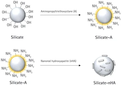
Nanotechnology involves the use or modification of 1 to 100- nm materials [7,73-75]. Major applications of nanotechnology in dentistry include surface modification of implants [76], enhanced polymer composites with nano-sized particles [74], and caries pre- vention [77]. Recent research shows that the addition of nanopar- ticles or nanoclusters increases the mechanical strength of tooth fillers such as resin composites [78-80]. Similar attempts have been made to improve the mechanical properties of the GIC using nanotechnology [67,81]. Introduction of nano-sized apatite not only maintains the mechanical properties of the GIC at all times, but also increases the release of fluoride ions [33,67]. Studies have also reported that GIC containing nano-sized apatite has better biocompatibility than conventional GIC [82,83]. Haider et al.

[83] reported that there are differences in biological properties de- pending on the shape of the nanoparticles incorporated into the nanofiber scaffold. In their experiment, nanorod HA showed a bet- ter biocompatibility than spherical HA. In the HA effect study on GIC, nanorod HA-fixed silicate showed better cellular compatibili- ty than the non-fixed silicate (Fig. 3).

OH OH

OH OH

Silicate

Silicate-A

Silicate-A

Silicate-nHA

OHOH

OH OH

NH2

NH2

NH2

NH2

NH2NH2 NH2

NH2

NH2

NH2

NH2

NH2

NH2

NH2

NH2

NH2

NH2

NH2 NH2

NH2

NH2

NH2

NH2

NH2

OHOH

Aminopropyltriethoxysilane (A)

Nanorod hydroxyapatite (nHA)

OH OH

Fig. 3. A schematic diagram that binds the nanorod hydroxyapatite (nHA) to the silicate surface. Aminopropyltriethoxysilane (A) is a

coupling agent used to conjugate amino groups to the glass surface. The amino acids introduced on the surface of the silicate can react

with nHA fixed carboxylic acid to produce silicate-nHA.

(6)

Apatite crystals increase the crystallinity of cured matrix, further stabilizing the hardening cement and improving the bond strength with the tooth structure [74,84,85]. Increasing fluoride release can reduce secondary caries around the restoration site [73,86]. How- ever, the possibility of interfacial failure of glass and bioceramic can be a problem that can affect the physical properties of the cured ce- ment [87]. The crystals of nano-HA preferentially remineralize enamel [7,88,89]. Recent reports suggest that the nano-HA-modi- fied resin composite has improved mechanical properties over the unmodified resin composite [7,90,91]. Similarly, adding nano-HA or nano-fluoroapatite to the powder content of GIC had a positive effect on compressive, tensile, and flexural strength of the cured ce- ment [67]. Fourier-transform infrared spectroscopy showed that adding apatite to GIC powder has been found to increase the crys- tallinity of cured GICs, which in turn improves chemical stability and water insolubility [67,92]. This results in a better survival rate than that observed with commercialized GICs [67].

The improved mechanical properties of GIC modified by HA are due to ionic bonds of polyacrylic acid and HA crystals [92]. As a strong ionic bond is formed between the calcium ion of the tooth structure and the crystal of the apatite of the cement, the GIC con- taining nano-HA is expected to strongly bond to the surface of teeth (Fig. 2) [33]. In addition, reducing the particle size of HA from a micrometer scale to a nanometer scale significantly increas- es the surface area, and improves infiltration into dentin and enam- el pores where crystals have been demineralized; this can improve bonding at the tooth-ionomer interface [93].

HA infiltered GIC, called glass carbomer, includes substances that are established by the acid-base reaction between the aqueous polymer acid and the ion leaching base glass, but they also include substances not commonly included in glass ionomer formulations [94]. As such, the bioactive component acts as a secondary filler.

According to solid state nuclear magnetic resonance spectroscopy, this filler is actually HA [95] and is included to promote the forma- tion of enamel-like substances in contact with the tooth, as previ- ously studied with GIC used as fissure sealants.

Since glass carbomers contain a higher proportion of glass than that in conventional GIC, as well as HA fillers, the set glass car- bomers are brittle. Silicone oil is added to overcome this problem [96]. It strengthens the material and remains bound by hydrogen bonding. The setting of glass carbomer involves two parallel reac- tions, one involving the glass plus polyacid and the other involving HA plus polyacid. Both are acid-base reactions, resulting in an ion- ic crosslinking polyacid matrix containing embedded filler. Howev- er, the filler is not only ion-depleted glass, but in this case also con- tains a partially reactive HA. Thus, the matrix is similar to that ob- tained using conventional GIC, except that it contains polydimeth-

ylsiloxane oil [97].

There are only preliminary studies on the clinical use of glass carbomer thus far; however, no long-term studies have been con- ducted for this material. Consequently, the durability of this mate- rial in the oral cavity of patients is not yet known.

Conclusion

Since the last decade, interest in the use of “smart” bioactive mate- rials has been growing in dentistry, especially with the aim of rem- ineralization of dentin. More predictable treatment results can be obtained with RMGI’s superior handling characteristics, com- bined quality during final overlay restoration, and possibility of im- mediate restoration placement. Therefore, future studies should focus on these materials, especially on their cytotoxicity, quality of induced dentin bridges, and protocols for higher bonding strength during final restoration.

Currently, nanotechnology is used to develop nanoscale glass filler to enhance biocompatibility. Furthermore, various studies are being conducted to develop a material that brings high biocompat- ibility and mineral inducing potential by adding biocompatible na- no-sized HA to RMGI. Irrespective of the clinical suitability of the material, clinicians will probably not select materials that are diffi- cult to handle. Thus, a more biocompatible material based on RMGI need to be developed for extensive clinical use in future.

Acknowledgments

Conflicts of interest

No potential conflict of interest relevant to this article was report- ed.

Author contributions

Conceptualization, Formal analysis, Resources, Supervision, and Validation: SK; Data curation: EYP; Writing-original draft: EYP, SK; Writing-review & editing: EYP, SK.

ORCID

Eun Young Park, https://orcid.org/0000-0002-1860-5425 Sohee Kang, https://orcid.org/0000-0002-3667-1952

References

1. Mount GJ. An atlas of glass-ionomer cements: a clinician’s guide. 2nd ed. Martin Dunitz: London; 2002.

2. Sidhu SK, Nicholson JW. A review of glass-ionomer cements

for clinical dentistry. J Funct Biomater 2016;7:16.

(7)

3. Khoroushi M, Keshani F. A review of glass-ionomers: from con- ventional glass-ionomer to bioactive glass-ionomer. Dent Res J (Isfahan) 2013;10:411–20.

4. Sidhu SK, Watson TF. Resin-modified glass ionomer materials:

a status report for the American Journal of Dentistry. Am J Dent 1995;8:59–67.

5. Wilson AD. Resin-modified glass-ionomer cements. Int J Prost- hodont 1990;3:425–9.

6. Nicholson JW. Polyacid-modified composite resins (“com- pomers”) and their use in clinical dentistry. Dent Mater 2007;

23:615–22.

7. Najeeb S, Khurshid Z, Zafar MS, Khan AS, Zohaib S, Marti JM, et al. Modifications in glass ionomer cements: nano-sized fillers and bioactive nanoceramics. Int J Mol Sci 2016;17:1134.

8. De Caluwe T, Vercruysse CW, Ladik I, Convents R, Declercq H, Martens LC, et al. Addition of bioactive glass to glass iono- mer cements: effect on the physico-chemical properties and biocompatibility. Dent Mater 2017;33:e186–203.

9. Berg JH, Croll TP. Glass ionomer restorative cement systems:

an update. Pediatr Dent 2015;37:116–24.

10. Wilson AD, Kent BE. A new translucent cement for dentistry:

the glass ionomer cement. Br Dent J 1972;132:133–5.

11. Williams JA, Billington RW, Pearson GJ. The comparative strengths of commercial glass-ionomer cements with and with- out metal additions. Br Dent J 1992;172:279–82.

12. Cho SY, Cheng AC. A review of glass ionomer restorations in the primary dentition. J Can Dent Assoc 1999;65:491–5.

13. Frankenberger R, Sindel J, Kramer N. Viscous glass-ionomer ce- ments: a new alternative to amalgam in the primary dentition?

Quintessence Int 1997;28:667–76.

14. Berg JH. The continuum of restorative materials in pediatric dentistry: a review for the clinician. Pediatr Dent 1998;20:93–

100.

15. Nicholson JW, Czarnecka B, Limanowska-Shaw H. The long- term interaction of dental cements with lactic acid solutions. J Mater Sci Mater Med 1999;10:449–52.

16. Mathis RS, Ferracane JL. Properties of a glass-ionomer/res- in-composite hybrid material. Dent Mater 1989;5:355–8.

17. Burgess J, Norling B, Summitt J. Resin ionomer restorative mate- rials: the new generation. J Esthet Dent 1994;6:207–15.

18. Xie D, Brantley WA, Culbertson BM, Wang G. Mechanical properties and microstructures of glass-ionomer cements. Dent Mater 2000;16:129–38.

19. Nagaraja UP, Kishore G. Glass ionomer cement: The different generations. Trends Biomater Artif Organs 2005;18:158–65.

20. Wiegand A, Buchalla W, Attin T. Review on fluoride-releasing restorative materials: fluoride release and uptake characteristics,

antibacterial activity and influence on caries formation. Dent Mater 2007;23:343–62.

21. Robertello FJ, Coffey JP, Lynde TA, King P. Fluoride release of glass ionomer-based luting cements in vitro. J Prosthet Dent 1999;82:172–6.

22. Tjandrawinata R, Irie M, Suzuki K. Marginal gap formation and fluoride release of resin-modified glass-ionomer cement: effect of silanized spherical silica filler addition. Dent Mater J 2004;23:305–13.

23. Musa A, Pearson GJ, Gelbier M. In vitro investigation of fluo- ride ion release from four resin-modified glass polyalkenoate ce- ments. Biomaterials 1996;17:1019–23.

24. Momoi Y, McCabe JF. Fluoride release from light-activated glass ionomer restorative cements. Dent Mater 1993;9:151–4.

25. Attar N, Turgut MD. Fluoride release and uptake capacities of fluoride-releasing restorative materials. Oper Dent 2003;28:

395–402.

26. Karantakis P, Helvatjoglou-Antoniades M, Theodoridou-Pahini S, Papadogiannis Y. Fluoride release from three glass ionomers, a compomer, and a composite resin in water, artificial saliva, and lactic acid. Oper Dent 2000;25:20–5.

27. Hayacibara MF, Ambrozano GM, Cury JA. Simultaneous re- lease of fluoride and aluminum from dental materials in various immersion media. Oper Dent 2004;29:16–22.

28. Yap AU, Tham SY, Zhu LY, Lee HK. Short-term fluoride release from various aesthetic restorative materials. Oper Dent 2002;

27:259–65.

29. Gao W, Smales RJ, Gale MS. Fluoride release/uptake from new- er glass-ionomer cements used with the ART approach. Am J Dent 2000;13:201–4.

30. Yli-Urpo H, Vallittu PK, Narhi TO, Forsback AP, Vakiparta M.

Release of silica, calcium, phosphorus, and fluoride from glass ionomer cement containing bioactive glass. J Biomater Appl 2004;19:5–20.

31. Osinaga PW, Grande RH, Ballester RY, Simionato MR, Delga- do Rodrigues CR, Muench A. Zinc sulfate addition to glass-ionomer-based cements: influence on physical and anti- bacterial properties, zinc and fluoride release. Dent Mater 2003;19:212–7.

32. Mazzaoui SA, Burrow MF, Tyas MJ, Dashper SG, Eakins D, Reynolds EC. Incorporation of casein phosphopeptide-amor- phous calcium phosphate into a glass-ionomer cement. J Dent Res 2003;82:914–8.

33. Lucas ME, Arita K, Nishino M. Toughness, bonding and fluo- ride-release properties of hydroxyapatite-added glass ionomer cement. Biomaterials 2003;24:3787–94.

34. Mousavinasab SM, Meyers I. Fluoride release by glass ionomer

(8)

cements, compomer and giomer. Dent Res J (Isfahan) 2009;

6:75–81.

35. Palmer G, Anstice HM, Pearson GJ. The effect of curing regime on the release of hydroxyethyl methacrylate (HEMA) from res- in-modified glass-ionomer cements. J Dent 1999;27:303–11.

36. Hamid A, Hume WR. Diffusion of resin monomers through human carious dentin in vitro. Endod Dent Traumatol 1997;

13:1–5.

37. Kan KC, Messer LB, Messer HH. Variability in cytotoxicity and fluoride release of resin-modified glass-ionomer cements. J Dent Res 1997;76:1502–7.

38. McLean JW, Nicholson JW, Wilson AD. Proposed nomencla- ture for glass-ionomer dental cements and related materials.

Quintessence Int 1994;25:587–9.

39. Meyer JM, Cattani-Lorente MA, Dupuis V. Compomers: be- tween glass-ionomer cements and composites. Biomaterials 1998;19:529–39.

40. Eliades G, Kakaboura A, Palaghias G. Acid-base reaction and fluoride release profiles in visible light-cured polyacid-modified composite restoratives (compomers). Dent Mater 1998;14:57–

63.

41. Ruse ND. What is a “compomer”? J Can Dent Assoc 1999;65:

500–4.

42. Young AM, Rafeeka SA, Howlett JA. FTIR investigation of monomer polymerisation and polyacid neutralisation kinetics and mechanisms in various aesthetic dental restorative materi- als. Biomaterials 2004;25:823–33.

43. Nicholson JW, Alsarheed M. Changes on storage of polyac- id-modified composite resins. J Oral Rehabil 1998;25:616–20.

44. Dahl JE, Li J, Ruyter IE. Long-term water uptake of compomers and its effect on mechanical properties. J Dent Res 1998;77(2 Suppl):657 (abstract 207).

45. Adusei GO, Deb S, Nicholson JW. A preliminary study of exper- imental polyacid-modified composite resins (‘compomers’) containing vinyl phosphonic acid. Dent Mater 2005;21:491-7.

46. Mendonca JS, Souza MH Jr, Carvalho RM. Effect of storage time on microtensile strength of polyacid-modified resin com- posites. Dent Mater 2003;19:308–12.

47. Bayindir YZ, Yildiz M. Surface hardness properties of res- in-modified glass ionomer cements and polyacid-modified composite resins. J Contemp Dent Pract 2004;5:42–9.

48. Loguercio AD, Reis A, Barbosa AN, Roulet JF. Five-year dou- ble-blind randomized clinical evaluation of a resin-modified glass ionomer and a polyacid-modified resin in noncarious cer- vical lesions. J Adhes Dent 2003;5:323–32.

49. Ermis RB. Two-year clinical evaluation of four polyacid-modi- fied resin composites and a resin-modified glass-ionomer ce-

ment in Class V lesions. Quintessence Int 2002;33:542–8.

50. Yli-Urpo H, Lassila LV, Narhi T, Vallittu PK. Compressive strength and surface characterization of glass ionomer cements modified by particles of bioactive glass. Dent Mater 2005;

21:201–9.

51. Yli-Urpo H, Narhi M, Narhi T. Compound changes and tooth mineralization effects of glass ionomer cements containing bio- active glass (S53P4), an in vivo study. Biomaterials 2005;

26:5934–41.

52. Xie D, Zhao J, Weng Y, Park JG, Jiang H, Platt JA. Bioactive glass-ionomer cement with potential therapeutic function to dentin capping mineralization. Eur J Oral Sci 2008;116:479–

87.

53. Ana ID, Matsuya S, Ohta M, Ishikawa K. Effects of added bioac- tive glass on the setting and mechanical properties of res- in-modified glass ionomer cement. Biomaterials 2003;24:

3061–7.

54. Hench LL. The story of Bioglass. J Mater Sci Mater Med 2006;17:967–78.

55. Mousavinasab SM, Khoroushi M, Keshani F, Hashemi S. Flex- ural strength and morphological characteristics of resin-modi- fied glass-ionomer containing bioactive glass. J Contemp Dent Pract 2011;12:41–6.

56. Khoroushi M, Mousavinasab SM, Keshani F, Hashemi S. Effect of resin-modified glass ionomer containing bioactive glass on the flexural strength and morphology of demineralized dentin.

Oper Dent 2013;38:E1–10.

57. Kim DA, Lee JH, Jun SK, Kim HW, Eltohamy M, Lee HH. Sol- gel-derived bioactive glass nanoparticle-incorporated glass ion- omer cement with or without chitosan for enhanced mechani- cal and biomineralization properties. Dent Mater 2017;33:805–

17.

58. Lee JH, Kang MS, Mahapatra C, Kim HW. Effect of aminated mesoporous bioactive glass nanoparticles on the differentiation of dental pulp stem cells. PLoS One 2016;11:e0150727.

59. Lee JH, El-Fiqi A, Jo JK, Kim DA, Kim SC, Jun SK, et al. Devel- opment of long-term antimicrobial poly(methyl methacrylate) by incorporating mesoporous silica nanocarriers. Dent Mater 2016;32:1564–74.

60. Padovani GC, Feitosa VP, Sauro S, Tay FR, Duran G, Paula AJ, et al. Advances in dental materials through nanotechnology:

facts, perspectives and toxicological aspects. Trends Biotechnol 2015;33:621–36.

61. Oliveira-Ogliari A, Collares FM, Feitosa VP, Sauro S, Ogliari FA, Moraes RR. Methacrylate bonding to zirconia by in situ silica nanoparticle surface deposition. Dent Mater 2015;31:68–76.

62. Mabrouk M, Selim MM, Beherei H, El-Gohary MI. Effect of in-

(9)

corporation of nano bioactive silica into commercial glassiono- mer cement (GIC). J Genet Eng Biotechnol 2012;10:113–9.

63. Choi JY, Lee HH, Kim HW. Bioactive sol-gel glass added iono- mer cement for the regeneration of tooth structure. J Mater Sci Mater Med 2008;19:3287–94.

64. Saravana KR, Vijayalakshmi R. Nanotechnology in dentistry.

Indian J Dent Res 2006;17:62–5.

65. Lee JH, Seo SJ, Kim HW. Bioactive glass-based nanocomposites for personalized dental tissue regeneration. Dent Mater J 2016;

35:710–20.

66. Park SJ, Gupta KC, Kim H, Kim S, Kang IK. Osteoblast be- haviours on nanorod hydroxyapatite-grafted glass surfaces. Bio- mater Res 2019;23:28.

67. Moshaverinia A, Ansari S, Moshaverinia M, Roohpour N, Darr JA, Rehman I. Effects of incorporation of hydroxyapatite and fluoroapatite nanobioceramics into conventional glass ionomer cements (GIC). Acta Biomater 2008;4:432–40.

68. Arita K, Yamamoto A, Shinonaga Y, Harada K, Abe Y, Nakaga- wa K, et al. Hydroxyapatite particle characteristics influence the enhancement of the mechanical and chemical properties of conventional restorative glass ionomer cement. Dent Mater J 2011;30:672–83.

69. Mitra SB, Wu D, Holmes BN. An application of nanotechnolo- gy in advanced dental materials. J Am Dent Assoc 2003;134:

1382–90.

70. Saunders SA. Current practicality of nanotechnology in dentist- ry. Part 1: Focus on nanocomposite restoratives and biomimet- ics. Clin Cosmet Investig Dent 2009;1:47–61.

71. Dorozhkin SV. Nanosized and nanocrystalline calcium ortho- phosphates. Acta Biomater 2010;6:715–34.

72. Ramesh N, Moratti SC, Dias GJ. Hydroxyapatite-polymer bio- composites for bone regeneration: a review of current trends. J Biomed Mater Res B Appl Biomater 2018;106:2046–57.

73. Hannig M, Hannig C. Nanomaterials in preventive dentistry.

Nat Nanotechnol 2010;5:565–9.

74. Najeeb S, Khurshid Z, Matinlinna JP, Siddiqui F, Nassani MZ, Baroudi K. Nanomodified peek dental implants: bioactive com- posites and surface modification-a review. Int J Dent 2015;

2015:381759.

75. Khurshid Z, Zafar M, Qasim S, Shahab S, Naseem M, AbuReqa- iba A. Advances in nanotechnology for restorative dentistry.

Materials (Basel) 2015;8:717–31.

76. Le Guehennec L, Soueidan A, Layrolle P, Amouriq Y. Surface treatments of titanium dental implants for rapid osseointegra- tion. Dent Mater 2007;23:844–54.

77. Hannig M, Hannig C. Nanotechnology and its role in caries therapy. Adv Dent Res 2012;24:53–7.

78. Curtis AR, Palin WM, Fleming GJ, Shortall AC, Marquis PM.

The mechanical properties of nanofilled resin-based compos- ites: the impact of dry and wet cyclic pre-loading on bi-axial flexure strength. Dent Mater 2009;25:188–97.

79. Terry DA. Direct applications of a nanocomposite resin system:

part 1-the evolution of contemporary composite materials.

Pract Proced Aesthet Dent 2004;16:417–22.

80. Chen MH. Update on dental nanocomposites. J Dent Res 2010;89:549–60.

81. Moshaverinia A, Roohpour N, Chee WWL, Schricker SR. A re- view of powder modifications in conventional glass-ionomer dental cements. J Mater Chem 2011;21:1319–28.

82. Kang IK, Park SJ, Kang SH, inventors; Kang SH, assignee. Glass based filler for dental restoration, method for manufacturing thereof, and dental restoration comprising thereof. Korea KR patent, 10-2020-0021006. 2020 Feb 20.

83. Haider A, Gupta KC, Kang IK. Morphological effects of HA on the cell compatibility of electrospun HA/PLGA composite nanofiber scaffolds. Biomed Res Int 2014;2014:308306.

84. Xia Y, Zhang F, Xie H, Gu N. Nanoparticle-reinforced res- in-based dental composites. J Dent 2008;36:450–5.

85. De Caluwe T, Vercruysse CW, Fraeyman S, Verbeeck RM. The influence of particle size and fluorine content of aluminosilicate glass on the glass ionomer cement properties. Dent Mater 2014;30:1029–38.

86. Ong JL, Chan DCN. A review of hydroxapatite and its use as a coating in dental implants. Crit Rev Biomed Eng 2017;45:411–

51.

87. Gu YW, Yap AUJ, Cheang P, Khor KA. Zirconia-glass ionomer cement–A potential substitute for miracle mix. Scr Mater 2005;

52:113–6.

88. Huang SB, Gao SS, Yu HY. Effect of nano-hydroxyapatite con- centration on remineralization of initial enamel lesion in vitro.

Biomed Mater 2009;4:034104.

89. Huang S, Gao S, Cheng L, Yu H. Remineralization potential of nano-hydroxyapatite on initial enamel lesions: an in vitro study.

Caries Res 2011;45:460–8.

90. Zakir M, Al Kheraif AA, Asif M, Wong FS, Rehman IU. A com- parison of the mechanical properties of a modified silorane based dental composite with those of commercially available composite material. Dent Mater 2013;29:e53–9.

91. Yap AU, Pek YS, Kumar RA, Cheang P, Khor KA. Experimental studies on a new bioactive material: HAIonomer cements. Bio- materials 2002;23:955–62.

92. Moshaverinia A, Ansari S, Movasaghi Z, Billington RW, Darr JA,

Rehman IU. Modification of conventional glass-ionomer ce-

ments with N-vinylpyrrolidone containing polyacids, nano-hy-

(10)

droxy and fluoroapatite to improve mechanical properties. Dent Mater 2008;24:1381–90.

93. Lee JJ, Lee YK, Choi BJ, Lee JH, Choi HJ, Son HK, et al. Physi- cal properties of resin-reinforced glass ionomer cement modi- fied with micro and nano-hydroxyapatite. J Nanosci Nanotech- nol 2010;10:5270–6.

94. Cehreli SB, Tirali RE, Yalcinkaya Z, Cehreli ZC. Microleakage of newly developed glass carbomer cement in primary teeth. Eur J Dent 2013;7:15–21.

95. Zainuddin N, Karpukhina N, Law RV, Hill RG. Characterisation

of a remineralising Glass Carbomer® ionomer cement by MAS- NMR spectroscopy. Dent Mater 2012;28:1051–8.

96. Hasan AMHR, Sidhu SK, Nicholson JW. Fluoride release and uptake in enhanced bioactivity glass ionomer cement (“glass carbomer™”) compared with conventional and resin-modified glass ionomer cements. J Appl Oral Sci 2019;27:e20180230.

97. Van Den Bosch W, Van Duinen RN, inventors; STICHTING GLASS FOR HEALTH, assignee. Self hardening glass carbom- er composition. United States patent US 20060217455 A1.

2006 Sep 28.

수치

Fig. 1.  Model of ionic bond formation with inorganic filler and  polyacid. When calcium fluoroaluminosilicate filler and polyacid  are mixed, the carboxyl ion of polyacid is ion-bonded with  aluminum and calcium ion in the silicate filler.
Fig. 2. Model of interaction between resin-modified glass ionomer (RMGI) and dental tissue
Fig. 3. A schematic diagram that binds the nanorod hydroxyapatite (nHA) to the silicate surface

참조

관련 문서