• 검색 결과가 없습니다.

Intentional partial odontectomy-a long-term follow-up study

N/A
N/A
Protected

Academic year: 2021

Share "Intentional partial odontectomy-a long-term follow-up study"

Copied!
5
0
0

로드 중.... (전체 텍스트 보기)

전체 글

(1)

R E S E A R C H Open Access

Intentional partial odontectomy —a long- term follow-up study

Hyun-Suk Kim 1 , Pil-Young Yun 1 and Young-Kyun Kim 1,2*

Abstract

Background: The surgical extraction of the third molar is the most frequently encountered procedure in oral and maxillofacial surgery and is related with a variety of complications. This study examined the efficacy of intentional partial odontectomy (IPO) in the third molars which have no periapical lesions and are located near important anatomical structures such as inferior alveolar nerve.

Methods: Seven patients (four males, three females, 39.1 ± 11.6 years), who received IPO to reduce the risk of inferior alveolar nerve injury (IANI), were followed long-term. The treated teeth were horizontally impacted third molars in the mandibular left ( n = 5) or mandibular right (n = 4) areas and were all ankylosed with the surrounding alveolar bone. During the IPO, the bone around the crown was removed to expose the crown, and then the tooth was resected at cement-enamel junction (CEJ). Any secondary trauma to the healthy root was minimized and remained intact after primary suture.

Results: The mean follow-up time was 63.2 ± 29.8 months, and all sites showed good bone healing after the crown removal. Also, sensory abnormality was not found in any patients after IPO. In one patient, the bone fragments erupted 4 months after IPO. In other patient, an implant placed on second molar site adjacent to the third molar that received IPO was explanted about 2 years after the patient ’s persistent discomfort.

Conclusions: In case where high risk of IANI exists, IPO may be chosen alternatively to surgical extraction to reduce the risk of nerve damage.

Keywords: Hypercementosis, Mandibular nerve, Tooth ankyloses, Tooth extraction

Background

The impacted mandibular third molar which could re- sult in pain or discomfort due to pericoronitis or dental caries needs to be extracted. Extracting the impacted mandibular third molar, however, is often challenging to dental surgeons because of related postoperative compli- cations. The complications are highly related with the depth and position of impaction (i.e., mesio-angular, horizontal, vertical, and disto-angular), and the proxim- ity to important anatomical structures such as the infer- ior alveolar nerve (IAN) canal [1]. Inferior alveolar nerve injury (IANI) is the most common complication during the extraction surgery [2].

In surgical extractions, the incidence of IAN damage varies from temporary paresthesia up to 8.1% and per- manent discomfort up to 3.6% [3]. In cases of patients with a high risk of IANI, intentional partial odontecomy (IPO) could be considered as an alternative treatment option to complete extraction. IPO, which was first suggested by Knutsson et al. in 1989, is a procedure designed to reduce the risk of IANI by removal only the crown portion of the tooth, leaving the root in situ [4].

Pericoronitis, one of the reasons for extracting the man- dibular third molar, is related with the persistence of the dental follicle that could pose as a source of infection. In such case, the removal of the crown portion with the fol- licles could subside inflammatory reaction occurred sur- rounding the tooth [3].

Despite the advantages that IPO could provide on an impacted tooth that is in a high risk of IANI, clinicians are reluctant to perform the procedure because residual

* Correspondence: [email protected]

1

Department of Oral and Maxillofacial Surgery, Section of Dentistry, Seoul National University Bundang Hospital, 300 Gumi-dong, Bundang-gu, Seongnam City, Gyunggi-do, Korea

2
Department of Dentistry and Dental Research Institute, School of Dentistry, Seoul National University, Daehak-ro 101, Jongno-gu, Seoul 03080, Korea

© The Author(s). 2017 Open Access This article is distributed under the terms of the Creative Commons Attribution 4.0

International License (http://creativecommons.org/licenses/by/4.0/), which permits unrestricted use, distribution, and

reproduction in any medium, provided you give appropriate credit to the original author(s) and the source, provide a link to

the Creative Commons license, and indicate if changes were made.

(2)

roots could become a source of postoperative infection in the future. Therefore, IPO has been a controversial operation since it lacks a long-term follow-up of retained roots.

This retrospective study was designed to examine the fate and complications of residual roots through long- term follow-ups and to inform whether the IPO could be suggested as an alternative treatment for the im- pacted third molars.

Methods Patients

In this study, seven patients (four males and three fe- males; age 39.1 ± 11.6 years), who underwent IPO be- cause of a high risk of intra- and postoperative complications, were followed long term. A total of nine mandibular third molars underwent IPO, five in the left and four in the right mandibles. The relation of the tooth to the ramus of the mandible, the rela- tive depth of the third molar in the bone, and the position of the tooth in relation to the long axis of the second molar were radiographically evaluated fol- lowing Pell & Gregory classification [5]. Based on this classification, the difficulty of extraction was evaluated using difficulty index described by Pederson [6].

Moreover, the preoperative status of the impacted tooth, such as ankylosis, hypercementosis, root shape, and IAN proximity, were recorded using radiographic views. Before the operation, all patients were fully given detailed accounts about the conventional extrac- tion and IPO, and all consented to receive the latter procedure.

Surgical procedure

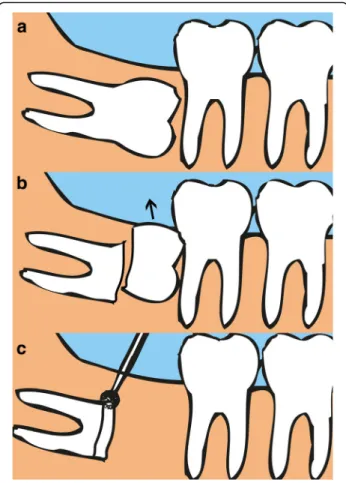
All IPOs were performed by a highly experienced oral and maxillofacial surgeon. Under local anesthesia with 2% lidocaine including 1:100,000 epinephrine, a full- thickness mucoperiosteal flap was elevated with a peri- osteal elevator. Alveolar bones around the impacted crown were removed with a surgical bur to expose the cementoenamel junction (CEJ) of the tooth, and the tooth was sectioned at the junction. After decrowning, the cutting margin was trimmed to 3 mm below the ad- jacent bone level (Fig. 1). The mobility of the remaining root was checked, and any secondary trauma to the healthy roots was minimized. The surgical area was irrigated with normal saline and then sutured primarily with 3-0 black silk (Ethicon Sutures, Ltd., USA).

Antibiotics (amoxicillin/clavulanate; Augmentin®, Ilsung Pharmaceuticals Co., Seoul, Korea) and a non-steroidal anti-inflammatory drug (talniflumate; Somalgen®, Kunwha Pharmaceutical Co., Seoul, Korea), and 100 mL of 0.1% chlorhexidine mouth gargling (Hexamedine®, Bukwang Pharm, Ansan, Korea) were prescribed for oral

hygiene maintenance. Sutures were stitched out 1 week after the surgery. Any complications related with sensory abnormality, pain, infection, migration of remained roots, and influence to the adjacent tooth was evaluated clinically and radiographically.

Results

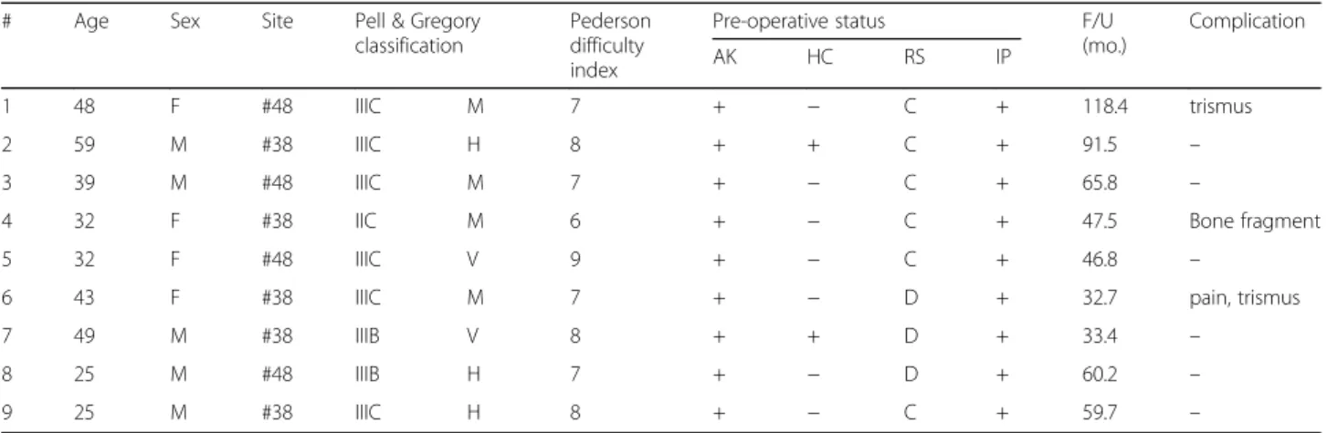
The mean follow-up period was 63.2 ± 29.8 months, and all operation sites were evaluated clinically and radio- graphically. According to Pell & Gregory classification, there were six IIIC, two IIIB, and one IIC. Out of the nine teeth, four were mesioangular, three horizontal im- pactions, and two vertical impactions. Applying Peder- son’s difficulty index, only one out of the nine cases was presumably “moderately difficult,” while the rest were

“very difficult” extractions. Considering that the diffi- culty index does not take ankylosis/hypercementosis into account, actual levels of extractions presented in this art- icle were very difficult even for a highly experienced oral surgeon (Table 1).

According to the panoramic views, all molars were im- pacted close to the white line of IAN canal (≤ 2 mm),

Fig. 1 Intentional partial odontectomy. a Pre-operative mandibular

third molar. b Coronectomy of the molar at cementoenamel junc-

tion (c). Trimming the cutting surface to 3 mm below the surround-

ing alveolar bone

(3)

and all roots lost lamina dura suggesting that the teeth are ankylosed with the surrounding alveolar bone. Also, two teeth showed signs of hypercementosis, and root shape of the three teeth was divergent.

On the recall check, none of the patients had sensory abnormality such as paresthesia and hypoesthesia related with the surgical sites. Moreover, all the operation sites were covered with the healthy gingiva without postoper- ative infection signs. Radiographically, none of the re- sidual roots that were surrounded by the intact bones migrated (Fig.2).

In one patient, a bony fragment erupted 4 months after IPO. The patient did not show any clinical symp- toms related to it, and the fragment was simply removed and it did not cause the failure of IPO.

In another patient, an implant placed on mandibular second molar area adjacent to the third molar that re- ceived IPO was explanted about 2 years after because of persistent discomfort. However, in this case, the bone around the resected root was intact, and there was not a sign of infection in the IPO site. Therefore, the reason for implant failure is considered as the peri-implantitis.

One out of nine cases reported severe pain (Visual Analog Scale of 6 out of 10) a day after IPO, which alle- viated after a week.

Discussion

To avoid complications when a mandibular third molar is impacted close to an IAN canal, IPO is considered as an alternative treatment to the surgical extraction [6]. A study about IANIs after the conventional extraction and IPO reported that the incidence of the damage to the IAN was higher in complete extraction (19% of 102 cases) compared with IPO (3% of 94 cases) [7]. Another case-control study also concluded that IANIs as a result of IPO was not reported at all, whereas the conventional surgical extraction resulted to 5% IANIs [8]. Despite these positive reports on IPO, many dental surgeons are still reluctant to remain the root portions and worry about the postoperative complications because of a lack of studies reporting the long-term follow-up results [9].

As of disadvantage of IPO, migration of residual roots could happen. A study mentioned that 30% of remnant roots migrated towards the superior border of the Table 1 Description of impacted third molars

# Age Sex Site Pell & Gregory

classification

Pederson difficulty index

Pre-operative status F/U

(mo.)

Complication

AK HC RS IP

1 48 F #48 IIIC M 7 + − C + 118.4 trismus

2 59 M #38 IIIC H 8 + + C + 91.5 –

3 39 M #48 IIIC M 7 + − C + 65.8 –

4 32 F #38 IIC M 6 + − C + 47.5 Bone fragment

5 32 F #48 IIIC V 9 + − C + 46.8 –

6 43 F #38 IIIC M 7 + − D + 32.7 pain, trismus

7 49 M #38 IIIB V 8 + + D + 33.4 –

8 25 M #48 IIIB H 7 + − D + 60.2 –

9 25 M #38 IIIC H 8 + − C + 59.7 –

Difficulty index 3 –4(slightly difficult), 5–6(moderately difficult), 7–10(very difficult)

AK ankylosis, HC hyper-cementosis, RS root shape, IP IAN proximity ( ≤ 2 mm), M mesio-angular, H horizontal, V vertical, C convergent, D divergent

Fig. 2 A panoramic view of pre- and post-IPO of the mandibular right third molar. a Pre-IPO. b 70 months after IPO

(4)

mandible in the first year, and additional surgery for removal of roots was required [10]. Another study re- ported 6% incidence of later root removal after the remained root migrated far from the IAN canal [11]. In other hand, Dolanmaz stated that none of the 43 patients who were treated by IPO surgery required additional removal resulting from the subsequent root migration [12]. In our study (n = 9), there was no radio- graphic evidence of migration of the residual roots. It seems that hypercementosised and ankylosed roots cov- ered with surrounding bone are not easily migrated from its original location.

IPO sometimes is known to cause an infection as one of the postoperative complications. IPO without complete removal of the dental follicle could lead to infection up to 5% [4]. Renton also reported 10–12%, relatively high incidence, of infection on operation sites after IPO [7]. Whereas, Dolonmaz showed that none of the 43 cases were related with postoperative infection, and Porgrel reported only one case of postoperative in- fection out of 50 IPO cases [13, 14]. In our study, post- operative infection was not observed in the IPO site.

After the long periods of healing, the operation sites were covered with healthy gingiva. In one case (case #2), an implant placed adjacent to the third molar that re- ceived IPO failed 2 years after implantation. In this case, the bone healing around the residual roots was normal, and the third molar area did not display any specific signs of abnormality upon visual exam and palpation tests. Therefore, in this case, the reason for implant fail- ure is not resulted by IPO, but possibly by peri- implantitis.

Another concern of performing IPO is postoperative pain. Hatano Y et al. had reported high incidence of postoperative pain in the coronectomy group compared to the conventional extraction group, which had dimin- ished within 1 week [9]. Hatano Y et al. proposed that possible reasons to the acute pain are a tight primary closure which could have caused high pressure inside the wound and a temporary pulpitis of the resected root.

According to O’Riordan study [3], the resected pulp could result in hyperemia or inflammatory edema which could develop into pulp pathogenesis. Therefore, ad- equate irrigation and avoidance of any manipulation of vital pulp that could facilitate dentinal bridge around pulp chamber are important. In our study, one patient complained of severe pain (VAS 6) a day after IPO. The pain was relieved within 1 week. Although histopatho- logic section was not performed on the symptomatic IPO tooth, our assumption is that the pulp had tempor- ary pulpitis which could have been caused by heat arose from inadequate coolant during the coronectomy.

In terms of risk of IANI, it is obvious that decrowning the highly risky, impacted mandibular third molar is

safer than the complete extraction. Therefore, when dental surgeons encounter the third molar extraction cases, they should initially evaluate difficulties of extrac- tion with radiographic findings. If the mandibular third molar is located too near an IAN canal, and if the roots have hypercementosis or ankylosis, a surgeon could con- sider IPO as a primary treatment option. However, pa- tients should be sufficiently informed of the advantages/

disadvantages of the IPO surgery and understand why this technique is necessary before the procedure starts.

In regard to IPO, a long-term follow-up is important to evaluate patient’s discomfort including neuropathy, post- operative infection, and development of any pathology.

In our study, the mean follow-up period was 61.7 ± 27.8 months, which was enough to assess any complication of IPO. However, the sample size of this study is relatively small as compared with other studies reporting the results of IPO. Moreover, our study has no control group to compare the effectiveness of conven- tional extraction with IPO. Therefore, it is suggested that a long-term study with more cases should be conducted to evaluate the benefits of IPO and to compare with the traditional extraction method.

Conclusions

In case where high risk of IANI exists, IPO may be chosen alternatively to surgical extraction to reduce the risk of nerve damage, but a larger sample size of similar follow-up period should be supplemented.

Additional file

Additional file 1: Case form and result of data. (XLSX 14 kb)

Abbreviations

CEJ: Cementoenamel junction; IAN: Inferior alveolar nerve; IANI: Inferior alveolar nerve injury; IPO: Intentional partial odontectomy

Acknowledgements Not applicable

Funding

There was no funding in support of this study.

Availability of data and materials

The dataset supporting the conclusions of this article is included within the article and Additional file 1.

Authors ’ contributions

KHS participated in the data collection and writing of the manuscript. YPY participated in the study design and performed the statistical analysis. KYK participated in the study design and coordination and helped to draft the manuscript. All authors read and approved the final manuscript.

Authors ’ information

All of the authors have no affiliations with or involvement in any

organization or entity with any financial interest or non-financial interest in

this manuscript. This manuscript represents original works and is not being

considered for publication elsewhere.

(5)

Ethics approval and consent to participate

This study was approved by the Institutional Review Board of Seoul National University Bundang Hospital (B-1707-406-104).

Consent for publication

Consent for publication was obtained.

Competing interests

No potential conflict of interest relevant to this article was reported.

Publisher ’s Note

Springer Nature remains neutral with regard to jurisdictional claims in published maps and institutional affiliations.

Received: 3 July 2017 Accepted: 4 September 2017

References

1. Vafaei N, Ferretti C (2008) Coronectomy: an alternative therapy for the symptomatic, impacted third molar report of 9 cases. Scientific 22:34 2. Leung YY, Cheung LK (2012) Coronectomy of the lower third molar is safe

within the first 3 years. J Oral Maxillofac Surg 70(7):1515 –1522 3. O'riordan BC (2004) Coronectomy (intentional partial odontectomy of lower

third molars). Oral Surg Oral Med Oral Pathol Oral Radiol Endod 98(3):274 –280 4. Sarwar H, Mahmood-Rao S (2015) Coronectomy; good or bad? Dent

Update 42:824 –828

5. Pell GJ, Gregory GT (1942) Report on a ten-year study of a tooth division technique for the removal of impacted teeth. Am J Orthod Oral Surg 28(11):

B660 –B666

6. Pederson GW (1988) Surgical removal of tooth. In: Pederson GW (ed) Oral surgery. WB Saunders, Philadelphia

7. Frafjord R, Renton T (2010) A review of coronectomy. Oral Surg 3(1 –2):1–7 8. Renton T, Hankins M, Sproate C, McGurk M (2005) A randomised controlled

clinical trial to compare the incidence of injury to the inferior alveolar nerve as a result of coronectomy and removal of mandibular third molars. Br J Oral Maxillofac Surg 43(1):7 –12

9. Hatano Y, Kurita K, Kuroiwa Y, Yuasa H, Ariji E (2009) Clinical evaluations of coronectomy (intentional partial odontectomy) for mandibular third molars using dental computed tomography: a case-control study. J Oral Maxillofac Surg 67(9):1806 –1814

10. Williams M, Tollervey D (2016) Lower third molar surgery –consent and coronectomy. Br Dent J 220(6):287 –288

11. Pogrel MA (2009) An update on coronectomy. J Oral Maxillofac Surg 67(8):

1782 –1783

12. Knutsson K, Lysell L, Rohlin M (1988) Postoperative status after partial removal of the mandibular third molar. Swed Dent J 13(1 –2):15–22 13. Dolanmaz D, Yildirim G, Isik K, Kucuk K, Ozturk A (2009) A preferable

technique for protecting the inferior alveolar nerve: coronectomy. J Oral Maxillofac Surg 67(6):1234 –1238

14. Pogrel MA, Lee JS, Muff DF (2004) Coronectomy: a technique to protect the

inferior alveolar nerve. J Oral Maxillofac Surg 62(12):1447 –1452

수치

Fig. 1 Intentional partial odontectomy. a Pre-operative mandibular third molar. b Coronectomy of the molar at cementoenamel  junc-tion (c)
Fig. 2 A panoramic view of pre- and post-IPO of the mandibular right third molar. a Pre-IPO

참조

관련 문서