• 검색 결과가 없습니다.

Evaluation of the Healing Process of Autogenous Tooth Bone Graft Material Nine Months after Sinus Bone Graft: Micromorphometric and Histological Evaluation

N/A
N/A
Protected

Academic year: 2021

Share "Evaluation of the Healing Process of Autogenous Tooth Bone Graft Material Nine Months after Sinus Bone Graft: Micromorphometric and Histological Evaluation"

Copied!
6
0
0

로드 중.... (전체 텍스트 보기)

전체 글

(1)

Case Report

RECEIVED May 5, 2013, REVISED May 22, 2013, ACCEPTED September 21, 2013 Correspondence to Young-Kyun Kim

Department of Oral and Maxillofacial Surgery, Section of Dentistry, Seoul National University Bundang Hospital 82 Gumi-ro 173 beon-gil, Bundang-gu, Seongnam 463-707, Korea

Tel: 82-31-787-7541, Fax: 82-31-787-4068, E-mail: kyk0505@snubh.org

CC

This is an open access article distributed under the terms of the Creative Commons Attribution Non-Commercial License (http://creativecommons.org/licenses/

by-nc/3.0) which permits unrestricted non-commercial use, distribution, and reproduction in any medium, provided the original work is properly cited.

Evaluation of the Healing Process of Autogenous Tooth Bone Graft Material Nine Months after Sinus Bone Graft:

Micromorphometric and Histological Evaluation

Young-Kyun Kim, Sang-Ho Jun 1 , In-Woong Um 2 , Sooyeon Kim 3

Department of Oral and Maxillofacial Surgery, Section of Dentistry, Seoul National University Bundang Hospital,

1

Department of Oral and Maxillofacial Surgery, Section of Dentistry, Korea University Anam Hospital,

2

R&D Institute Korea Tooth Bank,

3

KOREA INTERNATIONAL (GR.11)

Abstract

Micromorphometric and histological examinations were conducted with a collected tissue specimen nine months after sinus bone graft using autogenous tooth bone graft material (AutoBT). As a result of micro-computed tomographic analysis, the total bone volume (graft material+new bone) was 76.45%, and the proportion of new bone was 45.4%. The bone mineral density and the average Hounsfield Unit of new bone were 0.26 and 1,164.69, respectively. The histological examination showed that AutoBT particles were united well with new bone. AutoBT was considered to have excellent bone healing ability after sinus graft and bone density that can resist repneumatization.

Key words: Autogenous tooth, Bone, Sinus

Introduction

Any material whose resorption speed is not too high and whose bone healing process approximates that of au- togenous bone graft may be useful in maxillary sinus bone grafting. Likewise, a more excellent clinical outcome might be expected when this material is used in combination with other bone substitutes with slow resorption proper- ties[1-3]. Autogenous tooth bone graft material (AutoBT;

Korea Tooth Bank, Seoul, Korea) was proven to exhibit bone healing ability through osteoinduction and osteo- conduction, demonstrating a histological healing process similar to that of free bone grafting being resorbed over

3∼6 months. Accordingly, AutoBT is regarded as a poten- tial substitute when autogenous bone is needed for sinus bone graft, and it may have a useful effect on increasing the volume of bone graft materials and minimizing re- pneumatization[4]. AutoBT is a brand name of autogenous tooth bone graft material.

The authors sought to investigate the healing process

through micromorphometric and histological examination

with a collected tissue specimen nine months after sinus

bone graft using powder-type autogenous tooth bone graft

material. This histologic study and the consent forms were

approved by SNUBH IRB (E-1110-067-003).

(2)

Fig. 1. Initial panoramic radiograph. The residual bone height of the left maxillary posterior area is definitely insufficient, showing a progressive periodontal disease.

Fig. 2. Initial computed tomography. Mucosal thickening and dome-shaped radiopacity of the left maxillary sinus were observed.

Fig. 3. Computed tomography (CT) examination of sinus bone graft. (A) CT view before surgery. Dome-shaped radiopacity and sinus mucosal thick- ening were observed. (B) CT view nine months after sinus bone graft.

(C) CT view after implant placement.

(D) CT view seven months after im- plant placement (16 months after si- nus bone graft).

Case Report

1. Case report

A 45-year-old male patient visited Seoul National University Bundang Hospital for implant treatment on the edentulous first and second molar areas in the left maxilla.

The radiological examination revealed a mucous retention cyst in the left maxillary sinus; however, there were no clinical symptoms. The residual bone height to sinus floor

was definitely insufficient, therefore, sinus bone graft and

delayed implant placement were planned (Fig. 1, 2); #15,

16, and 17 teeth were extracted before surgery, and pow-

der-type autogenous tooth bone graft material was made

and stored. Sinus membrane elevation was performed

through the lateral window approach, and a sinus bone

graft was performed using AutoBT powder. After surgery,

the areas healed normally without occurrence of any specif-

ic complication. Mucoperiosteal flap elevation was per-

formed for implant placement after nine months. Two tis-

sue specimens were collected from the areas where the

(3)

Fig. 4. Biopsy specimen. (A) Two tissue specimens were collected from areas #26, 27. (B) Alveolar bone of approximately 1 mm thickness was observed, and osseous tissue showing a dense appearance was created upward and mixed with autogenous tooth bone particles.

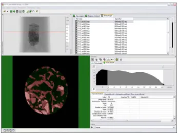
Fig. 5. The cylindrical specimen obtained was scanned using micro computed tomography (Skyscan 1172, Bruker-Microct, Kontich, Belgium). Three-dimensional image reconstruction was performed for the data, with two-dimensional and three-dimensional image analyses performed for the reconstructed data using the CTAn analysis program.

implant was to be placed (#26, 27) using a trephine bur with an external diameter of 3.0 mm. Two implants (Perio Implant; DIO, Busan, Korea) were then placed, and wound closure was performed. After a healing period of approx- imately seven months, the implants were exposed, and the healing abutment was connected through the second surgery (Fig. 3).

2. Micromorphometric and histologic examination

The tissues contained in the trephine bur were stored in formalin, and a block embedded with methacrylate-based chemical curing resin was made through the washing and dehydration process for tissue sample fabrication and micro CT scanning. Trephine bur and external resin were carefully removed using a diamond bur without causing damage to the tissues (Fig. 4). The cylindrical specimen obtained was scanned using micro computed tomography (MicroCT) (Skyscan 1172; Bruker-Microct, Kontich, Belgium).

Three-dimensional image reconstruction was performed for the reconstructed data, two-dimensional and three-dimen- sional image analyses were performed using the CTAn anal- ysis program (Fig. 5). Hounsfield unit (HU) and bone miner- al density (BMD) were calibrated using phantom in order to eliminate errors of each specimen. Regional objective image (ROI) was formed from the taken core sample, except that the autogenous bone and total HU and BMD were obtained from the binary image. HU more than 350 was regarded as new bone. HU of grafted material was obtained

through analysis of binary image peak and HU more than 2,100 was eliminated from the data. This analytical method was based on Tajima et al .’s method[5].

The specimen that had undergone MicroCT scanning was secondarily embedded using Technovit4000VLC, and a tis- sue sample of 30∼40 μm thickness was prepared by grind- ing the resin block. For the histological analysis, the healing status was evaluated through Masson trichrome staining, toluidine blue staining, and H&E staining.

3. Results

As a result of MicroCT analysis, the total bone volume (graft material+new bone) of the bone tissue sample in the maxillary sinus was 76.45%, and the proportion of new bone was 45.4%. Total core BMD was 0.35 and BMD of new bone was 0.26. Average HU of the total core was 1,555.07 and average HU of new bone was 1,164.69.

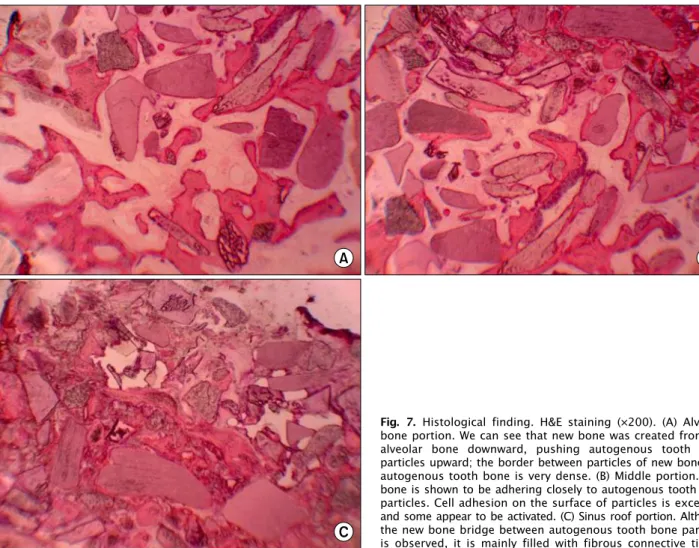
Average trabecular thickness of new bone was 0.08282 mm. On the other hand, based on the histological examina- tion, formation of new bone and rising toward the maxillary sinus from the left alveolar bone was observed, and new bone and AutoBT particles were mixed in the middle area.

Likewise, the examination showed a mixture of AutoBT

and fibrous connective tissue in the roof area of the maxillary

sinus (Fig. 6, 7).

(4)

Fig. 6. Histological finding. (A) Masson trichrome (MT) staining (×12.5), (B) MT staining (×200), (C) toluidine blue (TB) staining (×12.5), (D) TB staining (×200). Formation of new bone and rising toward the right maxillary sinus from the left alveolar bone area was observed (A, C) a mixture of new bone and autogenous tooth bone particles was observed in the middle area (B, D).

Discussion

Jeong et al .[6]. conducted a retrospective study on sinus bone graft using AutoBT. A total of 100 implants in 51 patients were selected; the patients underwent maxillary sinus augmentation and implant placement using AutoBT between July 2009 and November 2010. When AutoBT was used alone or together with other graft material, the implant survival rate was 96.15%. Based on the histo- morphological examination, AutoBT showed gradual re- sorption and new bone formation through osteoconduction and osteoinduction. They suggested that AutoBT materials are appropriate for use in maxillary sinus augmentation Lee et al .[7] conducted a study for histomorphometric evalu- ation and comparison of the efficiency of various bone graft materials and autogenous tooth bone graft material

used in the sinus bone graft procedure. All biopsy specimens were harvested four months after the procedure and divided into three groups according to bone graft material:

Autogenous tooth bone graft material as group 1, OrthoblastII

(Integra Lifescience Corp., Irvine, CA, USA)+Biocera

(Osscotec, Cheonan, Korea) as group 2, and DBX (Synthes,

West Chester, PA, USA)+BioOss (Geistlich Pharm AG,

Wolhusen, Switzerland) as group 3. New bone formation

was 52.5±10.7% in group 1, 52.0±23.4% in group 2, and

51.0±18.3% in group 3. The ratio of new bone to graft

material was 81.3±10.4% in group 1, 72.5±28.8% in group

2, and 80.3±24.0% in group 3. After a four-month healing

period, all groups showed favorable new bone formation

around the graft material and implant. They suggested that

autogenous tooth bone graft material may be used as novel

bone graft material for sinus bone graft. Kim et al .[8] and

(5)

Fig. 7. Histological finding. H&E staining (×200). (A) Alveolar bone portion. We can see that new bone was created from the alveolar bone downward, pushing autogenous tooth bone particles upward; the border between particles of new bone and autogenous tooth bone is very dense. (B) Middle portion. New bone is shown to be adhering closely to autogenous tooth bone particles. Cell adhesion on the surface of particles is excellent, and some appear to be activated. (C) Sinus roof portion. Although the new bone bridge between autogenous tooth bone particles is observed, it is mainly filled with fibrous connective tissue.

Lee et al .[7], who performed sinus bone graft and guided bone regeneration using autogenous tooth bone from hu- mans, took the tissue specimen two months and four months later for histomorphometric analysis. As a result, they found favorable new bone formation and suggested that autoge- nous tooth bone graft materials could be used in various bone grafts. Huang et al .[9] performed three-dimensional bone structure and BMD evaluations of autogenous bone graft after sinus augmentation. Iliac bone was mainly used for autogenous bone graft. After trephine core biopsy four∼

five months after bone graft, microcomputed tomography analysis was performed. The BMD of native bone was 0.22, and that of grafted bone was 0.12. Bone volume was reported to be 24.4% for native bone and 15.43%

for grafted bone. Lundgren et al .[10] performed histo- morphometric evaluation of the maxillary sinus augmenta- tion with particulated mandible (symphysis). Trephine core biopsy specimen was collected six months and 12 months

after sinus graft, and its bone volume was measured using MicroCT. The donor site bone volume was 58±19%, and that of residual alveolar bone was 45±15%. Six and twelve months later, 40±12% and 48±10%, respectively, were recorded. Chackartchi et al .[11] evaluated the healing proc- ess of sinus floor augmentation using large (1∼2 mm) or small (0.25∼1 mm) bovine bone mineral particles. After a tissue specimen was collected 6∼9 months later, residual graft material, new bone formation, and soft tissue ratio were measured using MicroCT and histomorphology. In the MicroCT examination, the residual graft material, bone, and soft tissue were 23.12%, 7.99%, and 68.89%, re- spectively, for large particles, and 22.89%, 14.64%, and 62.47%, respectively, for small particles. In the histo- morphology examination, residual graft material and bone were 33.71% and 27.14%, respectively, for large particles and 34.57% and 28%, respectively, for small particles.

According to their findings, it was difficult to distinguish

(6)

in the MicroCT image the exact borders between the new bone and graft particles, which are in direct contact and were found to exhibit a similar pattern. Therefore, they reported that the MicroCT examination showed low pro- portions of bone and graft material compared with the histological exam.

In this study, as a result of analysis of the healing status nine months after maxillary sinus bone graft using Micro CT, the total bone volume (graft material+new bone) was 76.45%, and the proportion of new bone was 45.4%. The BMD and the average HU of new bone were 0.26 and 1,164.69, respectively, which is D 2 bony quality. On the other hand, according to Chackartchi et al .[11], total bone volume would likely show a further increase in the histo- morphometric examination. In the histological examina- tion, the direct conglutination of new bone and AutoBT was observed, and there was no inflammatory response.

In other words, growth of new bone toward the graft mate- rial from the residual alveolar bone was observed nine months after the maxillary sinus bone graft, and the graft material remained. Likewise, the examination showed that bone healing was slowest in the sinus roof area. Sinus bone graft using AutoBT showed bone healing of D 2 bony quality in the histological examination nine months later; the effect of preventing sinus re-pneumatization due to residual bone graft could be assumed.

Acknowledgements

This study was supported by a grant from the Korean Health Technology R&D Project, Ministry of Health &

Welfare, Republic of Korea (A102065).

References

1. Hatano N, Shimizu Y, Ooya K. A clinical long-term radio- graphic evaluation of graft height changes after maxillary si- nus floor augmentation with a 2:1 autogenous bone/xenograft mixture and simultaneous placement of dental implants. Clin Oral Implants Res 2004;15:339-45.

2. Velich N, Németh Z, Tóth C, Szabó G. Long-term results with different bone substitutes used for sinus floor elevation. J Craniofac Surg 2004;15:38-41.

3. Garg AK. Current concepts in augmentation grafting of the maxillary sinus for placement of dental implants. Dent Implantol Update 2001;12:17-22.

4. Kim YK, Kim SG, Byeon JH, et al . Development of a novel bone grafting material using autogenous teeth. Oral Surg Oral Med Oral Pathol Oral Radiol Endod 2010;109:496-503.

5. Tajima N, Ohba S, Sawase T, Asahina I. Evaluation of sinus floor augmentation with simultaneous implant placement us- ing platelet-rich fibrin as sole grafting material. Int J Oral Maxillofac Implants 2013;28:77-83.

6. Jeong KI, Kim SG, Kim YK, Oh JS, Jeong MA, Park JJ. Clinical study of graft materials using autogenous teeth in maxillary sinus augmentation. Implant Dent 2011;20:471-5.

7. Lee JY, Kim YK, Kim SG, Lim SC. Histomorphometric study of sinus bone graft using various graft material. J Dental Rehabilitation Appl Sci 2011;27:141-7.

8. Kim SG, Kim YK, Lim SC, Kim KW, Um IW. Histomorpho- metric analysis of bone graft using autogenous tooth bone graft. Implantology 2011;15:134-41.

9. Huang HL, Chen MY, Hsu JT, Li YF, Chang CH, Chen KT.

Three-dimensional bone structure and bone mineral density evaluations of autogenous bone graft after sinus augmentation:

a microcomputed tomography analysis. Clin Oral Implants Res 2012;23:1098-103.

10. Lundgren S, Moy P, Johansson C, Nilsson H. Augmentation of the maxillary sinus floor with particulated mandible: a his- tologic and histomorphometric study. Int J Oral Maxillofac Implants 1996;11:760-6.

11. Chackartchi T, Iezzi G, Goldstein M, et al . Sinus floor aug-

mentation using large (1-2 mm) or small (0.25-1 mm) bovine

bone mineral particles: a prospective, intra-individual con-

trolled clinical, micro-computerized tomography and histo-

morphometric study. Clin Oral Implants Res 2011;22:473-80.

수치

Fig. 3. Computed tomography (CT) examination of sinus bone graft. (A) CT view before surgery
Fig. 4. Biopsy specimen. (A) Two tissue specimens were collected from areas #26, 27. (B) Alveolar bone of approximately 1 mm thickness was observed, and osseous tissue showing a dense  appearance was created upward and mixed with autogenous  tooth bone par
Fig. 6. Histological finding. (A) Masson trichrome (MT) staining (×12.5), (B) MT staining (×200), (C) toluidine blue (TB) staining (×12.5), (D) TB staining (×200)
Fig. 7. Histological finding. H&E staining (×200). (A) Alveolar  bone portion. We can see that new bone was created from the  alveolar bone downward, pushing autogenous tooth bone  particles upward; the border between particles of new bone and  autogen

참조

관련 문서

Clinical and histomorph ometric evaluation of extraction sockets treated with an autologous bone marro w graft... Barone A, Aldini NN, Fini M, Giardino R, Calvo Guirado

The OSFE (osteotome sinus floor elevation) technique has been used for maxillary sinus augmentation.. The implants were clinically and radiographically followed

5) After GBR, the membrane was removed i n i ni ti al ti me, the usage of nonabsorbabl e membrane and autogenous bone resul ted i n the mostfavorabl e bone formati

success rates of dental implants placed at the time of or after alveolar ridge augmentation with an autogenous mandibular bone graft and titanium mesh: a 3-to

In 4-week group, the group filled with bone graft with decortication revealed larger new bone formation area than shown in the group that had a defect area

Histologic evaluation of early human bone response to different implant surfaces2. Histologic evaluation of human bone integration on machined and

The result of using PLGA nanofiber membrane with bone graft material showed that the PLGA nanofiber membrane in the experimental group of 2 weeks were ten times more new

of mineralized cancellous bone allograft(Puros) and anorganic bovine bone matrix(Bio-oss) for sinus augmentation: Histomorphometry at 26 to 32 weeks after