Case Report
원고 접수일 2011년 3월 31일, 원고 수정일 2011년 6월 13일, 게재 확정일 2011년 6월 29일
책임저자 김수관
(501-759) 광주시 동구 서석동 375번지, 조선대학교 치의학전문대학원 구강악안면 외과학교실
Tel: 062-220-3815, Fax: 062-228-7316, E-mail: [email protected]
RECEIVED March 31, 2011, REVISED June 13, 2011, ACCEPTED June 29, 2011
Correspondence to Su-Gwan Kim
Department of Oral and Maxillofacial Surgery, School of Dentistry, Chosun University
375, Seosuk-dong, Dong-gu, Gwangju 501-759, Korea
Tel: 82-62-220-3815, Fax: 82-62-228-7316, E-mail: [email protected]
CC This is an open access article distributed under the terms of the Creative Commons Attribution Non-Commercial License (http://creativecommons.org/licenses/
by-nc/3.0) which permits unrestricted non-commercial use, distribution, and reproduction in any medium, provided the original work is properly cited.
Putty형 탈회동종골을 이용한 골유도 재생술: 증례보고
장한성ㆍ김수관ㆍ문성용ㆍ오지수ㆍ박진주ㆍ정미애1ㆍ양석진ㆍ정종원ㆍ김정선2
조선대학교 치의학전문대학원 구강악안면외과학교실, 1강원대학교 치위생학과, 2광주보건대학교 치위생과
Abstract
Guided Bone Regeneration Using a Putty-type Demineralized Bone Matrix: Case Report
Han-Seung Jang, Su-Gwan Kim, Seong-Yong Moon, Ji-Su Oh, Jin-Ju Park, Mi-Ae Jeong
1, Seok-Jin Yang, Jong-Won Jung, Jeong-Sun Kim
2Department of Oral and Maxillofacial Surgery, School of Dentistry, Chosun University,
1
Department of Dental Hygiene, Kangwon National University,
2
Department of Dental Hygiene, Gwangju Health College University
Allomatrix (Wright Medical Tech, Inc., Arlington, Tenn, USA), is a newly designed, injectable putty with a reliable demineralized bone matrix (DBM), derived from human bone. The compound contains 86% DBM and other bone growth factors such as bone morphogenic protein (BMP)-2, BMP-4, insulin-like growth factor (IGF)-1, and transforming growth factor (TGF)-β1.
It has excellent osteoinduction abilities. In addition, DBM is known to have osteoconduction capacity as a scaffold due to its collagen matrix. This product contains a powder, which is a mix of DBM and surgical grade calcium sulfate as a carrier.
A practitioner can blend the powder with calcium sulfate solution, making a putty-type material which has the advantages of ease of handling, better fixation, and no need for a membrane, because it can function as membrane itself. This study reports the clinical and radiographic results of various guided bone regeneration cases using Allomatrix, demonstrating its strong potential as a graft material.
Key words: Bone morphogenic protein, Demineralized bone matrix, Growth factor, Osteoinduction
서 론
구강악안면 영역에서 연령증가에 따르는 치조골 흡수는 환자의 기능회복을 목적으로 하는 치료 시 끊임없는 도전이 되어왔다.
이와 같은 문제를 해결하기 위해 현재 골유도 재생술이 이용해
왔으며, 골유도 재생술이란 골형성을 위한 공간 안으로 골형성에
관여하는 세포가 선택적으로 재군집되어 일정 부피의 신생골이
형성되는 것이다[1]. 세포가 재군집되거나 골형성을 돕는데 있어
서 신선 자가골을 이용하거나 동종골, 이종골, 합성골 등 다양한
특성의 골대체재가 개발되어 골유도 재생술의 임상적 효용성을
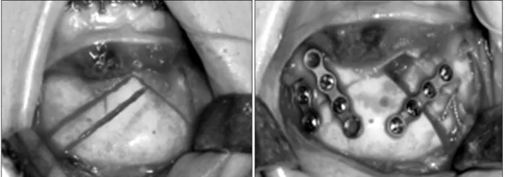
Fig. 1. The clinical photograph after flap elevation showing bone
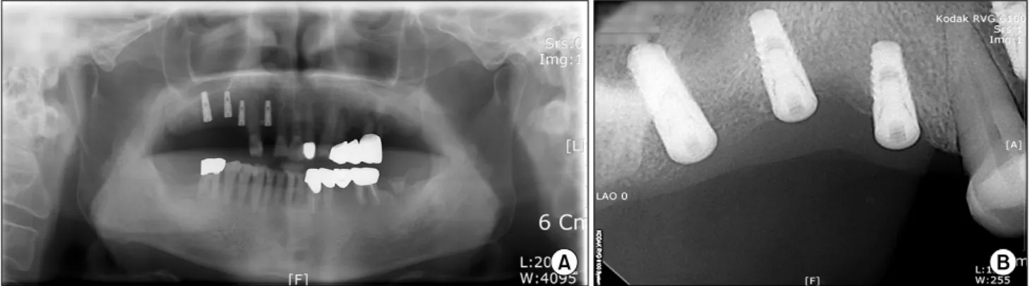
defect.Fig. 2. Initial panoramic radiograph.
Fig. 3. After implant installation on #14, 15, 16.
Fig. 5. The regenerated bone was maintained after 6 months. (A) panoramic radiograph, (B) standard radiograph.
Fig. 4. Guided bone regeneration was performed on peri-implant
bone defect area.높여주고 있다.
본 연구는 골유도 재생술을 시행하는데 있어 Allomatrix
TM(Wright Medical Tech, Arlington, TN, USA)를 사용한 환자의 임상적 효율성과 성공을 보고하는데 있다.
증례보고
1. 증례 1
78세 남자 환자로 상악 우측 측절치, 제1, 2소구치 및 제1대구
치의 무치악 부위에 임플란트를 식립하였다(Fig. 1, 2). Dentis
Haptite 제품의 임플란트를 상악 우측 측절치(4.3×10 mm),
제1, 2소구치(3.7×10 mm), 제1대구치(4.3×12 mm)에 식립하
였으며 식립부위의 심한 수직 골결손부가 존재하여(Fig. 3) 골결
손부에 Allomatrix
TM과 차단막을 이용하여 골유도 재생술을 시행
하였다(Fig. 4). 식립 당시 Osstell Mentor를 이용한 ISQ 값은
측절치부터 차례대로 66, 65.5, 63, 68이었으며 Periotest를 이용
Fig. 6. Genioplasty was performed.
Fig. 7. Guided bone regeneration was performed on inter-bony
gap.한 PTV는 3, 2, 9, 0으로 측정되었다. 술 후 6개월에도 이식된 골은 잘 유지되고 있었으며(Fig. 5) 9개월 후 2차 수술을 시행하였 고 2차 수술 시 ISQ 값은 77, 70, 60, 62이었으며 PTV는 5,
3, 6, 2로 측정되었다.
2. 증례 2
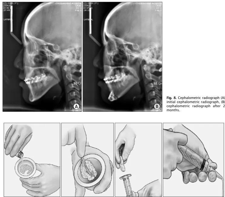
17세 여자 환자로 안면 비대칭으로 이부성형술을 시행하였으 며, 골절단술 후 우측이동에 따라 형성된 골 간극에 골절편 및 Allomatrix
TM를 이용하여 골유도 재생술을 시행하였다(Fig. 6, 7). 술 후 특별한 합병증은 발생하지 않았으며, 술 후 2개월 후 측면방사선 사진상 이식된 골은 잘 유지되었고 유의할 만한 골 흡수는 보이지 않았다(Fig. 8).
고 찰
치조골 결손은 발치 후 무치악 기간이 장기간 지속됨에 따라
심화되어 보철수복 혹은 임플란트 식립 시 수직적, 수평적인 측면 에서 한계를 드러낸다. 치조골의 결손에 대한 수복의 방법으로 골유도 재생술, 골신장술(Distraction Osteogenesis), 치조제 확 장술(ridge splitting technique) 등 다양한 술식이 이용되고 있 으며, 술식에 따라 장단점이 존재하므로 상황에 맞는 적용이 필요 하다[2-4]. 이 중 골유도 재생술은 임상에서 가장 많이 이용하는 술식으로 Hurley 등[5]에 의해 정형외과 영역에서 처음으로 보고 되었다. 그 후 치주조직 재생 시 이주세포의 이동속도 차이가 있다는 것과[6] 치주인대 세포들의 선택적 유도 증식이 가능하다 는 사실이 밝혀지고[7,8], 조직유도 재생술이 이용되면서 골유도 재생술의 기반이 되었다[9].
자가골은 골 이식재 중 가장 이상적인 것으로 알려져 있으나, 그 특성상 합병증이 발생할 수 있으며 골 채취량이 제한적이어서 이를 대체하기 위한 다양한 골 이식재가 개발되고 있다[10,11].
동종골의 경우 탈회의 시행 여부에 따라 물성이 달라지는데 탈회
를 시행하지 않은 경우는 무기질을 제거하는 초기 치유단계를
지연시켜 일정기간 물리적 강도가 요구되는 단계에 사용될 수
있으며, 탈회를 시행한 경우 구조적 지지대 역할을 하는 물리적
성분의 부재로 초기 물리적 강도는 약해지지만 골치유 과정에서
흡수단계가 생략되어 빠른 골형성이 가능한 장점이 있다[12]. 이
러한 탈회 여부에 따른 골형성 능력의 평가에 관한 많은 연구가
있었는데 Mellonig 등[13]은 탈회 동결건조골이 탈회시키지 않은
이식재보다 더 우수한 신생골 형성을 보였다고 보고했다. 이와는
반대로 Piattelli 등[14]은 조직학적 및 조직형태학적 분석시 탈회
하지 않은 동결 건조골이 더 우수한 골 전도성을 보인다고 하였고,
Meffert[15]은 상악동 골이식술 시 6개월 후에 탈회 동결건조골은
결합조직만 남았지만 탈회하지 않은 동결건조골은 신생골의 형성
을 보였다고 보고했다. Yukna와 Vastardis[16]은 탈회하지 않은
동결 건조골이 조기에 더 빠른 속도로 더 많은 양의 신생골을
재생할 수 있다고 발표하였다. 한편 Rummelhart 등[17]과 Ca-
Fig. 8. Cephalometric radiograph (A)
initial cephalometric radiograph, (B) cephalometric radiograph after 2 months.Fig. 9. Usage of Allomatrix.
mmack 등[18]은 유의한 차이가 없음을 보고한 바 있다.
탈회된 동종골은 물리적 강도가 약해져 있어 형태의 형성이 용이하지 않아 이식재의 손실이 발생할 수 있고, 원하는 형태로의 이식이 어렵다. Gel이나 Putty 형태로 시판된 이식재의 경우, 시술 시 조작성이 높아져 차폐막의 조작을 용이하게 하고, 시술 비용도 낮아지는 장점이 있다[19]. Kim 등[20]은 Gel/Putty 형태 의 탈회 동결건조골이 초기 이식 부위 공간 유지에 있어 입자형 골이식재에 비해 효과적이라고 하였다. Gel/Putty 형태의 이 같 은 조작성은 치조골을 증대시키는 술식의 종류에 제한받지 않고 다양하게 적용이 가능하다.
본 증례에 사용된 Putty 형태의 동종골인 Allomatrix
TM은 탈회 동종골에 carboxylmethylcellulose가 첨가되어 있는 형태로 calcium sulfate 용액과 혼합하여 사용함으로써 이식재의 조작이
용이하며 이식재의 안정성에 도움을 주게 되어 다양한 술식에 적용할 수 있다. 제품에서 제공되는 powder bottle과 liquid bottle에 있는 내용물을 mixing bowl에서 30∼60초간 혼합하면 레진의 dough stage와 유사한 점도를 보이게 되는데 약 10분 정도 이 상태를 유지하게 된다. 이 형태를 활용하게 되는데 바로 시술 부위에 적용하거나 syringe를 이용해 적용하는 것도 가능하 다(Fig. 9).
본 증례들은 임플란트 식립 시 결손부 및 이부성형술에 Allo-
matrix
TM를 이용하여 골유도 재생술을 시도하였고, 효과적인 골
재생을 보여주고 있다. 탈회여부에 따른 동종골의 골형성 능력에
대하여 여러 논의가 있었지만 본 증례에서는 임상적으로 성공적인
결과를 얻을 수 있었다.
References