총설
디프테리아 백신의 진화와 물리화학적, 분자생물학적, 면역학적 지식의 진보에 따른 새로운 백신의 개발에 관한 고찰연구
배경동*
The Evolution and Value of Diphtheria Vaccine
Bae, Kyung Dong*
접수: 2011년 12월 8일 / 게재승인: 2011년 12월 23일
© 2011 The Korean Society for Biotechnology and Bioengineering
Abstract:
1)This review article provides an overview of the evolution of diphtheria vaccine, its value and its future.Diphtheria is an infectious illness caused by diphtheria toxin produced by pathogenic strains of Corynebacterium diphtheriae.
It is characterized by a sore throat with membrane formation due to local tissue necrosis, which can lead to fatal airway obstruction; neural and cardiac damage are other common complications. Diphtheria vaccine was first brought to market in the 1920s, following the discovery that diphtheria toxin can be detoxified using formalin. However, conventional formalin-inactivated toxoid vaccines have some fundamental limitations. Innovative technologies and approaches with the potential to overcome these limitations are discussed in this paper. These include genetic inactivation of diphtheria toxoid, innovative vaccine delivery systems, new adjuvants (both TLR- independent and TLR-dependent adjuvants), and heat- and freeze-stable agents, as well as novel platforms for producing improved conventional vaccine, DNA vaccine, transcutaneous (microneedle-mediated) vaccine, oral vaccine and edible vaccine expressed in transgenic plants. These innovations target improvements in vaccine quality (efficacy, safety, stability and consistency), ease of use and/or thermal stability. Their successful development and use should help to increase global diphtheria vaccine coverage.
Vaccine Research Institute, Crucell/Berna Biotech Korea Adjunct Professor, Biological Engineering Department, Inha University Inharo 100, 402-715, Korea
Tel: +82-(0)10-2434-4029, Fax: +82-32-232-3029 e-mail: [email protected], [email protected]
Keywords: Diphtheria vaccine, TLR-dependent adjuvant,
TLR-independent adjuvant, vaccine delivery system, DNA vaccine, heat-stable and freeze-stable agents
1. Introduction
Diphtheria is an acute infectious illness caused by the local and systemic effects of diphtheria toxin produced by toxigenic strains of Corynebacterium diphtheriae. Respiratory diphtheria presents as a sore throat with an adherent pseudomembrane of the tonsils, pharynx or nose. Common complications include myocarditis, polyneuritis and airway obstruction, which are fatal in 5-10% of cases. Complications or deaths are less frequent in cases of cutaneous diphtheria, which presents as infected skin lesions.
Diphtheria toxin can cause death in humans by a very simple and powerful biochemical mechanism: it prevents the activity of elongation factor 2 (EF-2), which is responsible for protein synthesis in a host cell [25,56,98]. Therefore, diphtheria was a potential threat to the survival of human beings until both an antitoxin and a preventive vaccine were developed.
Vaccination against toxigenic Corynebacterium diphtheriae strainsis regarded as one of the most beneficial biopharmaceutical interventions developed in modern times. The introduction of diphtheria vaccine into the WHO Expanded Program on Immunization (EPI) in the 1970s markedly improved the global infant immunization rate and the vaccine was shown to induce protection against the diphtheria toxin through
targeted activation of the human immune system [129].
Nevertheless, occasional diphtheria outbreaks are still reported in both developing and developed countries [128]. Based on the published reports, it appears that the main causes of outbreaks are insufficient vaccination coverage, a low antibody level in serum and a lack of periodic booster following primary vaccination. Economic problems and a lack of medical infrastructure appear to be the main drivers of outbreaks in developing countries, whereas in developed countries the inadequate use of booster vaccine is thought to be the principal underlying factor. Innovative vaccines based on cutting-edge technologies need to be developed as soon as possible to overcome these limitations.
In the manufacture of conventional diphtheria vaccines, crude or partially purified toxin isdetoxified following the culture of toxigenic Corynebacterium diphtheriae in order to reduce the risks associated with handling toxin during the purification process [75,103]. This approach leads to chemical inconsistency among toxoid batches and an impure protein profile, which together result in variable vaccine efficacy and a relatively broad variation of product quality, as well as injection-site reactions when periodic booster shots are administered to adolescents and adults [75,78]. Therefore, numerous scientists have called for the development of a genetically detoxified vaccine, which would not have these drawbacks [73].
A number of cutting-edge vaccine technologies are being considered for use in combination with genetically detoxified antigen. All are aimed at eliciting sufficient immunity with much lower antigen content, thereby reducing the vaccine price and making vaccination easier. These innovative technologies include novel vaccine delivery systems, newly developed TLR-independent and TLR-dependent adjuvants, plasmid DNA technology, and heat- and freeze-stable agents, among others [13,19,21,92].
Novel vaccine technologies that make use of recent advances in molecular biology, immunology and biotechnology will enable us to develop diphtheria vaccines that can be produced more efficiently and are easier to use, less expensive and more stable. These improvements will lead to a significant increase in diphtheria vaccine coverage in both developing and developed countries.
2. General description of Corynebacterium diphtheriae
Corynebacterium diphtheriae, first described by Edwin Klebs
in 1883, is an aerobic gram-positive bacillus and is classified into four strains: gravis, intermedius, mitis and belfanti [82].These strains can be divided into two types according to toxigenicity: onetype is non-toxigenic whereas the other-
known to us as the etiological agent of human diphtheria-has toxicity because it carries a lysogenic bacteriophage with the gene encoding diphtheria exotoxin [36]. Both types are communicable. A diphtheria outbreak happens when a toxigenic strain is mass-transmitted within a community and/or a non-toxigenic strain growing in humans is infected by the phage carrying a diphtheria toxin gene and subsequently lysogenized [85]. In addition, the toxigenicity of the strains can vary due to differences in their biological capabilities such as a growth rate and toxin production rate [82].
Humans are the only known reservoir of Corynebacterium
diphtheriae, and both transmission and infection occur via
the respiratory tract and skin in humans [82,129]. Therefore, transmission, infection and subsequent disease outbreak occur through direct and indirect human contact, for example by sneezing, coughing and physical contact with skin lesions [10,29,35,131]. In addition, it has been reported that foods prepared with contaminated milk may be a potential transmission source [62]. The toxigenic Corynebacterium diphtheriae grows well at infection sites and subsequently produces the diphtheria toxin, which causes serious and ultimately deadly damage to local and remote cells. Toxigenic strains are therefore associated with relatively high case fatality rates of between 5% and 10% in the general population. In children under 5 years, the elderly and immunocompromised individuals, the case fatality rate can reach approximately 20% [17,70,129].Toxigenic Corynebacterium diphtheriaehas virtually disappeared from industrialized countries with a high level of routine immunization of infants and children, although occasional outbreaks of diphtheria due to waning immunity have occurred in relatively developed countries, such as in Russia from 1990 to 1995 [96]. Currently, over half of the adults in developed countries do not have protective antibody levels despite their childhood diphtheria vaccination [45,129].
Serum antibody levels over 0.01 IU/mL are considered protective [9,30].
Across Africa and southeast Asia, diphtheria remains prevalent due to inadequate immunization coverage. According to a recent WHO report, an estimated 23.2 million of the approximately 130 million surviving infants born worldwide in 2009 did not receive primary vaccination against diphtheria. Of the infants under 12 months who in 2009 did not receive any or all three doses of DTP (diphtheria-tetanus-pertussis) vaccine in accordance with WHO recommendations, 18.1 million lived in southeast Asia or Africa [130].
3. How Corynebacterium diphtheriae causes illness Over 300 protein toxins have been discovered so far. They are divided into three groups: exotoxin, intracytoplasmic
toxin and cell-surface-associated toxin. Diphtheria toxin, found by Roux and Yersin in 1888 and produced by toxigenic
Corynebacterium diphtheriae, belongs to the exotoxin
group [18,25,55].Diphtheria toxin is a single polypeptide of 535 amino acids and a molecular mass of 58.4 kDa, divided into two subunits- subunit A (catalytic domain) and subunit B (binding domain)- which are linked by a disulfide bond [81]. Because of this two-component structure it is structurally classified as an A-B type toxin. Diphtheria toxin is translocated by receptor- mediated endocytosis when subunit B binds to its specific cell-surface receptor, which is a receptor for heparin-binding epidermal growth factor (EGF)-like growth factor [81,84,90].
The presence or absence of this receptor is dependent on the cell type [54,57,81]. Once the diphtheria toxin has entered the endosome, the disulfide bond connecting subunits A and B is reduced and subunit A is released into the cytosol [60].
Thus, whole diphtheria toxin is inactive until it is split into its two components.
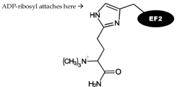
It is well established that the biochemical function of subunit A in the cytoplasm is to inhibit protein synthesis by irreversibly inactivating EF-2, an essential factor in the protein synthesis machinery of eukaryotic cells, through ADP-ribosylation [55,132]. EF-2 is an important factor in the elongation step for protein translation and the attachment of an ADP-ribosyl group to EF-2 renders it inactive.
Attachment of the ADP-ribosyl group occurs at an unusual derivative of histidine, called diphthamide (3-carboxyamino- 3(trimethylamino)propyl) histidine (see Fig. 1). The chemical reaction is as follows:
NAD + EF-2 → ADP-ribose-diphthamide histidine of EF-2 + nicotinamide + H+
As a result, protein synthesis is completely halted in the cells attacked by diphtheria toxin, which do not survive [24,88].
Fig. 1. Diphthamide histidine residue of EF-2.
When Corynebacterium diphtheriae infects humans, it generally has an incubation period of 2 to 5 days [129]. It typically infects anterior nasal, tonsillar and pharyngeal, laryngeal, cutaneous, ocular and genital areas, which means that
it can grow in any mucous membrane and in the skin [52,82].
As well as destroying local cells at infection sites, the diphtheria toxin can also enter the bloodstream and reach out to organ cells that have its receptor, typically heart and nerve cells. As a result, infection with a toxigenic strain can lead to systemic fever, myocarditis and neuritis, as well as the characteristic diphtheria symptoms of a sore throat and a grayish pseudomembrane on the mucosal areas, including the upper respiratory tract.
It has long been described that the most common complications of diphtheria are myocarditis and neuritis, because heart and nerve cells have relatively large numbers of receptors for the subunit B of diphtheria toxin on their cell membrane surface [52,53]. This explains the relatively high case fatality rates of between 5% and 10%. However, no toxigenic bacillus is found in the blood.
4. Conventional technologies for formalin-detoxified vaccine
Edwin Klebs described Corynebacterium diphtheriaein 1883, and its toxin was identified as the causative agent of diphtheria by Pierre Roux and Alexandre Yersin in 1888. Several decades later, in 1929, Gaston Ramon became the first to develop an effective diphtheria toxoid vaccine against four strains of toxigenic Corynebacterium diphtheriae. The vaccine elicits a common immune response against the toxins expressed from four different strains because these strains produce the same diphtheria toxin [82]. Ernst Lowenstein and Alexander Glenny paved the way for the development of this vaccine by inventing a way to convert tetanus and diphtheria toxins into toxoids using formaldehyde, which eliminates the toxicity of the toxins while preserving their antigenicity [47].
The following subsections describe the conventional technologies used to produce the current formalin-detoxified diphtheria vaccine.
4.1. Conventional production platform: chemical detoxification of the supernatant from culture broth
The conventional manufacture of diphtheria toxoid involves a combination of relatively simple methods, summarized in Fig. 2.
First, the aerobic fermentation of toxigenic Corynebacterium
diphtheriae is conducted under specific conditions such as
low inorganic iron concentration in the culture medium (iron deficiency conditions) in order to make the bacteria produce diphtheria toxin efficiently. Iron deficiency enhances the production of toxin because iron is required for the functioning of a repressor protein that blocks the transcription of the diphtheria toxin gene [55,83].The supernatant from the culture broth, containing diphtheria toxin and various impurities, is then incubated for 4 to 6 weeks at 37°C to detoxify the diphtheria toxin [103].
Subsequently, the various chemical forms of the toxoid formed by a chemical link (methylene bridge) between impurities and diphtheria toxin, or between diphtheria toxins, is purified using a combination of different purification methods such as ultrafiltration/diafiltration, salting-out, size exclusion chromatography, ion exchange, etc., with the choice depending on the manufacturer [5,16,86]. After a series of purification steps, the diphtheria toxoid has a purity of around 2,000 to 3,000 limits of flocculation/mg protein nitrogen (Lf/mgPN) [112].
Finally, the purified diphtheria toxoid in various chemical forms is adsorbed onto alum gel to increase the expression of IgG1 against the toxin by enhancing the Th2 immune response.
Fig. 2. Conventional manufacture of diphtheria toxoid.
As mentioned, the first detoxification method-using formaldehyde- was invented by Ernst Lowenstein in 1909 for inactivating crude tetanus toxin and byAlexander Glenny in 1921 for inactivating crude diphtheria toxin. This method was directly applied to the supernatant of culture broth due to the risk associated with handling purified toxins [49,67,95].
The drawback is that detoxification of the supernatant results in lot-to-lot inconsistency in the chemistry of the purified toxoid, and the differences in protein impurity profiles may lead to variability in vaccine quality and efficacy [24].
The chemical mechanism of inactivating diphtheria toxin present in the supernatant of culture broth is quite complex because the supernatant produced by current manufacturers contains many impurity proteins and peptides. Fortunately, the presence of high concentrations of impurity proteins, peptides and amino acids in the supernatant prevents toxoid polymerization and aggregation by formaldehyde [101]. The chemical reaction of crude diphtheria toxin and formaldehyde forms a connecting molecule called a methylene bridge between diphtheria toxin and impurity peptides. The mechanism of this reaction is simple, as shown in Fig. 3.
Fig. 3. Chemical reaction of crude diphtheria toxin and formaldehyde (HCHO).
However, this toxoid vaccine manufactured by the chemical reaction of crude diphtheria toxin and formaldehyde has some limitations. For example, its biochemical complexity due to reaction with many impurity proteins and peptides can lead to IgE-mediated immediate hypersensitivity when a booster is injected during adulthood [42,105]. To address this problem, a highly pure toxoid vaccine that does not induce this adverse event has been developed and brought to market. It is detoxified by the chemical reaction of purified diphtheria toxin, formaldehyde and amino acid [42,105].
4.2. Conventional production platform: chemical detoxification of purified diphtheria toxin
Numerous manufacturers have been producing diphtheria toxoid vaccine made by detoxifying purified diphtheria toxin with formaldehyde and amino acid (usually lysine), which is considered safer for vaccine recipients. This approach does potentially carry the risk that production workers might be exposed to the toxin during purification [104]. However, it is deemed that this risk can be eliminated by fully complying with the procedures described in current Good Manufacturing Practice (cGMP) guidelines.
The detoxification of highly purified toxin proteins using formaldehyde alone has two well-recognized limitations. First, the chemical toxoiding results in polymerization and aggregation due to the formation of methylene bridges among toxoid proteins [1,32,91]. Second, the resultant toxoid can reverse into a partially toxic state as time goes by [1,32,91]. After a long search by many scientists for ways to prevent this toxoid aggregation, in 1962 Linggood and colleagues found that toxoiding purified diphtheria toxin using the combination of formaldehyde and lysine not only prevents the synthesis of any toxoid aggregates but also produces a soluble diphtheria toxoid that does not revert into toxin over time [1,32,91]. Another surprising benefit of Linggood’s toxoiding method is that it can enhance the in vivo immunologic efficacy of the diphtheria toxoid. This novel toxoiding reaction is shown in Fig. 4.
Fig. 4. Toxoiding reaction of purified diphtheria toxin with formaldehyde plus lysine.
The diphtheriavaccine produced by this novel toxoiding reaction contains highly pure diphtheria toxoid with very low levels of process-related peptide and protein impurities.
The toxoid has a purity of around 3,300 Lf/mgPN, which means that the drug substance is almost completely pure.
However, even this method still has several disadvantages.
First, the random chemical reaction leads to an unpredictable location and number of methylene bridges, and therefore toxoid heterogeneity, which may become a quality issue with respect to the International Conference on Harmonization (ICH) guidelines for product characterization. For this reason, scientists have been trying to develop a genetically detoxified diphtheria vaccine, which would eliminate any heterogeneity issue. Otherdisadvantages are that the chemical detoxification process generally takes several weeks and may pose a potential safety risk to production workers [76,93].
Having said that, diphtheria vaccine manufactured by the chemical detoxification of purified toxin does offer benefits to vaccine recipients. It does not sensitize vaccinated individuals to various impurities and consequently does not induce side effects like IgE-mediated acute hypersensitivity when booster vaccines are injected in adulthood.
5. Genetically detoxified vaccine
Scientists have long seen a need for a genetically detoxified diphtheria vaccine, for several reasons. First, genetic detoxification would completely eliminate the chemical reaction step from the manufacturing process and speed up the process of manufacturing diphtheria vaccine [94]. Second, it would eliminate the risk of production workers being exposed to diphtheria toxin during the purification process. Third, it would eliminate some potential side effects, including IgE- mediated hypersensitivity, without requiring the cumbersome chemical detoxification step.
Hence, it is well recognized that the development of a genetically detoxified diphtheria vaccine is of great importance for all stakeholders, including vaccine manufacturers and production workers, healthcare professionals and vaccine recipients.
5.1. Genetically derived diphtheria toxoid
The genetic detoxification of diphtheria toxin was first discovered in 1971 by Tsuyoshi Uchida and Alwin M.
Pappenheimer Jr., who reported the random mutagenesis of the bacteriophage gene coding for diphtheria toxin [124].
Three types of randomly inactivated toxoids called CRM (cross-reacting material) were selected and investigated for their cytotoxicity, enzymatic activity and receptor-binding activity [60,61,71,101,126]. One type shows an amino acid substitution in subunit A only; it is represented by the mutants CRM176 and CRM197, which have G128D and G52E mutations, respectively. Another type displays a substitution in
subunit B only; its randomly established mutants are CRM103 and CRM107, which have S508F and S525F mutations, respectively. A third type with several mutations along subunit A and subunit B is represented by the randomly established mutant CRM228, which has G79D, E162K, S197G, P378S and G431S mutations [63].
Of all the mutants mentioned above, the most promising for vaccine development appeared to be CRM197, in which glycine located at the 52nd amino acid position of the native diphtheria toxin is substituted by glutamic acid. This mutant does not show any cytotoxicity or enzymatic activity and retains complete receptor-binding activity [46]. However, CRM197 turned out to have lower immunogenicity than formalin-inactivated toxoid, which is a practical obstacle toits use in the vaccine field [50,97].
Nonetheless, several studies have explored the potential use of CRM197 as a diphtheria vaccine. For instance, a CRM197- based nasal vaccine with chitosan as an adjuvant was reported to induce a Th2-biased immune response in mice and guinea pigs, and results from the mice study indicate it could have value as a booster for adolescents and adults previously immunized with the conventional diphtheria toxoid vaccine adsorbed to alum [73,74]. Similarly, another study reported that the transcutaneous immunization of CRM197 alone or with a mucosal adjuvant (a partially detoxified mutant of heat-labile toxin) shows potential for use as a booster [121].
Another research group showed that an alum-absorbed CRM197 vaccine injected intramuscularly elicited high IgG antibody titers in guinea pigs, while a spray-dried CRM197 nanoparticle vaccine delivered through a pulmonary route elicited high IgA antibody titers in this animal model [89].
Based on these positive findings, several clinical studies using CRM197 to substitute for the conventional toxoid vaccine have been carried out and have shown some positive results in humans [11,73,79,111]. However, no diphtheria vaccine based on CRM197 has been commercialized to date.
A look at the supportive use of CRM197 reveals that this mutant has been widely used (in the research, clinical development and commercial stages of vaccine innovation) as an efficient carrier protein for the conjugation of polysaccharide antigens in conjugate vaccines, including pneumococcal vaccine,
Haemophilus influenzae type b vaccine, meningococcal
vaccine, typhoid vaccine (Vi CPS) and cholera vaccine (CPS of V. cholerae O139) [31,39,48,69,77]. Generally, CRM197 is recovered from the culture broth of Corynebacteriumdiphtheriae C7 (β197) grown in the complex medium containing
casamino acid and yeast extract, and highly purified through the process combination of ultrafiltration, ammonium sulfate precipitation, and ion-exchange chromatography [101]. It is then directly conjugated with polysaccharide antigens by reductive amination [4,28,106].CRM197 has been reported to have anti-tumor activity by enhancing cellular and inflammatory immune responses. For example, it has been associated with significant increases in levels of circulating neutrophils and TNF-α, directed especially against cancers such as breast cancer, neuroblastoma and lung cancer, in which heparin-binding EGF-the binding receptor of diphtheria toxin- is overexpressed as a tumor-associated antigen [14,123]. Moreover, recently published clinical findings indicate that CRM197 may significantly reduce atherosclerotic stenosis [15]. Interestingly, researchers have discovered that CRM197 has very weak EF-2 ADP ribosylation activity, in contrast to the widely held belief that its enzymatic activity has been lost [64]. This finding would appear to explain the
in vivo mechanism of an antitumor effect of CRM197.
In summary, as CRM197 has been used successfully in the conjugate vaccine field, it is expected to provide a range of medical benefits to humans in the near future.
5.2. Plasmid DNA vaccines
Since the advent of molecular biology techniques in the 1980s, many researchers have explored the advantages of plasmidDNA vaccines. First, this type of vaccine provides humans with superior protection against pathogens because it makes target epitopes bind to major histocompatibility complex (MHC) class I and class II molecules, and subsequently elicits both Th1 and Th2 immune responses at the same time.
Consequently, infected cells and pathogens can be much more easily removed by the cellular and humoral immune responses induced by DNA vaccines [117]. Second, DNA vaccine is thought to induce a long-lasting immune response, meaning that a single shot hopefully can completely immunize vaccine recipients [51]. Third, a DNA vaccine is very convenient when it comes to storage because it does not need a cold chain [125].
Finally, it is very easy to manufacture, which leads to lower costs [115].
Given the extracellular growth characteristics of toxigenic
Corynebacterium diphtheria strains, a DNA vaccine against
diphtheria is thought to be particularly useful for countries without an adequate cold chain infrastructure.Indeed, the development of a DNA vaccine against diphtheria would seem to be a necessity for protecting populations in developing regions against this disease.No studies of a DNA vaccine against diphtheria have been published so far. However, there are some published in
vivostudies of DNA vaccine against tetanus, of which the
toxin has same A-B structure as diphtheria toxin, where subunit A (zinc endopeptidase) is responsiblefor toxicity and subunit B binds to a cell surface receptor (ganglioside GM1) on the neuronal membrane [3,122]. There is also one published report on the use of DNA coding for subunit A of diphtheria toxin as a treatment for pancreatic cancers overexpressingmesothelin as a tumor-associated antigen [116].
Despite the lack of research into a DNA vaccine against diphtheria, this is still thought to have high potential to one day replace the current diphtheria toxoid vaccine, due to the various advantages mentioned earlier.
6. Cutting-edge technologies for innovative vaccines
6.1. Oral and transcutaneous vaccine delivery systems
Parenteral immunization has drawbacks for vaccine recipients, healthcare workers and public health. In addition to its potential to cause discomfort or pain, it requires considerable medical training and carries therisk of potential safety issues such as needle-handling mistakes [117]. From the immunologic point of view, oral immunization appears to be more effective because it can elicit mucosal (IgA antibody) immunity as well as systemic (IgG antibody) immunity. This is important considering that the respiratory tract is a major infection route of Corynebacterium diphtheria.In relation to oral immunization, Peyer’s patch M cells were reported to be able to transport biodegradable microspheres less then 5 μm in diameter [37]. Therefore, the development of efficient needle-free, oral vaccine delivery systems-a hope long held by all involved with immunization- needs to consider the particle-transporting capability of the M cell.
Recent advances in biochemistry- and molecular biology-based technologies have sparked research into innovative vaccine delivery systems of this kind. For instance, microparticles or nanoparticles using starch, modified dextran, modified polylactide (PLA) and/or modified poly-lactic glycolic acid (PLGA), as well as liposomal particles dressed with chitosan and poly-vinylic alcohol (PVC), may offer improved stability of vaccine antigens and convenient oral immunization. Edible vaccines based on transgenic plants (tomato, banana, potato etc.) may also provide humans with much more convenient administration through the oral route. Other researchers have reported that unmethylated CpG motifs may facilitate the oral delivery of protein antigens. Finally, amicroneedle- mediated vaccine delivery system is thought to warrant further investigation as a convenient method of transcutaneous immunization, although it does not induce an IgA immune response [2,8,33,34,59,65,72,107,108,110,117-121].
An oral diphtheria vaccine based on formaldehyde- and lysine-inactivated CRM197 conjugated with a starch microparticle was trialed in humans some years ago, following successful tests in mice. Human antisera were obtained according to clinical protocol and assayed for anti-diphtheria toxin antibodies using enzyme-linked immunosorbent assay (ELISA) and for diphtheria toxin-neutralizing antibodies using the Vero cell method. However, the clinical trial failed,
Immunization route
Representative
delivery systems Application Current status or
potential
Oral
Starch-based
microparticle Conjugation with CRM197 Success in mice; failure
in clinical trials PLGA or PCL
nanoparticle Encapsulation of diphtheria toxoid No further study
Liposome with
chitosan and PVA Encapsulation of diphtheria toxoid Interesting technology with potential Edible tomato Transfection of fusion gene comprising various immunodominant
epitopes of diphtheria, tetanus and pertussis toxins
Interesting technology with potential
CpG motif Oral delivery of tetanus toxoid antigen No further study
Skin
(Transcutaneous)
Microneedle- mediated
Diphtheria toxoid-containing liposome or vesicle together with microneedle arrays
Interesting technology with potential Table 1. Summary of research into oral or transcutaneous diphtheria vaccines
presumably due to the instability of the starch microparticle in the human gastrointestinal tract and the different structure of gut and Peyer’s patches between mice and humans [109,110,111]. Further efforts are clearly needed to discover novel polymers that are suitable for use in the human gut and Peyer’s patches.
Another approach to oral delivery systems for convenient immunization against diphtheria uses nanoparticles of modified PLA and PLGA, or poly-epsilon-caprolactone (PCL) and PLGA copolymer, in which diphtheria toxoid is encapsulated by spray drying. In vitro results indicate that PCL nanoparticles have the best capability for transporting diphtheria toxoid into Caco-2 cells, a human colon carcinoma cell line [59,119].
Liposomal technology has recently been tried for efficient oral immunization against diphtheria. A liposome containing diphtheria toxoid is combined with chitosan and PVA in order to enhance particle stabilization (by preventing particle aggregation) and promote its affinity with mucin.
Because both chitosan and PVA are well-known as mucoadhesive polymers and the resultant particle size was estimated to be around 383 nm, in vitrostudies showed that the liposomal particles were well adsorbed by mucin and diphtheria toxoid was released in a controlled manner [107].
Although this liposome-chitosan-PVA particle has not yet been tested in animals or humans, it is thought to have good potential for the oral administration of diphtheria toxoid.
Research has been conducted into a range of edible vaccines based on transgenic plants, including plants expressing diphtheria antigen as a component of the triple-antigen DTP combination. Of the various approaches studied, one that uses the recombinant fusion gene coding for protective epitopes of the three toxins (diphtheria, tetanus and pertussis toxins) looks very interesting for the development of an edible tomato vaccine platform and shows considerable promise due to the consistent expression of six antigenic epitopes (two epitopes from each toxin) by the fused gene
integrated into the tomato chromosome [120].
An interesting study in mice around 10 years ago investigated the oral administration of tetanus toxoid vaccine containing unmethylated CpG motif, and showed this to be an effective oral delivery system [72]. It was the first reported use of unmethylated CpG motif in orally delivered protein antigens.
Although this approach has not been tried using diphtheria toxoid, it seems likely that unmethylated CpG motif has a potential application for the oral delivery of diphtheria vaccine.
In addition to oral immunization, transcutaneous vaccine delivery has long been the subject of research. Transcutaneous immunization has been recognized as one of the best ways to elicit a strong systemic immune response because of the presence of specialized dendritic cells (Langerhans cells) in the epidermis. Commercialization of transcutaneous immunization can therefore offer several advantages including convenient administration and strong immune response.
Since research into epidermal powder immunization has been stopped-despite successful results-due to the complexity and high investment costs associated with its use in humans, attention has focused on microneedle-mediated vaccine delivery systems [19]. The latest of these studies explored the use of diphtheria toxoid-containing cationic liposomes or anionic surfactant-based vesicles, together with subunit B of cholera toxin (CTB) as an adjuvant [33]. However, neither the liposome nor the vesicle performed strongly in terms of eliciting anti- diphtheria toxin IgG antibodies and neutralizing antibodies.
Diphtheria toxoid plus CTB without liposome or vesicle elicited an immune response similar to that achieved with conventional alum-adjuvanted vaccine immunized subcutaneously after 2nd boost. Further study of liposomes and vesicles for transcutaneous immunization appears to be warranted.
In summary, various studies of oral and microneedle- mediated immunization against diphtheria have been conducted, and some show interesting potential for future research and development, as shown in table 1. However, no
results applicable to clinical trials have emerged so far.
6.2. Heat- and freeze-stable technology
Unlike chemical medicines, many vaccines require cold-chain storage in order to maintain the biologically active status of their main ingredients (vaccine antigens) until use. This reflects the thermal instability of most vaccine antigens, mainly due to their particular biochemical and structural characteristics [12,27,43]. In addition, alum-adjuvanted vaccines- such diphtheria toxoid vaccine-need to avoid freezing due to the strong tendency to agglomeration peculiar to aluminum salts (alum) in a freeze-thaw event [13,20,23,68].
In spite of longstanding recognition of the thermal instability of vaccine components, a substantial proportion of vaccine supplies are wasted or unintentionally administered in suboptimal condition due mainly to a shortage or lack of cold chain facilities, especially in the developing world [6,80,113].
Therefore, developing and commercializing technologies to increase the thermal stability of vaccine antigens and alum is important for all stakeholders in the immunization field, including vaccine recipients, vaccine manufacturers and healthcare providers.
There have been many studies aimed at discovering affordable, nontoxic and efficient excipients or novel agents for use as a thermal stabilizer. Recently, various polyols such as glycerin, PEG300 and propylene glycol have been tested as potential excipients for preventing the agglomeration of alum-adjuvanted combination vaccines containing diphtheria toxoid during freeze-thaw cycles [13]. The results of this research suggest that such agglomeration is completely preventable when an appropriate concentration of propylene glycol, glycerin or PEG300 is added to the vaccine formulation. Although the ability of these polyols to prevent agglomeration was assessed by particle size analysis and not in vivo testing, this technology is thought to be very promising for developing a freeze-stable alum-adjuvanted diphtheria toxoid vaccine.
Despite the importance of vaccine heat stability due to the difficulties developing countries face in maintaining the cold chain, no studies on the use of excipients as a heat stabilizer have been performed for alum-adjuvanted diphtheria toxoid vaccine to date. However, according to a recent study of the heat stability of alum-adjuvanted hepatitis B vaccine, vaccine containing 40 mM histidine and 40 mM phosphate at relatively low pH (5.2) as excipients was stable with respect to particle size and in vivo potency when stored at 37°C and 45°C for 6 months [58]. Hence, combining polyols against freeze with both histidine and phosphate against heat shows potential for developing an alum-adjuvanted diphtheria toxoid vaccine with heat and freeze stability.
One interesting study aimed at overcoming cold chain insufficiency by developing a heat-stable diphtheria toxoid
vaccine made use of a protein-coated microcrystal (PCMC) technology first developed in 2003 [87]. The vaccine, consisting of diphtheria toxoid-coated microcrystals prepared according to the procedure suggested by Parker, was tested for its heat stability at a range of temperatures (e.g. 4, 25, 37 and 45°C) and investigated for its ability to elicit an IgG immune response in Balb/c mice after intramuscular injection. The study found that the immunogenicity of the vaccine was not affected by 2 days of exposure to 45°C prior to administration.
In spite of some disadvantages, such as incompatibility with alumsalts and preparation complexity, PCMC technology is thought to be promising in the light of the current trend to develop novel adjuvants and the progress of related technologies.
6.3. Novel adjuvants
An adjuvant is defined as a vaccine component that enhances the immune response to the vaccine’s antigen. The term
‘adjuvant’ was coined by Ramon in 1926 to reflect its immunologic functions [100]. These functions include depot generation at the injection site; targeting antigen to innate immune cells such as dendritic cells and macrophages;
antigen presentation modulation from MHC class II to MHC class I, or vice versa; immune modulation from Th2 immune response to Th1 immune response, or vice versa, and from Th-independent immune response to Th-dependent immune response; and preferential stimulation of humoral or cytotoxic T lymphocyte (CTL) immune response [22,26,41,114]. In addition to these immunologic functions, an adjuvant generally has a dose-sparing effect, which may lower the vaccine price.
Adjuvants can be broadly grouped into three classes based on toll-like receptor (TLR) independence, dependence or both.
TLR-dependent adjuvants have a pathogen-associated molecular pattern (PAMP), whereas TLR-independent adjuvants do not;
some adjuvants are a combination of TLR-independent and TLR-dependent components.
Commercialized TLR-independent adjuvants include conventional alum salts, which is approved by national regulatory authorities worldwide; the virosome (a kind of liposome developed by Crucell) which is currently approved by most regulatory authorities except for the US Food and Drug Administration (FDA); and two oil-in-water emulsions (MF59 from Novartis and AS03 from GSK), which are approved by the European Medicines Agency (EMA).
Representative TLR-dependent adjuvants are TLR4-binding momophosphoryl lipid A (MPL, Dynavax), which is approved for use in Argentinaand is undergoing clinical evaluation in the United States; TLR5-activating flagellin (Vaxinnate), which is in clinical trials; and TLR9-stimulating unmethylated CpG motifs (Dynavax, Pfizer, Intercell). Examples of combination TLR-independent/TLR-dependent adjuvants are the EMA-
approved adjuvant AS04 (MPL + alum salts, GSK); and two combinations under clinical development by GSK: AS01 (MPL + liposome + QS21) and AS02 (MPL + oil-in-water emulsion + QS21) [7,8,38,40,44]. The best characterized adjuvants are conventional alum salts, oil-in-water emulsion, liposome and QS-21, among TLR-independent adjuvants;
and PAMPs such as unmethylated CpG motifs, flagellin and MPL among TLR-dependent adjuvants.
To date, all diphtheria toxoid vaccines have been manufactured with their toxoids adsorbed onto alum salts. The main functions of alum salts are to induce a long-lasting immune response by forming diphtheria toxoiddepot at the injection site, to elicit a Th2 immune response and to help target diphtheria toxoid towards dendritic cells and macrophages. One of the disadvantages of alum salts as an adjuvant is that it can stimulate an IgE immune response, which may lead to anaphylaxis in susceptible vaccine recipients, especially those given low-priced diphtheria toxoid vaccines that have many impurities due to their simple manufacturing process.
Incorporating novel adjuvants (already commercialized and under development) into relatively impure diphtheria toxoid vaccines may therefore be important for preventing IgE- mediated immediate hypersensitivity reactions. Good candidates may be MPL, unmethylated CpG motifs and oil-in-water emulsion, which are relatively inexpensive compared to other PAMPs. MPL, a TLR4 ligand, is known to induce humoral and cellular immune responses. Unmethylated CpG motif, a TLR9 ligand, can elicit humoral and cellular immune responses simultaneously when fused to a protein antigen, as reported in a study involving hepatitis B surface antigen (HBsAg).
Oil-in-water emulsion has been shown to stimulate the humoral immune response [66,99,127].
All three adjuvants look very promising for use in diphtheria toxoid vaccine, as the humoral immune response is important for the neutralization of diphtheria toxin.
7. Perspectives
Diphtheria toxoid vaccine has been incorporated into a number of combination vaccines, including Td (tetanus toxoid and diphtheria toxoid for adults), DTaP (diphtheria toxoid, tetanus toxoid and acellular pertussis protein antigens), DTwP (diphtheria toxoid, tetanus toxoid and whole-cell pertussis antigen) and DTwP-HepB-Hib (pentavalent vaccine comprising diphtheria toxoid, tetanus toxoid, whole-cell pertussis antigen, hepatitis B surface antigen and Haemophilus influenzae type B antigen, manufactured by Crucell, Panacea Biotech and Shantha Biotechnics).
The novel technologies discussed in this paper that show potential for improving the diphtheria toxoid vaccine, as
well as vaccines against other diseases, should be worth applying to combination formulations containing diphtheria toxoid antigen. Future technologies that are thought to be applicable are discussed below.
7.1. Current vaccines
Conventional diphtheria vaccines are likely to remain in use for some time because they have been shown to be effective in preventing diphtheria-providing people are immunized according to the vaccination schedule recommended by the World Health Organization. However, many developing countries face major obstacles to effective immunization. A shortage or lack of essential infrastructure and resources such as the cold chain, vaccination clinics and trained healthcare providers has resulted in intermittent diphtheria outbreaks.
Therefore, of all the innovative technologies discussed in this paper, adding heat- and freeze-stable agents into current vaccine formulations, including combination vaccines, appears to be the most appropriate, rational and urgent step towards ensuring that all people worldwide are protected against diphtheria. The introduction of heat- and freeze-stable vaccines would substantially reduce vaccine wastage and the unintentional use of vaccines in suboptimal condition, especially in the developing world.
7.2. Innovative vaccine platforms
Many novel vaccine technologies have been studied and developed since the 1920s, when the first diphtheria vaccine was brought to market. These innovations address several fundamental limitations of the first-generation vaccine.
Ongoing progress with the exploration and application of these technologies is likely to lead to the development of various novel vaccines, which have genetically detoxified diphtheria toxoid or DNA plasmid as their active pharmaceutical ingredient, contain novel adjuvants that enhance their protective efficacy, and include heat- and freeze-stable agents that make them easier to store and use. These new-generation vaccines will also make use of innovative delivery systems, so that they can be administered transcutaneously, orally or in an edible form.
Considering the future of technology platforms now in research and development, innovative vaccines seem to be developing in three directions.
First, the DNA plasmid vaccine with a fusion gene comprising various immunodominant epitopes of diphtheria, tetanus and pertussis toxins appears to be a good candidate for further development (It would be technologically difficult for this vaccine to also contain immunodominant epitopes of HBsAg, the hepatitis B surface antigen, due to the virus-like characteristics of this antigen). A combination DTaP vaccine based on DNA plasmid technology would not require a cold chain and would be relatively low in cost-potential benefits
that are particularly attractive for developing countries.
Second, a genetically detoxified vaccine combined with several other novel technologies-such as an innovative vaccine delivery system, novel TLR-independent or TLR-dependent adjuvants, and heat- and freeze-stable agents-seems a good candidate to attract strong attention from vaccine makers. It also seems likely that industry will want to further develop this platform, making the vaccine even more convenient by adopting technologies that enable it to be administered transcutaneously ororally. It is thought that these vaccine technologies based primarily on genetic detoxification can be applied to the development of a combination DTaP vaccine containing diphtheria toxoid, tetanus toxoid and acellular pertussis antigens.
Third, edible vaccine technology is thought to have strong potential as a platform for a new-generation DTaP vaccine, considering its benefits like ease of vaccination and no need for the cold chain. In addition, the technology would allow HBsAg to be added to this vaccine combination.
In conclusion, several innovative technologies that are currently under investigation and development open up the possibility of developing vaccines that will overcome all the limitations of the current diphtheria vaccine. These new- generation vaccines will offer benefits in terms of quality, thermal stability and ease of immunization. The successful pursuit of these technological opportunities can be expected to lead to a significant increase in global immunization against diphtheria-usually combined with tetanus and pertussis-in the near future.
Acknowledgements
This work was supported by Crucell and Crucell/Berna Biotech Korea. I would also like express my deep appreciation to Dr Giuseppe Marzio and Ms Andrea Dingemans of Crucell, and Mr DongUk Kim of Crucell/Berna Biotech Korea for their help with preparing this review article.
References
1. Akama, K., S. Kameyama, S. Otani, S. Sadahiro, and R. Murata (1971) Reversion of toxicity of diphtheria toxoid. Japanese Journal of Medical Science & Biology 24: 183-187.
2. Amorij, J. P., T. A. Westra, W. L. Hinrichs, A. Huckriede, and H. W. Frijlink (2007) Towards an oral influenza vaccine:
comparison between intragastric and intracolonicdelivery of influenza subunit vaccine in a murine model. Vaccine 26:
67-76.
3. Anderson, D. G., W. Peng, A. Akinc, N. Hossain, A. Kohn, R.
Padera, R. Langer, and J. A. Sawicki (2004) A polymer library approach to suicide gene therapy for cancer. Proceedings of
the National Academy of Sciences of the United States of America 101: 16028-16033.
4. Anderson, P., J. Treanor, S. Porcelli, and M. Pichichero (2003) Non-interference between two protein carriers when used with the same polysaccharide for pneumococcal conjugate vaccines in 2-year-old children. Vaccine 21: 1554-1559.
5. Antoni, G., M. Bigio, G. Borri, M. C. Casagli, and P. Neri (1983) Purification of diphtheria toxin by chromatography on cibacron blue-sepharose. Experientia 39: 885-886.
6. Arya, S. C. (1994) Human immunization in developing countries: practical and theoretical problems and prospects.
Vaccine 12: 1423-1435.
7. Atmar, R. L. and W. A. Keitel (2009) Adjuvants for pandemic influenza vaccines. Current Topics in Microbiology and Immunology 333: 323-344.
8. Bae, K., J. Choi, Y. Jang, S. Ahn, and B. Hur (2009) Innovative vaccine production technologies: the evolution and value of vaccine production technologies. Archives of Pharmacal Research 32: 465-480.
9. Bell, F., A. Martin, C. Blondeau, C. Thornton, J. Chaplais, and A. Finn (1996) Combined diphtheria, tetanus, pertussis, and Heamophilus influenzae type b vaccines for primary immunization. Archives of Disease in Childhood 75: 298-303.
10. Belsey, M. A. (1970) Isolation of Corynebacterium diphtheriae in the environment of skin carriers. American Journal of Epidemiology 91: 294-299.
11. Botet Asensi, F. I., A. Veronese, M. Del Carmen Otero, M.
Desamparados Tamarit Pérez, J. L. Hontangas Lopez, and S.
Viviani (2003) Immunogenicity and safety in infants of a DTwPHib full liquid vaccine. Acta Paediatrica 92: 541-545.
12. Brandau, D. T., L. S. Jones, C. M. Wiethoff, J. Rexroad, and C. R. Middaugh (2003) Thermal stability of vaccines. Journal of Pharmaceutical Sciences 92: 218-231.
13. Braun, L. J., A. Tyagi, S. Perkins, J. Carpenter, D. Sylvester, M. Guy, D. Kristensen, and D. Chen (2009) Development of a freeze-stable formulation for vaccine containing aluminum salt adjuvants. Vaccine 27: 72-79.
14. Buzzi, S., D. Rubboli, G. Buzzi, A. M. Buzzi, C. Morisi, and F.
Pironi (2004) CRM197 (nontoxic diphtheria toxin): effects on advanced cancer patients. Cancer Immunology, Immunotherapy 53:1041-1048.
15. Buzzi, S., G. Buzzi, A. M. Buzzi, and S. Martini (2007) CRM197:
reduction of atherosclerotic stenoses in carotids of three elderly patients. Therapy 4: 293-298.
16. Carroll, S. F., J. T. Barbieri, and R. J. Collier (1988) Diphtheria toxin: purification and properties. Methods in Enzymology 165: 68-76.
17. Atkinson, W., J. Hamborsky, L. McIntyre, and S. Wolfe (2007) Epidemiology and Prevention of Vaccine-preventable Diseases (the pink book), 10th ed., pp. 59-70. Centers for Disease Control and Prevention, Public Health Foundation, Washington DC, USA.
18. Cerdeno-Tarraga, A. M., A. Efstratiou, L. G. Dover, M. T.
Holden, M. Pallen, S. D. Bentley, G. S. Besra, C. Churcher, K.
D. James, A. De Zoysa, T. Chillingworth, A. Cronin, L. Dowd, T. Feltwell, N. Hamlin, S. Holroyd, K. Jagels, S. Moule, M. A.
Quail, E. Rabbinowitsch, K. M. Rutherford, N. R. Thomson, L.
Unwin, S. Whitehead, B. G. Barrell, and J. Parkhill (2003) The complete genome sequence and analysis of Corynebacterium diphtheriae NCTC13129. Nucleic Acids Research 31: 6516-6523.
19. Chen, D., C. A. Erickson, R. L. Endres, S. B. Periwal, Q. Chu,
C. Shu, Y. F. Maa, and L. G. Payne (2001) Adjuvantation of epidermal powder immunization. Vaccine 19: 2908-2917.
20. Chen, D., A. Tyagi, J. Carpenter, S. Perkins, D. Sylvester, M.
Guy, D. D. Kristensen, and L. J. Braun (2009) Characterization of the freeze sensitivity of a hepatitis B vaccine. Human Vaccines 5: 26-32.
21. Chen, W. C. And L. Huang (2005) Non-viral vector as vaccine carrier. Advances in Genetics 54: 315-337.
22. Chodaczek, G. (2004) Adjuvants as factors improving efficiency of vaccination. Postȩy higieny i medycyny doświadczalnej 58: 47-59.
23. Clausi, A., J. Cummiskey, S. Merkley, J. F. Carpenter, L. J.
Braun, and T. W. Randolph (2008) Influence of particle size and antigen binding on effectiveness of aluminum salt adjuvants in a model lysozyme vaccine. Journal of Pharmaceutical Sciences 97: 5252-5262.
24. Collier, R. J. (1975) Diphtheria toxin: mode of action and structure. Bacteriological Reviews 39: 54-85.
25. Collier, R. J. (2001) Understanding the mode of action of diphtheria toxin: a perspective on progress during the 20th century. Toxicon 39: 1793-1803.
26. Cooper, P. D. (1994) The Selective Induction of Different Immune Responses by Vaccine Adjuvants. pp. 125-158. In: G.
L. Ada (eds.). Strategies in Vaccine Design, Landes, Austin, TX, USA.
27. Corbel, M. J. (1996) Reasons for instability of bacterial vaccines.
Developments in Biological Standardization 87: 113-124.
28. Crane, D. T., B. Bolgiano, and C. Jones (1997) Comparison of the diphtheria mutant toxin, CRM197, with a Haemophilus influenzae type-b polysaccharide-CRM197 conjugate by optical spectroscopy. European Journal of Biochemistry 246:
320-327.
29. Crosbie, W. E. and H. D. Wright (1941) Diphtheria bacilli in floor dust. Lancet 237: 656-659.
30. Danilova, E., A. Shiryayev, V. Skogen, E. K. Kristoffersen, and H. Sjursen (2005) Short-term booster effect of diphtheria toxoid in initially long-term protected individuals. Vaccine 23: 1446-1450.
31. Deeks, E. D. (2010) Meningococcal quadrivalent (serogroups A, C, w135, and y) conjugate vaccine (Menveo): in adolescents and adults. Bio. Drugs 24: 287-297.
32. Del Giudice, G., M. Pizza, and R. Rappuoli (1998) Molecular basis of vaccination. Molecular Aspects of Medicine 19: 1-70.
33. Ding, Z., S. M. Bal, S. Romeijn, G. F. Kersten,W. Jiskoot, and Bouwstra (2011) Transcutaneous immunization studies in mice using diphtheria toxoid-loaded vesicle formulations and a microneedle array. Pharmaceutical Research 28: 145-158.
34. Ding, Z., E. Van Riet, S. Romeijn, G. F. Kersten, W. Jiskoot, and J. A. Bouwstra (2009) Immune modulation by adjuvants combined with diphtheria toxoid administered topically in BALB/c mice after microneedle array pretreatment.
Pharmaceutical Research 26: 1635-1643.
35. Efstratiou, A. and R. C. George (1999) Laboratory guidelines for the diagnosis of infections caused by Corynebacterium diphtheriae and C. ulcerans. Communicable Disease and Public Health 2: 250-257.
36. Efstratiou, A., K. H. Engler, and A. De Zoysa (1998) Diagnosis and epidemiology of diphtheria. Methods in Molecular Medicine 15: 191-212.
37. Eldridge, J. H., R. M. Gilley, J. K. Staas, Z. Moldoveanu, J. A.
Meulbroek, and T. R. Tice (1989) Biodegradable microspheres:
vaccine delivery system for oral immunization. Current Topics in Microbiology and Immunology 146: 59-66.
38. El Sahly, H. (2010) MR59™ as a vaccine adjuvant: a review of safety and immunogenicity. Expert Review of Vaccines 9:
1135-1141.
39. Eskola, J., T. Kilpi, A. Palmu, J. Jokinen, J. Haapakoski, E.
Herva, A. Takala, H. Käyhty, P. Karma, P., R. Kohberger, G.
Siber, and P. H. Mäkelä (2001) Efficacy of a pneumococcal conjugate vaccine against acute otitis media. The New England Journal of Medicine 344: 403-409.
40. Felnerova, D., J. F. Viret, R. Glück, and C. Moser (2004) Liposomes and virosomes as delivery systems for antigens, nucleic acids and drugs. Current Opinion in Biotechnology 15: 518-529.
41. Flebbe, L. M. and H. Braley-Mullen (1986) Immunopotentiating effects of the adjuvants SGP and Quil A. I. Antibody responses to T-dependent and T-independent antigens. Cellular Immunology 99: 119-127.
42. Fratelli, F., J. Abrahão-Neto, A. T. Caricati, M. M. Borges, R. Guidolin, and C. P. Caricati (2011) An alternative method for purifying and detoxifying diphtheria toxin. Toxicon 57:
1093-1100.
43. Galazka, A., J. Milstien, and M. Zaffran (1998) Thermostability of vaccines, World Health Organization, Geneva. Switzerland.
44. Garçon, N., P. Chomez, and M. Van Mechelen (2007) GlaxoSmithKline Adjuvant Systems in vaccines: concepts, achievements and perspectives. Expert Review of Vaccines 6:
723-739.
45. Gardner, P. (2001) Issues related to the decennial tetanus- diphtheria toxoid booster recommendations in adults. Infectious Disease and Clinics of North America 15: 143-153.
46. Giannini, G., R. Rappuoli, and R. Ratti (1984) The amino-acid sequence of two non-toxic mutants of diphtheria toxin:
CRM45 and CRM197. Nucleic Acids Research 12: 4063-4069.
47. Glenny, A. T. and H. J. Sudmersen (1921) Notes on the production of immunity to diphtheria toxin. The Journal of Hygiene 20: 176-220.
48. Godefroy, S., M. Peyre, N. Garcia, S. Muller, D. Sesardic, and C. D. Partidos (2005) Effect of skin barrier disruption on immune responses to topically applied cross-reacting material, CRM(197) of diphtheria toxin. Infection and Immunity 73:
4803-4809.
49. Grabenstein, J. D. (2010) Toxoid Vaccines, pp. 105-124.
In: A. W. Artenstein (eds.). Vaccines: A Biography. Springer, NY, USA.
50. Gupta, R. K., R. J. Collier, R. Rappuoli, and G. R. Siber (1997) Differences in the immunogenicity of native and formalinized cross reacting material (CRM197) of diphtheria toxin in mice and guinea pigs and their implications on the development and control of diphtheria vaccine based on CRMs. Vaccine 15: 1341-1343.
51. Gurunathan, S., C. Y. Wu, B. L. Freidag, and R. A. Seder (2000) DNA vaccines: a key for inducing long-term cellular immunity. Current Opinion in Immunology 12: 442-447.
52. Hadfield, T. L., P. McEvoy, Y. Polotsky, V. A. Tzinserling, and A. A. Yakovlev (2000) The pathology of diphtheria. The Journal of Infectious Diseases 181: S116-S120.
53. Havaldar, P. V., M. N. Sankpal, and R. P. Doddannavar (2000) Diphtheritic myocarditis: clinical and laboratory parameters of prognosis and fatal outcome. Annals of Tropical Paediatrics 20: 209-215.