DOI:10.5125/jkaoms.2010.36.4.320
Abstract (J Korean Assoc Oral Maxillofac Surg 2010;36:320-4)
Ⅰ.
서 론하악 우각부 골절은 제2대구치의 후방에서 하악지 (ramus)와 하악체(body)를 연결하는 부위의 골절을 말한다
1. 하악 골절 중에서 20%를 차지하며, 과두부(36%)와 체부
(21%) 다음으로 흔하게 발생한다
2. 이에 대한 치료로 다른
하악 골절 부위와 마찬가지로 관혈적 정복과 함께 open reduction and internal fixation (ORIF)을 추천하는데 이는 하 악골의 조기 가동을 가능하게 하여 환자의 불편을 크게 줄
여줄 수 있다
3. 이에 하악골 우각부 골절의 ORIF에서 어떤 고정판을 어디에 어떻게 식립해야 하는지에 대한 많은 논 의가 있어 왔다
4,5. 근래에는 분쇄 골절이나 심한 변위를 보 이는 불리 골절(unfavorable fracture)의 경우가 아니면 Champy’s line을 따라 miniplate를 식립하는 것이 우각부 골 절의 표준적인 ORIF 방법이 되었다. 이후 1개 또는 2개의 miniplate를 식립해야하는지 논의가 이어지고 있는데 최근 에는 2개의 miniplate를 구내 접근과 트로카를 이용하여 고 정 하 는 방 법 을 선 호 하 고 있 다
6,7. 하 지 만 transbuccal approach는 0.5 cm 크기의 피부 절개가 필요하여 반흔 형성 과 안면신경손상 가능성이 있어 주의를 요한다. 이에 본 교 실에서는 하악 우각부 골절 환자의 합병증을 최소로 할 수 있는 구내 접근만을 이용한 2개의 miniplate 식립과 하악골 조기 가동 protocol을 제안하였고, 이 protocol이 우각부 골 절 환자에게 효과적인 방법인지 조사하기 위해 예비 임상 연구를 시작하였다.
양 병 은
431-070 경기도 안양시 동안구 평안동 896 한림대학교 의과대학 구강악안면외과학교실 Byoung-Eun Yang
Department of Oral and Maxillofacial Surgery, School of Medicine, Hallym University
896 Pyeongandong, Dongangu, Anyang, Gyeonggi, 431-070, Korea Tel: +82-31-380-3875 Fax: +82-31-380-3872
E-mail: [email protected]
하악 우각부 골절 시 2개의 miniplate를 이용한 관혈적 정복술에 대한 전향적 예비 임상연구
양승빈∙장창수∙김주원∙임진혁∙김좌영∙양병은
한림대학교 의과대학 구강악안면외과학교실, 한림대학교 임상치의학대학원 구강악안면임프란트학과
The prospective preliminary clinical study of open reduction and internal fixation of mandibular angle fractures using 2 miniplates
Seung-Bin Yang, Chang-Su Jang, Ju-Won Kim, Jin-Hyuk Yim, Jwa-Young Kim, Byoung-Eun Yang Department of Oral and Maxillofacial Surgery, School of Medicine, Hallym University, Anyang, Korea
Department of Oral and Maxillofacial Implantology, Graduate School of Clinical Dentistry, Hallym University, Chuncheon, Korea
Introduction: The placement of a single miniplate is not sufficient to achieve rigid fixation in mandibular angle fractures. It often causes difficulties in reducing the intermaxillary fixation (IMF) period. Consequently, the placement of 2 miniplates is preferable. The intraoral approach in an open reduction and internal fixation (ORIF) of a mandibular angle fracture with 2 miniplates is often challenging. Accordingly, an alternative of transbuccal approach is performed. However, this method leaves a scar on the face and can result in facial nerve injury. This clinical study suggests a protocol that can maintain rigid fixation without a transbuccal approach in mandibular angle fractures.
Materials and Methods: The subjects were 7 patients who sustained fractures of the mandibular angle and treated at Department of Oral and max- illofacial surgery, Sacred Heart Hospital, Hallym University. ORIF under general anesthesia was done using the intraoral approach. One miniplate was inserted on external oblique ridge of the mandible, and the other was placed on lateral surface of the mandibular body with contra-angle drill and driver. A radiographic assessment and occlusal contact point examination was carried out before surgery, and 2, 4 and 6 weeks after surgery.
Results: The mean operation time was 80 minutes. Regarding the occlusion state, the number of contact points increased after surgery. Paresthesia and infection were reported to be complications before surgery.
Conclusion: The placement of 2 miniplates using contra-angle drill for ORIF of mandibular angle fractures allows early movement of the mandible without IMF. We propose this approach to reduce the patients’discomfort and simplify the surgical procedure.
Key words: Mandible, Angle fracture, Contra-angle drill, Intra-oral approach
[paper submitted 2010. 4. 14 / revised 2010. 5. 11 / accepted 2010. 6. 10]
1. 연구대상
본 연구대상은 2009년 11월 1일부터 2010년 1월 31일까 지 3개월간 한림대학교 성심병원 구강악안면외과를 내원 한 하악 우각부 골절 환자로, 모두 7명이었다. 전원 남성이 었으며, 평균 23.4세였다. 특별한 질환이 없는 American Society of Anaesthesiologist (ASA) 신체등급 1급 환자였으 며, 악간고정을 문제없이 시행할 수 있는 유치악 환자였다.
골절양상은 7명 전부 선상 골절(linear fracture)이었다. 또한 제3대구치가 연관된 복합 골절(compound fracture)이었다.
5명은 비변위, 2명은 변위 골절이었고, 7명 중 1명만 다른 부위 골절이 동반되었다.(Table 1)
2. 연구방법
1) 임상적 평가
주관적 평가와 객관적 평가로 나누어 교합에 대하여 평 가하였다. 주관적 평가는 수술 전과 수술 후, 술후 관찰기 간 동안 질문을 통해 이루어졌다. 객관적 평가는 수술 전과 수술 후 관찰기간 동안 shim stock을 이용해 접촉점 개수 (number of contact point)를 확인하는 것으로 이루어졌다.
환자의 파노라마 방사선사진을 술전과 수술 직후, 2, 4, 6 주 후에 촬영하여, 하악골의 치유 양상과 골절편의 변위 유 무, 기타 방사선학적으로 확인할 수 있는 합병증을 조사하 였다.
3. 외과적 술식
악간고정술을 하악은 arch bar를, 상악은 skeletal anchor- age screw (SAS) (20-JA-006H, Dual-Top, Jeil Medical, Seoul, Korea)를 이용하여 수술 1일 전이나 수술 중에 시행하였다.
구강 내 접근 및 하악 우각부의 관혈적 정복술은 전신마 취하에 숙련된 구강악안면외과 의사가시행하였다. 교합면 상방 10 mm 위치의 하악지 전연에서 외방 5 mm 지점부터 시작하여 제2대구치 원심 부위까지 점막절개를 시행하였 다. 제3대구치가 부분 맹출하여 구강 내로 노출된 경우에 는 제3대구치를 절개선에 포함하였다. 골막을 박리하여 골 절 부위를 노출시킨 후 제3대구치의 발치를 시행하였다.
이후 악간고정을 0.4 mm 강선으로 시행하고 골절선을 정 복하였다. 우선 외사선을 따라 miniplate 1개를 식립하였다.
주로 4 hole straight type을 이용하였으나 경우에 따라 4 hole bridge type을 이용하기도 하였다.(Le Forte System, Jeil Medical, Seoul, Korea, or Leibinger Mandible Fracture,
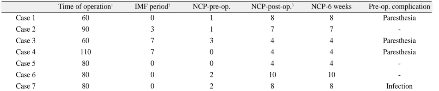
Table 1. Summary of patient received ORIF by this protocol
Site of Fracture Type of fracture (1) Type of fracture (2)
Case 1Left angle Compound Non-displaced
Case 2 Left angle Compound Non-displaced
Case 3 Left angle Compound Displaced
Case 4 Left angle and right parasymphysis Compound Non-displaced
Case 5 Right angle Compound Displaced
Case 6 Left angle Compound Non-displaced
Case 7 Left angle Compound Non-displaced
(ORIF: open reduction and internal fixation)
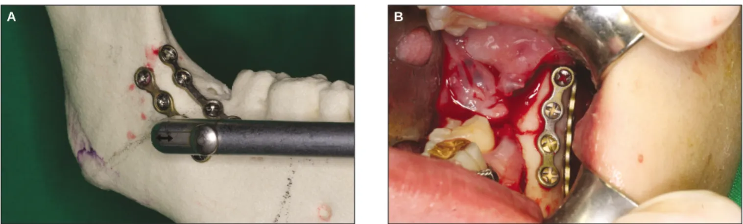
Fig. 1. Internal fixation on mandibular angle was performed using contra-angle drill laterally.
A. Approach of contra-angle drill, B. Clinical view after internal fixation in case 6.
A B
Freiburg, Germany) 다른 1개의 고정판(Leibinger Mandible Fractutre, Freiburg, Germany)은 contra-angle drill과 driver (Luhr-Fritzemeier, 65147102, StrykerLeibinger, Vienna, Austria)를 이용하여 하악골 외면에 적용하였다.(Figs. 1. A, B) 술후 1일째부터 악간고정 없이 하악을 조기 가동하였으 며, 2주간의 유동식을 권장하였다.
Ⅲ.
결 과치료결과는 Table 2와 같다. 수술은 평균 80분이 소요되 었으며 7명의 환자 중 4명을 악간고정 없이 경과 관찰하였 다. 한 명은 3일 동안 악간고정하였으며, 교합이 불안정했 던 2명은 7일 동안 악간고정을 유지하였다. 술전 접촉점 검 사에서 구치부 조기 접촉이나 통증으로 인해 0-3개의 접촉 점을 나타냈다. 하지만 술후 검사에는 4-10개의 접촉점을 나타냈다. 술후 6주 후의 개수는 접촉점이 더 늘어나지 않 는 것을 확인한 최종 개수이다. 환자가 느끼는 교합이 모두 양호한 상태였으며, 저작, 발음, 연하에 별다른 증상을 호 소하지 않았다.
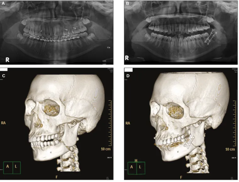
술전, 술후 방사선사진의 평가에서는 변위된 골절편이 제자리로 정복되어 하악연의 연속성이 크게 손상되지 않 은 상태에서 고정된 것을 확인할 수 있었다. 또한 6주간의 경과 관찰에서 방사선사진으로 감염에 의한 골절편의 흡 수나 조기 가동으로 인한 골절편의 이개 등이 없는 것을 확 인할 수 있었다.(Figs. 2. A-D)
술후 특별한 합병증은 발생하지 않았다. 단 3명의 환자에 서, 수상 당시부터 발생한 하순의 감각 이상(paresthesia)을 수술 직후까지 호소하였으나, 6주 후에는 정상으로 회복되 었다. 이외 1명이 골절 후 감염이 되어 내원하였는데, 충분 한 항생제 투약 후에 조절이 되어 별다른 문제없이 수술을 진행하였다.
Ⅳ.
고 찰1970년대 이후 Michelet 등
8과 Champy 등
9이 발전시킨 adaptive miniplate osteosynthesis은 하악 우각부 골절 환자 의 ORIF에 있어서 표준적인 치료방식이 되었다. Champy
등
9은 reconstruction plate나 dynamic compression plate 대신 하악의 Champy’s line을 따라 monocortical screw를 이용한 작고, 얇으며, 구부러지기 쉬운 miniplate을 식립하는 것으 로 충분한 고정을 이룰 수 있다고 주장하였다. 하지만 그 이후로는 miniplate를 몇 개 식립해야 하는지 논의되었다.
Ellis
4는 후향적 연구를 이용하여 2개의 non-compression miniplate (2.0 mm)를 이용하여 우각부 골절을 치료한 경우 의 합병증 발생률이 23%였다고 보고하였다. 이에 반해 1개 의 non-compression miniplate (2.0 mm)를 이용한 경우와 1 개의 malleable non-compression miniplate를 이용한 경우에 각각 2.5%, 0%의 합병증 발생률을 보고하였다. Mehra 등
10은 1개의 miniplate를 식립하였을 때 낮은 감염률(2.04%)를 보고하였고, Barry 등
11도 후향적 연구를 통하여 1개의 miniplate를 식립하였을 때 비교적 낮은 합병증 발생률(12%) 을 보고하였다. Schierle 등
12도 30명을 대상으로 한 전향적 비교연구에서, 두 군간의 감염과 감각신경손상, 부정교합 발생률의 차이가 통계적으로 유의하지 않으므로, 굳이 2개 를 식립할 필요가 없다고 하였다. Seemann 등
13은 10년 동안 의 후향적 연구를 통해 골유합 실패율을 비교 조사하였는 데, 2가지 방법이 비슷한 실패율을 보였다고 보고하였다.
이와는 반대로 하악 하연이나 우각부 협측에 miniplate를 추가하는 것이 더 효율적이며 신뢰성이 높다고 Fox 등
6과
Levy 등
14이 주장하였다. 이들은 후향적 비교연구를 통하여
2개의 miniplate를 우각부 골절에 식립하였을 때 감염률이 더 낮다고(2.9%, 3.1%) 보고하였다. 비유합률을 조사한 Levy 등
14의 같은 연구에서는 1개의 miniplate를 식립한 경 우가 5.3%, 2개의 miniplate를 식립한 경우 0%의 비유합 발 생률을 보였다. Choi 등
15은 하악이 기능적인 하중을 받는 동안 하악 하연을 따라 식립한 2번째 miniplate가 고정이 안 정화되는 것을 보조한다는 실험실 연구로 입증하였다. Fox 등
6은 2개의 non-compression miniplate를 식립했을 때 유난 히 높은 감염률을 보였던 Ellis
4의 연구에 주목했는데, Ellis 는 하악 하연의 miniplate를 bicortical technique을 이용하여 식립했으므로 직접적인 비교가 어렵다고 주장하였다.
Sugar 등
7은 140명의 randomized controlled trial을 통하여 2 가지 방법을 비교하였는데, 2개의 miniplate를 식립한 경우
Table 2. Treatment results of patient received ORIF by this protocol
Time of operation
1IMF period
2NCP-pre-op. NCP-post-op.
3NCP-6 weeks Pre-op. complication
Case 160 0 1 8 8 Paresthesia
Case 2 90 3 17 7 -
Case 3 60 7 3 4 4 Paresthesia
Case 4 110 7 0 4 4 Paresthesia
Case 5 80 0 0 4 4 -
Case 6 80 0 2 10 10 -
Case 7 80 0 2 8 8 Infection
(
1: unit is min,
2: unit is day,
3: NCP post-op. is result of 1 weeks postoperatively, IMF: intermaxillary fixation, NCP: number of contact point)
J Korean Assoc Oral Maxillofac Surg 2010;36:320-4
가 더 효율적이며, 합병증이 낮다고 보고하였다.
한 개의 miniplate만 식립하였을 때 가장 큰 문제점 중 하 나는 1개의 miniplate로 골절 부위가 단단하게 고정되지 않 기 때문에 골절편에 비틀림(torsional movement)이 일어날 수 있다는 점이다. 다시 말해 골절편의 전후방적인 운동은 허용하지 않지만 내외측 방향의 운동은 막을 수 없다
5. 따 라서 우각부 골절에 1개의 miniplate를 식립하여 ORIF를 하 는 경우, 2개를 식립하는 것보다 골절편 이개로 인한 비유 합, 부정유합 가능성이 높아진다. 따라서 이런 합병증을 예 방하기 위해 통상 1주 이상의 악간고정 기간을 갖는다.
본 연구에서는 이러한 이론에 근거하여 2개의 miniplate 를 식립하고 악간고정을 하지 않는 방법을 채택하였다. 하 지만 추가의 miniplate를 식립하기 위해서 transbuccal approach를 이용해야 하는데 이 방법은 피부로 접근하는 단점을 가지고 있다. 또한 hole 형성을 위한 기구조작 (drilling) 시 주의하지 않으면 monocortical technique이 되지 않고, 하치조신경이나 인접치의 치근을 손상시킬 위험이
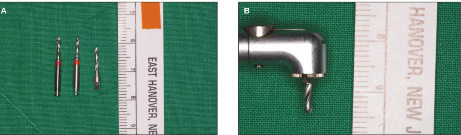
있다. 이에 구내 접근만으로 2개의 miniplate를 식립할 수 있는 방법을 고안하게 되었다. 하지만 contra-angle drill의 drill bit은 기성 제품(6016908, Stryker Leibinger, Vienna, Austria)의 전체 길이가 19 mm, 날길이가 8 mm로 길어 drilling 시 drill head를 골면에 수직으로 접합시키기 어렵 고, drilling 시 transbuccal approach와 마찬가지로 하치조신 경이나 제2대구치의 치근을 침범할 수 있다고 생각되어 날 길이가 5 mm인 것으로 새로 제작하였다.(Figs. 3. A, B)
새로운 방법으로 식립한 본 연구의 7명 환자 모두 성공적 으로 ORIF가 시행되었다. 수술 시간 면에서 평균 80분을 기록하였는데 구내 접근만으로 contra-angle drill을 이용하 여 하악골 측면에 ORIF을 하는 것은 시야 확보의 어려움으 로 시간이 늘어나는 것이라고 유추해볼 수 있다.
술후 악간고정이 없이 조기 가동했던 환자는 4명, 악간고 정을 3일이 1명, 7일이 2명이었다. 원래 목표는 술후 전부 악간고정 없이 경과를 관찰하려 했으나 교합이 불안정하 여 수술 후 다시 arch bar를 고정하고 악간고정을 시행했던
Fig. 2. Radiograph of case 3.
A. Preoperative panoramic view, B. Postoperative panoramic view after 6 weeks, C. Preoperative computed tomography (CT) view, D. Postoperative CT view.
A B
C D
2명이 악간고정을 7일 동안 유지하였다. 3일 동안 유지했 던 환자는 술후에 이환측 구치부가 약간 개교합이었으나 3 일 후 해소되어 바로 풀었다.
수술 이후 6주까지 이어진 접촉점 검사를 통하여 교합 안 정성을 확인하였다. 접촉점 개수가 술전보다 술후 1주 후의 검사에서 크게 늘어나는 것을 확인하였다. 이 개수는 6주의 최종 검사에서도 그대로 유지되어 교합이 안정됨을 확인할 수 있었다. 저작되지 않아 불편함을 호소하는 환자 역시 없 었다. 이와 같은 결과로 2개의 고정판을 이용하여 ORIF할 경우 악간고정 과정이 크게 중요하지 않고, 견고성에 있어 서 새로운 방법이 transbuccal approach를 이용한 것과 차이 가 없다고 추론할 수 있었으나, 이에 대해서는 더 많은 환자 를 대상으로 하는 연구가 필요할 것으로 생각한된다.
이와 같이 구내 접근만으로 contra-angle drill을 이용하여 2개의 miniplate를 식립하는 것은 합병증 없이, 하악골의 조 기 가동을 가능하게 하면서 피부에 반흔도 남기지 않는 우 수한 방법이 될 수 있다고 생각한다. 하지만 조사된 환자의 수가 부족하고, 추적조사 기간이 짧기 때문에 이 protocol이 효과적이라는 결론을 내기에는 아직 부족하다. 또한 대상 환자 모두 제3대구치가 연관된 복합 골절이었나 전부 선상 골절이었으며, 전부 유치악이었기 때문에 분쇄 골절이나 무치악 환자, 심하게 변위된 불리 골절 환자에게도 똑같이 적용될 수 있는 protocol인지 연구가 필요하다. 더 많은 증 례를 모으고 장기간의 추적조사를 통하여 이 protocol에 대 한 신뢰성을 높이는 일이 앞으로 중요할 것이다.
Ⅴ. 결 론