https://doi.org/10.6111/JKCGCT.2021.31.5.228
Synthesis and characterization of CaZrO 3 :Pr 3+ phosphor by skull melting method
Hyunmin Choi, Youngchool Kim and Jeongwon Seok*
,†Hanmi Gemological Institute & Laboratory (HGI, GIG), Seoul 03139, Korea
*Department of Advanced Materials & Energy Engineering, Dongshin University, Naju 58245, Korea (Received August 23, 2021)
(Revised September 7, 2021) (Accepted September 9, 2021)
Abstract Ca
1xZrO
3:xPr phosphor with perovskite structure was successfully synthesized by using skull melting method.
The crystal structure, morphology and optical properties of synthesized phosphor were characterized using X-ray diffraction (XRD), scanning electron microscopy (SEM), ultraviolet fluorescence reaction and photoluminescence. The XRD results indicated that single crystals of CaZrO
3:Pr
3+belongs to orthorhombic perovskite system. The synthesized phosphor could be excited by UV light (254 nm) and the emission spectra results indicated that green luminescence of CaZrO
3:Pr
3+due to charge transfer transition
3P
0
3H
4,
3P
1
3H
5and
3P
0
3H
5at 506, 536 and 548 nm was dominant.
Key words Skull melting method, CaZrO
3:Pr
3+, Phosphor, Luminescence, Emission spectra
스컬용융법에 의한 CaZrO
3
:Pr3+
형광체 합성 및 특성분석최현민, 김영출, 석정원*
,†(주)한미보석감정원, 서울, 03139
*동신대학교 신소재에너지전공, 나주, 58245 (2021년 8월 23일 접수)
(2021년 9월 7일 심사완료) (2021년 9월 9일 게재확정)
요 약 페로브스카이트 구조를 가지는 Ca
1xZrO
3:xPr 형광체를 스컬용융법으로 합성하였다. 합성한 형광체의 결정구조, 형태 및 광학적 특성은 XRD, SEM, 자외선 형광반응 및 광발광을 통해 분석하였다. XRD 측정에서는 CaZrO
3:pr
3+의 단결정 이 페로브스카이트 구조의 사방정계로 분석되었다. 합성된 형광체는 254 nm의 UV 광에 의해 여기 될 수 있고 방출 스펙트 럼 결과는 506, 536 및 548 nm에서 전하전이
3P
0
3H
4,
3P
1
3H
5및
3P
0
3H
5로 인해 CaZrO
3:Pr
3+는 녹색 발광이 우세 하였다.
1. 서 론
페로브스카이트 구조를 가지는 CaZrO
3는 다층 세라믹 수소센서, 전계방출 디스플레이(FED), 캐퍼시터, 고체전 해질 및 결정질 형광체 재료용 호스트 등에 사용되고 있 으며[1,2], 특히 Mn, Cu, Nd, Eu, Tb 및 Dy 등을 부활 제(activator) 및 공부활제(co-activator)로 첨가하여 적색, 청색 또는 녹색 계열의 형광체를 합성하는 연구가 꾸준 히 진행되고 있다[3,4]. CaZrO
3를 호스트로 하는 형광체 합성은 CaCO
3와 ZrO
2를 혼합 후 1300~1400
oC로 가열
하여 합성시키는 고상반응법[5,6] 및 Ca(NO
3)
2∙4H
2O과 ZrO(NO
3)
2∙XH
2O 또는 ZrN
2O
7를 반응시켜 합성하는 졸- 겔법[7,8] 등으로 가능하다.
본 연구에서 사용된 Skull melting 법은 주로 YSZ(yttria stabilized zirconia)합성에 응용되고 있으며[9,10], 유리의 용융실험[11,12] 및 금속의 합금에도 응용이 되고 있으 나[13,14] CaZrO
3를 호스트로 하는 녹색 형광체의 용융 실험에 응용된 예는 전무한 실정이다. 이 방법은 고주파 에 의한 유도가열 원리를 이용함으로써 유도가열이 가능 한 원료의 용융에 있어 비교적 짧은 시간에 고온을 얻을 수 있으므로 고융점 산화물 또는 금속 용융 실험에 있어 매우 효율적인 방법이라 할 수 있다. 특히 산화물 용융 시 도가니 내벽에 융액과 동일성분의 얇은 crust(self 도
†
Corresponding author
†
E-mail: [email protected]
가니)가 형성됨으로 도가니로부터의 오염을 방지할 수 있어 고순도를 요구하는 산화물 용융실험에 매우 유용한 방법이라 할 수 있다. 또한 용융 시 융액 내에서는 열대 류보다 자장에 의한 대류가 우선함으로 일반적으로 형광 체 합성에 응용되고 있는 고상법에 비해 사전 출발원료 의 준비시간을 단축시킬 수 있다. 따라서 본 연구에서는 출발원료 준비시간 단축 및 비교적 짧은 시간에 고온을 얻을 수 있어 기존 방법에 비해 제조 공정시간을 단축시 키며 대량생산이 가능한 스컬용융법을 응용하여 형광체 를 합성하고자 하였다.
2. 실험 방법
형광체 합성을 위한 출발원료는 CaCO
3, ZrO
2및 Pr
6O
11을 사용하였다. CaCO
3는 1000
oC에서 2시간 동안 하소 (calcination)하여 CaO로 준비하였으며, Ca
1xZrO
3:xPr
3+조성을 가지는 형광체 합성을 위해 Ca:Zr:Pr = 0.95:1.00:
0.05 mol%가 되도록 CaO:ZrO
2:Pr
6O
11= 863.85:1998.12:
138.03 g으로 하여 총 3 kg을 4시간 동안 ball mill하여 준비하였다. 혼합된 원료를 내경 12 × 높이 15 cm인 냉 각도가니(cold crucible)에 충진 후 고주파로 유도가열하 여 용융시켰다. 원료를 냉각도가니에 충진 할 때 고주파 에 의해 상온에서도 유도가열이 가능한 카본 링을 충진 원료의 중앙에 위치시켰는데, 이는 상온에서 고주파 유 도가열이 되지 않는 출발원료를 가열시키기 위해서다.
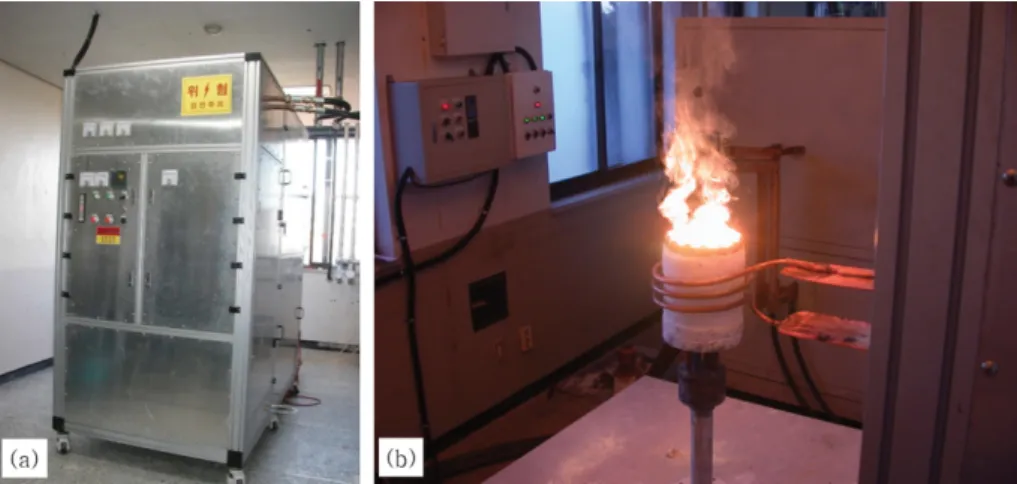
산화물의 비저항은 온도와 반비례 관계이며, 일정 온도 로 가열이 되면 비저항이 낮아져 고주파에 의한 유도가 열이 가능해지게 된다. Figure 1(a)는 본 연구에 사용된 고주파 유도 발생기이며, (b)는 냉각도가니 내에서 원료 가 용융되고 있는 사진이다. 이 스컬용융시스템은 출력 주파수가 3.02~8.00 MHz까지 가능하도록 직접 설계 및 제작하였으며, 본 실험에서는 3 .4 MHz 주파수를 사용하
Fig. 1. Photographs of skull melting system. (a) is high frequency (HF) inductive generator, (b) is work coil and cold crucible.
Fig. 2. Flow chart for the experimental procedure.
였다. 충진된 원료의 커플링(원료의 용융이 시작되는 시 점)은 고주파 유도가열기에 파워를 인가하고 20분 후에 시작되었으며, 커플링 후 3 kg의 충진원료를 완전 용융 시키는데 40분 정도의 시간이 소요되었다. 충진원료를 완전히 용융하고 융액 상태를 2시간을 유지한 후 고주 파 유도가열기의 파워를 끄고 자연냉각 시켰다. 합성한 잉곳(ingot)은 XRD, SEM, UV 및 PL 측정을 통해 특 성을 분석하였다. Figure 2에는 실험과정을 나타내었다.
3. 결과 및 고찰
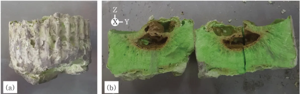
Figure 3(a)는 용융시킨 원료를 냉각도가니 내에서 자
연 냉각시킨 후 도가니로부터 분리시킨 잉곳으로서 크기
는 직경 10.5 cm, 높이 6.6 cm 정도이다. (b)는 (a)의 잉
곳을 절반으로 분리시킨 것으로서 분리된 단면을 보면
전형적인 단결정의 군집성장(group growth)의 형태를 보
인다. 핵은 도가니 내벽에서부터 자발적으로 형성되어 도가니 중앙부분으로 단결정이 성장하였음을 볼 수 있으 며, 도가니 내벽과 가까운 부분 보다는 중앙부분으로 갈 수록 단결정의 크기는 점점 더 커짐을 볼 수 있다. 이는 도가니 내벽과 가까운 부분 보다는 중앙부분에서 과냉각 (supercooling)이 서서히 이루어 졌기 때문이다. 이 사진 에서 편의상 잉곳의 상부 방향을 Z축, 정면을 X축 및 우측 방향을 Y축으로 정하였다.
Figure 4(a)와 (b)는 Fig. 3 (b) 잉곳의 X축의 단면부분 (정면 부분에 결정이 성장된 부분)과 Z축 표면(단결정 성장이 종료된 부분)을 SEM으로 촬영한 사진으로서 150~
200 m 정도 크기의 각진 주상 형태의 단결정이 성장되 었음을 알 수 있다.
Figure 5는 합성한 잉곳을 미분쇄하여 XRD를 측정한 결과로서 페로브스카이트 구조를 가지는 orthorhombic 결정상으로 Pcmn 공간군에 속하는 CaZrO
3로 분석되었 다. 이는 ICDD의 PDF Card No. 01-076-2401 데이터 와 정확히 일치하였으며 다른 결정상은 측정되지 않은 것으로 보아 완전한 CaZrO
3가 합성되었음을 알 수 있다.
CaZrO
3호스트에 도핑 된 Pr
3+(113pm)는 이온 반경이 작은 Zr
4+(72 pm)의 자리에 치환될 가능성도 있지만 이 온 반경이 유사한 Ca
2+(114 pm) 자리에 대부분 치환된다
[15]. 이 과정에서 3개의 Ca
2+자리 중 2개의 자리에 Pr
3+가 대체되고 나머지 1개는 공공(vacancy, V''
Ca)이 형 성된다[16,17]. 즉 3 Ca
2++ 2Pr
3+ 2Pr
*Ca+ V''
Ca의 식으로 나타낼 수 있다.
Figure 6은 합성한 CaZrO
3:Pr
3+잉곳을 미세한 분말로 만든 다음 자외선 형광기에서 형광 반응을 촬영한 사진 이다. (a)는 일반광에서 촬영한 사진으로서 옅은 녹황색 Fig. 3. Photographs of CaZrO
3:Pr
3+ingot; (a) before split and (b) cross section of the synthetic ingot by skull melting method.
Fig. 4. SEM micrographs of the CaZrO
3:Pr
3+ingot; (a) is the plane in the X-axis direction (×100), (b) is the plane in the Z-axis direction (×100).
Fig. 5. X-ray diffraction pattern of the CaZrO
3:Pr
3+phosphor.
을 보이며, (b)는 254 nm 자외선 파장 하에서 촬영한 사 진으로서 청녹색 형광을 보이며, (c)는 3 65 nm 파장의 자외선 하에서 촬영한 사진으로서 형광반응은 나타나지 않았다.
Figure 7(a)는 PL 여기 스펙트럼(excitation spectra) 데 이터로서 방사파장(emission wavelength,
em)은 536.4 nm 조건에서 측정하였다. 255 nm 영역의 피크는 O
2 Pr
3+의 전하이동에 의한 밴드(charge transfer transition;
CTT)로서 산소원자의 2p 오비탈에서 Pr
3+의 비어 있는 4f-5d 오비탈로의 전이에 의한 것이다[18]. Figure 7(b) 는 PL 발광 스펙트럼(emission spectra) 데이터로서 여 기파장(excitation wavelength,
ex)은 254 nm 조건에서 측정하였다. 측정된 데이터에서는
3P
0
3H
J(J = 4, 5) 및
3
P
1
3H
J(J = 4, 5)에 의한 방사스펙트럼으로서
3P
0
3
H
4,
3P
1
3H
5및
3P
0
3H
5는 각각 506, 536 및 548 nm 파장에 해당되며[19]. 이 스펙트럼은 모두 녹색파장 영역에 해당됨으로 본 실험에서 합성된 형광체는 녹색 파장을 발광하게 된다.
4. 결 론
본 연구에서는 기존의 형광체 합성법에 비해 합성시간 을 단축시킬 수 있고(원료 준비 및 용융시간 등) 대량 생산이 가능한 스컬용융법을 응용하여 형광체를 합성하 였다. 출발원료로 CaCO
3, ZrO
2및 Pr
6O
11를 사용하였으 며, CaCO
3는 1000
oC에서 2시간 동안 하소(calcination) 하여 CaO로 준비하였다. 출발원료의 혼합은 Ca:Zr:Pr = 0.95:1:0.05 mol%가 될 수 있도록 CaO:ZrO
2:Pr
6O
11= 863.85:1998.12:138.03 g으로 하여 총 3 kg을 준비하였다.
준비된 원료는 스컬용융법으로 용융시켰으며, 용융된 상 태를 2시간 유지후 냉각도가니에서 자연냉각 시켰다. 냉 각도가니에서 분리시킨 잉곳은 직경 10.5 cm, 높이 6.6 cm 정도로 전형적인 단결정의 군집성장 형태를 보였다. 잉 곳 내부에 성장된 단결정들은 SEM을 통해 직경 150~
200 m 정도 크기의 각진 주상 형태임을 확인하였으며, 단결정들은 작은 물리적 충격으로 각각 분리시킬 수 있 었다. 결정구조 분석을 위해 파우더 상태로 XRD를 측 정한 결과 페로브스카이트 구조를 가지는 orthorhombic 상의 CaZrO
3임을 확인하였다. 자외선 형광기에서는 254 nm 파장에서는 청녹색의 형광반응을 보였으며, 365 nm 파장에서는 형광반응이 없었다. PL에서의 여기 스펙트럼 은 53 6.4 nm 방사파장 조건에서 O
2 Pr
3+에 의한 CTT ( = 255 nm)가 측정되었으며, 발광 스펙트럼은 254 nm 여기파장 조건에서 녹색파장에 해당되는
3P
1
3H
J(J = 4, 5) 및
3P
0
3H
J(J = 4, 5)에 의한 스펙트럼이 측정되 었다.
References