Copyright ⓒ 2013, The Microbiological Society of Korea
Dual Coating Improves the Survival of Probiotic Bifidobacterium Strains during Exposure to Simulated Gastro-Intestinal Conditions
Joo Yeon Kang1, Do Kyung Lee2, Jae Eun Park1, Min Ji Kim2, Joong-Su Lee3, Jae-Gu Seo3, Myung Jun Chung3, Hea Soon Shin1, and Nam Joo Ha2*
1College of Pharmacy, Duksung Women’s University, Seoul 132-714, Republic of Korea
2College of Pharmacy, Sahmyook University, Seoul 139-742, Republic of Korea
3R&D Center, Cell Biotech Co., Ltd., Gimpo 415-871, Republic of Korea
위장관내 조건에서 이중코팅 처리 된 프로바이오틱 비피도박테리움의 생존력 향상
강주연1․이도경2․박재은1․김민지2․이중수3․서재구3․정명준3․신혜순1․하남주2*
1덕성여자대학교 약학대학, 2삼육대학교 약학대학, 3㈜쎌바이오텍 연구소
(Received June 14, 2013 / Accepted September 25, 2013)
Probiotics have been reported to benefit human health by modulating immunity, lowering cholesterol, improving lactose tolerance, and preventing some cancer. Once ingested, probiotic microorganisms have to survive harsh conditions such as low pH, protease-rich condition, and bile salts during their passage through the gastro-intestinal (GI) tract colonize and proliferate to exert their probiotic effects. The dual coating technology, by which the bacteria are doubly coated with peptides and polysaccharides in consecutive order, was developed to protect the ingested bacteria from the harsh conditions. The aim of the study was to evaluate the viable stability of a doubly coated blend of four species of Bifidobacterium by comparing its bile/acid resistance and heat viability in vitro with that of the non-coated blend. After challenges with acid, bile salts, heat, and viable cell counts (VVCs) of the dual coated and non-coated blend were determined by cultivation on agar plates or flow cytometric measurement after being stain with the BacLigtht kitTM. The results showed that the dual coated blend was much higher resistant to the acidic or bile salt condition than the non-coated blend and heat viability was also higher, indicating that the dual coating can improve the survival of probiotic bacteria during their transit through the GI tract after consumption.
Keywords: Bifidobacterium, dual coating technology, duolac, probiotics
*For correspondence. E-mail: [email protected]; Tel.: +82-2-3399-1607;
Fax: +82-2-3399-1617
In today’s society, there has been increasingly interested in their personal health and functional food. Probiotic products are an important functional food as they represent about 65% of the world functional food market, and the market for probiotic products continues to expand (Agrawal, 2005; Jankovic et al., 2010). Probiotics are defined as ‘live microorganisms which, when administered in adequate amounts, confer a health benefit on the host’ (FAO/WHO, 2002). Researchers have reported to play a therapeutic role by modulating immunity, lowering cholesterol, improving lactose tolerance and preventing some cancer (Kailasapathy and Chin, 2000; Sanders et al., 2007).
Probiotics are orally administrated and are available in various forms such as food products, capsules, sachets, or tablets.
Ingested probiotics have to survive adverse conditions such as low pH, protease-rich condition, and bile salts during their passage through the gastro-intestinal (GI) tract to be able to influence the human gut microflora (Weichselbaum, 2009; Burgain et al., 2011). However some probiotic bacteria are sensitive to oxygen, and many require media ingredients or modified gas environments to enable their growth (Dave and Shah, 1996;
Talwalkar and Kailasapathy, 2004). Especially, Bifidobacterium strain that are the most widely used probiotic bacteria and are included in many products and functional foods vary greatly in their sensitivity to the harsh acidic environment of the stomach and many foods (Clark and Martin, 1994; Lankaputhra and
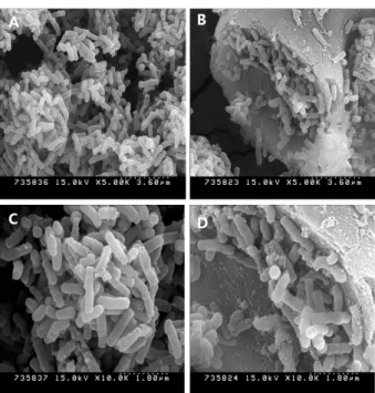
Fig. 1. Matrix structure of dual coated bacteria. Fig. 2. Field emission scanning electron microscope images of the non-coating (A, C) and dual coating (B, D) Bifidobacterium blend.
Shah, 1997; Charteris et al., 1998; Truelstrup et al., 2002; Guarner and Malagelada, 2003; Annan et al., 2008). For this reason, many researchers reported that there is poor survival of probiotic bacteria in products containing free probiotic cells (De Vos et al., 2010). Various technologies of encapsulation such as emulsification, spray drying, spray cooling, and freeze drying have been developed for protection live cells in the food industry (De Vos et al., 2010;
Burgain et al., 2011), and it has been reported these technologies of encapsulation improved the viability of probiotic bacteria in the GI tract (Krasaekoopt et al., 2003; Picot and Lacroix, 2004;
Sohail et al., 2011; Su et al., 2011; Saarela et al., 2011).
Dual coating technology is so-called fourth generation coating technology for protection of LAB during the passage through the GI tract and manufacturing process and was patented in Korea (patent no. 0429495), Japan (patent no. 3720780), and Europe (patent no. 1514553B) (Burgain et al., 2011; Cha et al., 2011). The technology was developed to protect the ingested bacteria from the harsh conditions. In the technology, bacteria cells are coated with peptides and subsequently with polysaccharide matrix (Figs. 1 and 2). A polypeptide behaves in a pH-dependent way, and there exists a pH gradient along the stomach and intestine.
Thanks to the coating layers, the doubly coated cells remain mostly uncoated at pH 4.0, which is the pH of stomach after meal and begin to be released from the coats at pH 6.0 and are fully released at pH 7.0, the pH in the intestine. The polysaccharide matrix protects from moisture, heat, and physical pressure, and so stability is increased. Therefore the dual coated bacteria after ingestion are able to reach the intestine alive and in good condition to colonize and proliferate while uncoated bacteria that is prone to be damaged in the gastro-intestinal environment.
In this study, we evaluated in vitro acid/bile resistance and heat viability of a probiotic blend consisting of four Bifidobacterium species which were doubly coated or non-coated and compared the resistances of the dual coated blend with those of the non- coated blend.
Materials and Methods
Bacteria
Dual coated Bifidobacterium bifidum BF3 (KCTC 12199BP),
B. infantis BT1 (KCTC 12859BP), B. longum BG7 (KCTC 12200BP), and B. rhamnosus BR3 (KCTC 12201BP) were blended in equal proportions and tested to assess the resistance to acid or bile salts. The same tests were applied to a blend containing the same kind of species of non-coated Bifidobacterium. Comparison of acid, bile, and heat resistance between the two formulations was made.
Resistance to acid and bile salt
For acid tolerance test, the blend of dual coated Bifidobacterium or non-coated Bifidobacterium was inoculated in BL broth (around 107-8 CFU/ml) which was adjusted to pH 2.0, 3.0, 5.0, or 7.0 using 0.1 M HCl or 0.1 M NaOH. Samples were taken at various time points (0, 0.5, 1, 3, and 8 h) and subjected to viable cell count using BL agar plate with 0.005% bromocresol purple or LIVE/DEAD BacLight kitTM (Invitrogen, USA).
For bile tolerance test, the blends were inoculated into acidified BL broth (pH 4.0) (approximately 107 CFU/ml) containing oxgall (BD, USA) at the concentration of 0, 0.1, 0.3, or 1% (w/v).
Samples were taken at various time points (0, 3, 6, 9, 12, and 24 h) and subjected to viable cell count with the same methods, as described above.
Staining of bacterial cells
Cells collected at the time points were adjusted to be 106-7 CFU/ml in potassium phosphate buffer (PBS, pH 7.2), treated with the reagents in the BacLight kit as recommended by the manufacturer, and gently shaken for 15 min in dark condition.
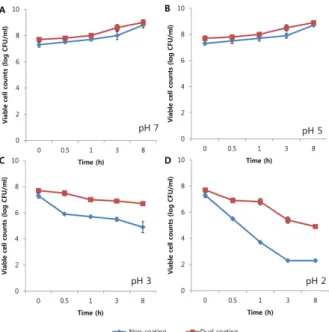
Fig. 3. Time course analysis of viable cell counts of the dual coated Bifidobacterium and non-coated Bifidobacterium blend under different acidic conditions (pH 7.0, 5.0, 3.0, and 2.0). Viable cell counts were determined by collecting cells at the time points, which were then plated on BL agar plates and incubated at 37℃
anaerobically.
First labeling was confirmed by the use of a fluorescent microscopy (Olympus, Japan) set to accept fluorescence intensity at a wavelength (emission 1; green and emission 2; red).
Flow cytometric measurements (FCM)
Flow cytometric measurements were performed on a FACSCalibur flow cytometer (Becton Dickinson Immunocytometry Systems, USA) equipped with a 15-mW, 488-nm, air-cooled argon ion laser and a cell-sorting catcher tube. Cell samples were diluted to approximately 106 cells/ml and delivered at the low flow rate, corresponding to 150 to 500 cells/sec. FSC, SSC, and three fluorescence signals were measured. A band pass filter of 530 nm (515 to 545 nm) was used to collect the green fluorescence (FL1), a band pass filter of 585 nm (564 to 606 nm) was used to collect the yellow-orange fluorescence (FL2), and a long pass filter of 670 nm was used to collect the red fluorescence (FL3). FSC was collected with a diode detector. SSC and the three fluorescence signals were collected with photomultiplier tubes. All signals were collected by using logarithmic amplifications.
A combination of FSC and SSC was used to discriminate bacteria from background.
Heat stability
For stability test, we studied the differences in the viability values between non-coated and dual coated Bifidobacterium during 2 weeks storage period at 40℃. The blend of dual coated
Bifidobacterium or non-coated Bifidobacterium were kept for 2 weeks at 40℃ for subsequent microbial counts. Later, samples were randomly taken before and after 1, 3, 7, and 14 days during the 2 weeks storage period. Test samples were rehydrated to the original volume with 0.1% peptone for 10 min at room temperature, and appropriated dilutions were poured in BL agar (Difco, USA). Plates were incubated anaerobically at 37℃ for 48 h.
After that, the number of CFUs was counted.
Results
Acid tolerance
To assess acid tolerance of the four bifidobacterial species in each blend, the cells were challenged with various acidic conditions (pH 2.0, 3.0, 5.0, or 7.0) up to for 8 h and sampled at 0, 0.5, 1, 3, and 8 h after incubation. There was little difference in viable cell counts (VCCs) determined using agar plate between the blends of dual coated or non-coated bifidobacteria under conditions of pH 7.0 and 5.0 over the test period (Figs.
3A and 3B). Differences in acid tolerance between them began to appear when incubated for 30 min at pH 3.0 during which the VCCs of the non-coated blend dropped by 1.4 log-fold compared to the initial counts whereas the dual coated blend showed a small decrease in the VCCs, from 7.7±0.12 log- CFU/ml to 7.5±0.14 log-CFU/ml (Fig. 3C). Incubation at pH 2.0 severely affected the VCCs of both blends. For the non-coated blend, the initial viable counts dropped from 7.3±0.19 log- CFU/ml to 5.3±0.11 log-CFU/ml (2 log-fold drop) only after 30 min and to under the lower detection limit, 2.3±0.0 log- CFU/ml, after 8 h (5 log-fold drop or greater). By contrast, the dual coated blend was much less affected under the same pH condition. Its VCCs were determined to be 6.9±0.19 log- CFU/ml after 30 min incubation, showing a small decrease, and 4.9±0.06 log-CFU/ml even after 8 h incubation (Fig. 3D).
LIVE/DEAD BacLight kitTM was used to monitor visually the fraction of live or dead bacterial cells in the test samples as live and dead cells fluoresce green and red lights, respectively, when stained with the kit. When the cells in the dual coated or non-coated blend were incubated for 3 h in pH 7.0 condition, most of the cells were live (green) and no distinct differences between the samples were observed (Figs. 4A and 4B). However, most of the non-coated bacteria incubated for 3 h in pH 2.0 condition were severely damaged (yellow and orange) or dead (red). By contrast, half of the dual coated bacteria were visually found to be live (Figs. 4C and 4D).
In order to quantify the live and dead cells in the samples, the green and red bacterial counts were analyzed by FCM (Fig. 5).
The percentages of green and red bacteria for the non-coated blend at 3 h in pH 3.0 condition were 28.3% (live cell) and
Fig. 5. Flow cytometric analysis of bifidobacterial cells incubated in acidic conditions. All bacteria were previously stained with STYO Green I and propidium iodide. Gates indicate the position and concentration of intact cells on the plots. The strains of non-coated Bifidobacterium blend incubated at pH 3.0 (A) or at pH 7.0 (B) for 3 h. The dual coated Bifidobacterium blend incubated at pH 3.0 (C) or at pH 7.0 (D) for 3 h. Q1 and Q2, dead cell; Q3, injured cell or un-staining cell; Q4, live cell. The counts of live cells (E) and dead cells (F) for the dual coated blend were compared with non-coated blend.
A B
C D
Fig. 4. Fluorescent microscopic images of cells of the non-coated Bifidobacterium blend or dual coated Bifidobacterium blend stained with the Live/Dead Baclight kit after challenge with acid. The non-coated bacteria (A, C) and dual coated bacteria (B, D) were incubated at pH 7.0 (A and B, respectively) or at pH 2.0 (C and D, respectively) for 3 h. Live cells, green; dead cells, red; yellow and orange, injured.
64.3% (dead cell), respectively (Fig. 5A). Under the same condition, live and dead cells for the dual coated blend were determined to be 53.7% and 34.7% respectively (Fig. 5C). In the 3 h samples at pH 7.0, the percentages of live and dead cells in the non- coated blend were 87.1% (live cell) and 2.5% (dead
cell), respectively (Fig. 5B). On the other hand, the respective percentages for the dual coated blend were 89.0% for live cells and 0% for dead cells (Fig. 5D). Therefore the result further supports that the dual coated bacteria are more resistant to acid than the non-coated bacteria.
Bile tolerance
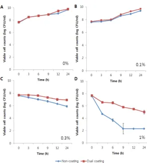
Bile tolerance of the bacteria in the non-coated or dual coated blend was investigated by incubating them in BL broth (pH 4.0) containing oxgall at the final concentration of 0, 0.1, 0.3, or 1.0% (w/v). The pH of BL broth was adjusted to 4.0 because such acidic condition is assumed to be closer to the human proximal intestinal condition (Succi et al., 2005). At 0.1% oxgall, bacterial cells of both blends proliferated over time, and no large differences were observed in the VCCs between the blends (Fig. 6). However, the growth of the non- coated bacteria began to be inhibited at 0.3% oxgall, and the VCCs dropped by 1.7 log fold after 24 h incubation while the growth of the dual coated bacteria was barely affected (Fig.
6C). Distinct differences in the VCCs were seen when they were incubated at 1.0% oxgall. Under the condition, the viability of the non-coated bacteria dropped sharply from 7.6±0.10 log-CFU/ml to 2.3±0.0 log-CFU/ml after 24 h incubation whereas the dual coated bacteria were less affected and their VCCs after 24 h were much higher than those of non-coated bacteria (5.0±0.28 log-CFU/ml vs 2.3±0.0 log-CFU/ml) (Fig. 6D).
Bacterial cells in the non-coated or dual coated blend incubated at 1.0% oxgall for 3 h were subjected to staining with the BacLight kit and examined under the fluorescent microscope.
Fig. 8. Flow cytometric analysis of bifidobacterial cells incubated under different bile salt conditions. All bacteria were previously stained with STYO Green I and propidium iodide. Gates indicate the position and concentration of intact cells on the plots. The non-coated bacteria incubated at 0% oxgall (A) or at 1.0% oxgall (B) for 3 h. The dual coated bacteria incubated at 0% oxgall (C) or at 1.0% oxgall (D) for 3 h. P1, total percents (live, dead, injured cell, and debris); P2, total percents (live, dead, and injured cell); Q1 and Q2, dead cell; Q3, injured cell or un-staining cell; Q4, live cell. The counts of live cells (E) and dead cells (F) for the dual coated blend were compared with non-coated blend.
Fig. 6. Time course analysis of viable cell counts of the non-coated Bifidobacterium blend and dual coated Bifidobacterium blend incubated at different concentrations of oxgall (0, 0.1, 0.3, and 1%).
Viable cell counts were determined by collecting cells at the time points, which were then plated on BL agar plates and incubated at 37℃ anaerobically.
A B
Fig. 7. Fluorescent microscopic images of the dual coated and non-coated bacteria stained with Live/Dead Baclight kit after challenge with oxgall. The non-coated bacteria (A) and dual coated bacteria (B) were incubated at 1.0% oxgall for 3 h. Live cells, green; dead cells, red; yellow and orange, injured.
The result was that a great number of the non-coated bacteria were found to be dead (red in color) from the fluorescent observation while half of the dual coated bacteria were found to be live (green) (Fig. 7).
When the stained bacteria were further analyzed by FCM,
the percentages of live and dead cells of the non-coated Bifidobacterium blend incubated at pH 4.0 and 0% oxgall for 3 h were 71.5% (live cell) and 20.3% (dead cell), respectively (Fig. 8A). Under the same condition, live and dead cells of the dual coated blend were 78.2% and 11.3%%, respectively (Fig.
8C). In the 3 h samples at 1.0% oxgall, the percentages of live and dead cells of the non-coated blend were 26.2% and 66%, respectively (Fig. 8B). By contrast, the respective percentages of the dual coated bacteria were 67.5% for live cells and 21.0%
for dead cells (Fig. 8D).
Heat stability
To measure the heat resistance, non-coated or dual coated bacteria were kept at 40℃ for 14 days. Figure 9 shows the viability of non-coated or dual coated bacteria at the end of storage at 40℃ for 14 days. The initial counts of non-coated
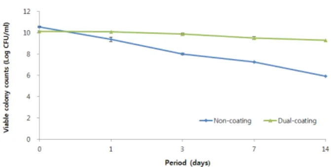
Fig. 9. Survival of non-coated and dual coated Bifidobacterium blend during 2 weeks storage period at 40℃.
and dual coated bacteria ranged from 10.57±0.08 log-CFU/ml and 10.13±0.05 log-CFU/ml and there was little difference in viable cell counts between them (Fig. 9). However, non-coated bacteria showed a tendency to decrease highly throughout the storage period. After 1 day, non-coated blend counts decreased from 10.57±0.08 log-CFU/ml to 9.38±0.2 log-CFU/ml and kept decreasing at the end of storage period (Fig. 9). The viable counts of non-coated blend declined by about 44% after 2 weeks compared to the initial viable count. By contrast, dual coated blend showed a small decrease. Its VCCs were determined to be 10.10±0.02 log-CFU/ml after 1day and 9.31±0.11 log-CFU/ml even after 2 weeks (Fig. 9). These results suggested that dual coated bacteria are more resistant to heat than non-coated bacteria.
Discussion
The viability of probiotics in functional foods is the most important requirement, because it has a direct bearing on effects of functional foods. For this reason, many researchers studied methods for protection of live cells and various technologies of encapsulation have been developed. The demands of successful encapsulation are protection against adverse environmental conditions such as low pH, biliary salts, and proteases during passage through the GI tract and efficient release of the probiotics bacteria. The encapsulation technologies depend on the capsule material, particle size, and bacterial strain (Burgain et al., 2011).
It has been reported that various probiotics encapsulation technologies improve the viability of bacteria during exposure to simulated gastro-intestinal conditions such as alginate-coated gelatin microspheres (Annan et al., 2008), alginate–human-like collagen (Su et al., 2011) and in alginate gel microbeads (Sohail et al., 2011). Alginate is a representative encapsulating material and extensively used, but they are sensitive to the acidic environment and very difficult to scale up (Mortazavian et al., 2008). Many researchers have attempted to remedy disadvantages in various materials and ways. Among the probiotic
encapsulation technologies, the spray coating technology is easy to scale up and is adapted to give multilayer coatings (Burgain et al., 2011).
We have developed the dual coating technology which is so-called fourth generation coating technology. The dual coating system is based on a pH-dependant release mechanism which protects the cells against acidic environments in the stomach and releases the bacteria from coating in the neutral pH environment of the intestines. The dual coated blend consisting of B. bifidum BF3 (KCTC 12199BP), B. infantis BT1 (KCTC 12859BP), B.
longum BG7 (KCTC 12200BP), and B. rhamnosus BR3 (KCTC 12201BP) was found to be highly resistant to acid or bile salt compared to the non-coated counterpart in this study. These results indicate that the dual coating technology can improve the survival of probiotic bacteria during their transit through the GI tract after consumption. In the case of probiotic encapsulation, the objective is not only to improve the survival of probiotic bacteria through the GI tract, but also to protect the cell against adverse environment. It is estimated that daily consumption of 107 CFU/ml of live probiotic cells are needed to confer health benefits to the consumer (Ouwehand and Salminen, 1998; Shah, 2000). However, probiotic is affected by storage environments such as temperature, and humidity, and it might contribute to reductions in viable cell counts during storage period. Weinbreck et al. (2010) reported that viable counts of unencaposulation bacteria declined dramatically during 2 weeks when stored at 37℃, whereas encapsulation bacteria were much less affected under the same condition. Likewise, the role of the encapsulation also is important at the end of storage period. The dual coating technology improved the viability of Bifidobacterium blend during 2 weeks storage period at 40℃ compared to the non- coated counterpart in this study. These findings demonstrate the dual coating technology can improve stability and viability of probioitc bacteria more effectively from harsh environments.
적 요
프로바이오틱 박테리아는 면역력 활성 조절, 콜레스테롤 수 치 억제, 유당내성 강화, 항종양 활성 등의 다양한 생리활성 기능 으로 건강 증진 효과가 있는 것으로 보고되고 있다. 프로바이오 틱 박테리아는 일단 섭취하게 되면 위장관을 통과하는 동안 산 도가 낮거나 단백질분해 효소가 많은 열악한 환경에서 생존해야 하며 프로바이오틱 효과를 발휘하기 위해 증식해야 한다. 이중 코팅 기술은 펩타이드와 다당류의 이중코팅으로 섭취된 프로바 이오틱 박테리아를 열악한 조건으로부터 보호하기 위해 개발되 었다. 본 연구에서는 이중 코팅 된 4종의 비피도박테리움 혼합물 의 생존 안정성을 평가하기 위해 코팅이 되지 않은 비피도박테 리움 혼합물과 담즙, 산 저항성 및 열 안정성을 비교⋅평가하였 다. 이중 코팅 된 균주와 코팅이 되지 않은 균주를 산과 담즙 조
건 및 40℃에 노출 시킨 후 한천배지에 배양하여 생존생육 세포 수를 측정하였으며, BacLigtht 키트를 이용하여 염색 한 후 유세 포 분석기를 이용하여 생균과 사균의 세포수를 평가하였다. 이 중코팅 된 균주 혼합물의 경우 코팅이 되지 않은 균주 혼합물 보 다 산, 담즙 내성이 더 높았으며, 열 안전성 또한 코팅 되지 않은 균주 혼합물보다 높은 것으로 나타났다. 이 같은 결과들로 이중 코팅 기술은 프로바이오틱 박테리아의 안정성 및 섭취 후 위장 관 트랙을 통과하는 동안 균주의 생존률을 향상시킬 수 있음을 확인하였다.
Acknowledgements
The authors are grateful to the financial support by Sahmyook University Research Fund. This work (Grants No.000449270111) was supported by Business for Cooperative R&D between Industry, Academy, and Research Institute funded by the Korea Small and Medium Business Administration.
References
Agrawal, R. 2005. Probiotics: an emerging food supplement with health benefits. Food Biotechnol. 19, 227–246.
Annan, N.T., Borza, A.D., and Truelstrup Hansen, L. 2008.
Encapsulation in alginate-coated gelatin microspheres improves survival of the probiotic Bifidobacterium adolescentis 15703T during exposure to simulated gastro-intestinal conditions. Food Res.
Int. 41, 184–193.
Burgain, J., Gaiani, C., Linder, M., and Scher, J. 2011. Encapsulation of probiotic living cells: From laboratory scale to industrial applications. J. Food Engineer. 104, 467–483.
Cha, M.K., Chung, M.J., Kim, J.E., Lee, K.O., and HA, N.J. 2011.
Comparison of dual coated(DuolacTM) and uncoated lactic acid bacteria from potential probiotics. Biotechnol. Biotechnol. Eq. 25, 2489–2493.
Charteris, W.P., Kelly, P.M., Morelli, L., and Collins, J.K. 1998.
Development and application of an in vitro methodology to determine the transit tolerance of potentially probiotic Lactobacillus and Bifidobacterium species in the upper human gastrointestinal tract. J. Appl. Microbiol. 84, 759–768.
Clark, P.A. and Martin, J.H. 1994. Selection of bifidobacteria for use as dietary adjuncts in cultured dairy foods: III – Tolerance to simulated bile concentrations of human small intestines. Cultur. Dairy Products J. 29, 18–21.
Dave, R.I. and Shah, N.P. 1996. Evaluation of media for selective enumeration of Streptococcus thermophilus, Lactobacillus delbrueckii ssp. bulgaricus, Lactobacillus acidophilus, and Bifidobacteria. J. Dairy Sci. 79, 1529–1536.
De Vos, P., Faas, M.M., Spasojevic, M., and Sikkema, J. 2010.
Encapsulation for preservation of functionality and targeted delivery of bioactive food components. Int. Dairy J. 20, 292–302.
FAO/WHO (Food and Agriculture Organization of the United Nations/World Health Organization) 2002. Guidelines for the Evaluation of Probiotics in Food. London, Ontario, Canada. April 30 and May 1, 2002.
Guarner, F. and Malagelada, J.R. 2003. Gut flora in health and disease.
Lancet 361, 512–519.
Jankovic, I., Sybesma, W., Phothirath, P., Ananta, E., and Mercenier, A.
2010. Application of probiotics in food products – challenges and new approaches. Curr. Opin. Biotechnol. 21, 175–181.
Kailasapathy, K. and Chin, J. 2000. Survival and therapeutic potential of probiotic organisms with reference to Lactobacillus acidophilus and Bifidobacterium spp.. Immunol. Cell Biol. 78, 80–88.
Krasaekoopt, W., Bhandari, B., and Deeth, H. 2003. Evaluation of encapsulation techniques of probiotics for yoghurt. Int. Dairy J. 13, 3–13.
Lankaputhra, W.E.V. and Shah, N.P. 1997. Improving viability of Lactobacillus acidophilus and bifidobacteria in yogurt using two step fermentation and neutralized mix. Food Australia 49, 363–366.
Mortazavian, A.M., Azizi, A., Ehsani, M.R., Razavi, S.H., Mousavi, S.M., Sohrabvandi, S., and Reinheimer, J.A. 2008. Survival of encapsulated probiotic bacteria in Iranian yogurt drink (Doogh) after the product exposure to simulated gastrointestinal conditions.
Milchwissenschaft 63, 427–429.
Ouwehand, A.C. and Salminen, S.J. 1998. The health effects of cultured milk products with viable and non-viable bacteria. Int. Dairy J. 8, 749–758.
Picot, A. and Lacroix, C. 2004. Encapsulation of Bifidobacteria in whey protein-based microcapsules and survival in stimulated gastrointestinal conditions and in yoghurt. Int. Dairy J. 14, 505–515.
Saarela, M., Alakomi, H.L., Mättö, J., Ahonen, A.M., Puhakka, A., and Tynkkynen, S. 2011. Improving the storage stability of Bifidobacterium breve in low pH fruit juice. Int. J. Food Microbiol. 149, 106–110.
Sanders, M.E., Gibson, G., Gill, H.S., and Guarner, F. 2007. Probiotics:
their potential to impact human health. CAST issue paper No. 36, October 2007.
Shah, N.P. 2000. Probiotic bacteria: Selective enumeration and survival in dairy foods. J. Dairy Sci. 83, 894–907.
Sohail, A., Turner, M.S., Coombes, A., Bostrom, T., and Bhandari, B.
2011. Survivability of probiotics encapsulated in alginate gel microbeads using a novel impinging aerosols method. Int. J. Food Microbiol. 145, 162–168.
Su, R., Zhu, X.L., Fan, D.D., Mi, Y., Yang, C.Y., and Jia, X. 2011.
Encapsulation of probiotic Bifidobacterium longum BIOMA 5920 with alginate–human-like collagen and evaluation of survival in simulated gastrointestinal conditions. Int. J. Biol. Macromol. 49, 979 –984.
Succi, M., Tremonte, P., Reale, A., Sorrentino, E., Grazia, L., Pacifico, S., and Coppola, R. 2005. Bile salt and acid tolerance of Lactobacillus rhamnosus strains isolated from Parmigiano Reggiano cheese.
FEMS Microbiol. Lett. 244, 129–137.
Talwalkar, A. and Kailasapathy, K. 2004. A review of oxygen toxicity in probiotic yogurts: influence on the survival of probiotic bacteria and protective techniques. Compr. Rev. Food Sci. Food Safety 3, 117 –124.
Truelstrup Hansen, L., Allan-Wojtas, P.M., Jin, Y.L., and Paulson, A.T.
2002. Survival of Ca-alginate microencapsulated Bifidobacterium spp. in milk and simulated gastrointestinal conditions. Food Microbiol. 19, 35–45.
Weichselbaum, E. 2009. Probiotics and health: a review of the evidence.
Nutrition Bulletin 34, 340–373.
Weinbreck, F., Bodnár, I., and Marco, M.L. 2010 Can encapsulation lengthen the shelf-life of probiotic bacteria in dry products? Int. J.
Food Microbiol. 136, 364–367.