Naegleria fowleri and Acanthamoeba spp. are free-living amoebas that are widely distributed in natural environments. Although uncommon, infection with these protozoans can cause fatal disease in humans and animals. In this study, in order to select the appropriate method to survey Naegleria fowleri and Acanthamoeba spp. in water samples, four molecular biology techniques and one commercially available kit for real-time PCR were compared.
The results indicated that the duplex real-time PCR was the most sensitive, and could be used to simultaneously detect two different free-living amoebas. Using the duplex real-time PCR approach, the two free-living amoebas were surveyed in three local streams in Daejeon, Republic of Korea. The concentrated free-living amoebas were inoculated onto non-nutrient agar plates which had been spread with heat-inactivated Escherichia coli and incubated for 5~7 days. After incubation, gDNA was extracted and used as the template for amplification by duplex real-time PCR. Acanthamoeba spp. and N. fowleri was detected from ten (83.3%) and two (16.6%) of the twelve samples, respectively. As these two free-living amoebas can be fatal, continuous surveillance is needed to track their distribution in the aquatic environment for the drinking water safety.
Keywords: Acanthamoeba spp., Naegleria fowleri, duplex real-time PCR
Free-living amoebas (FLAs), also known as amphizoic amoebas, are largely distributed in natural environments, including water and soil, throughout the world (da Rocha-Azevedo et al., 2009). FLAs include four genera that infect humans or other animals: Naegleria (N. fowleri only), Acanthamoeba, Balamuthia, and Sappinia (Schuster and Visvesvara, 2004; da Rocha-Azevedo et al., 2009). Unlike general parasites, these pathogenic FLAs can freely survive in natural environments without using human or animal hosts.
N. fowleri, known popularly as the ‘brain-eating amoeba’, is a thermophilic FLA. It is commonly distributed in warm water (lakes, rivers, and hot springs) and soil but does not exist in seawater (De Jonckheere, 2012). N. fowleri is recognized as the sole species that causes infections in humans, among the more than 30 presently characterized species of Naegleria (De Jonckheere, 2004; Visvesvara et al., 2007). Although N. fowleri is free-living in natural environments and survives through
Surveillance of Acanthamoeba spp. and Naegleria fowleri in environmental water by using the duplex real-time PCR
Min-jeong Kim
1,2, Gyu-Cheol Lee
1, Kunwoo Kim
1, Hyunji Lee
1, Min Young Kim
1, Dae Keun Seo
3, Jeong Yeob Lee
1, and Young-Cheol Cho
2*
1
Water Quality Research Center, K-water, Daejeon 34350, Republic of Korea
2
Department of Environmental Engineering, Chungbuk National University, Cheongju 28644, Republic of Korea
3
Drinking Water Quality and Management Team, K-water, Daejeon 34350, Republic of Korea
Duplex real-time PCR을 이용한 수계 중 가시아메바와 파울러자유아메바 조사
김민정
1,2・ 이규철
1・ 김건우
1・ 이현지
1・ 김민영
1・ 서대근
3・ 이정엽
1・ 조영철
2*
1
한국수자원공사 수질연구센터,
2충북대학교 환경공학과,
3한국수자원공사 수돗물품질부
(Received March 23, 2018; Revised April 3, 2018; Accepted April 4, 2018)
*For correspondence.
E-mail: choy@chungbuk.ac.kr;Tel.: +82-43-261-3577; Fax: +82-43-264-2465
predating bacteria, it may cause a fatal acute infection in a human host after traveling from the nose to the brain. This infection, known as primary amoebic meningoencephalitis, is contracted through human aquatic activities (Martinez and Visvesvara, 1997).
Acanthamoeba spp. are pathogenic in healthy people, causing serious amoebic keratitis (AK) in users of contact lenses (Marciano-Cabral and Cabral, 2003). Cases of keratitis caused by Acanthamoeba spp. have increased explosively in the past 20 years, in general due to increases in contact lens use and improper sanitary control of the lens (Seal and Hay, 1994; Awwad et al., 2007). In addition to AK, Acanthamoeba spp. may cause the opportunistic infection granulomatous amoebic encephalitis (GAE) in patients who have low immunity (Siddiqui and Khan, 2012). In Republic of Korea, two cases of Acanthamoeba- induced GAE have been seen. In both cases, the patients died because of brain inflammation (Im and Kim, 1998).
Therefore, the surveillance of these harmful N. flowleri and Acanthamoeba spp. in aquatic environments is important.
Various molecular techniques such as PCR, nested PCR, loop- mediated isothermal amplification (LAMP), and real-time PCR have been developed for the detection of N. floweri (Marciano- Cabral et al., 2003; Qvarnstrom et al., 2006; Madarova et al., 2010; Mahittikorn et al., 2015) and Acanthamoeba spp. (Mathers et al., 2000; Schroeder et al., 2001; Yang et al., 2013; Derda et al., 2014). In addition, a real-time PCR kit for detecting N.
fowleri and Acanthamoeba spp. is commercially available.
However, there have been few reports on the development of such detection methods and their use in surveillance of FLAs in the Republic of Korea. Jung et al. (2008) reported only the survey results for Acanthamoeba spp. in raw, settled, filtered, and treated water, according to the water treatment process in Busan metropolitan city.
Although it is important to investigate the presence of two FLAs in aquatic environments, there are no applicable or suitable national standard methods to survey two FLAs in Republic of Korea. Therefore, one commercially available and four previously described non-commercial molecular methods were compared in order to establish an optimal detection method for Acanthamoeba spp. and N. fowleri in environmental water samples. In addition, the surveillance of two FLAs was conducted for the first time in Korean aquatic environments.
Materials and Methods
Reference strains
Naegleria fowleri Carter (ATCC 30215) and Acanthamoeba castellanii (Douglas) Page (ATCC 30011) were purchased from the American Type Culture Collection. The N. fowleri and A. castellanii were incubated at 30°C for 4~5 days in Nelson’s medium (Qvarnstrom et al., 2006) and fresh water amoeba medium (ATCC Medium 997), respectively, in order to maintain active states.
Samples
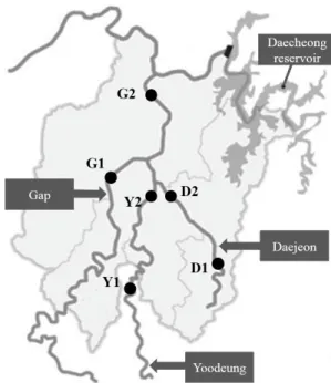
Six sampling locations were selected from Gap stream, Yoodeung stream, and Daejeon stream, all of which are located in Daejeon, Republic of Korea (Fig. 1). At September and October in 2016, one liter of water was collected from each of six locations, in duplicate. The raw water samples, 970 ml each, were filtered using the polycarbonate filters (Isopore Membrane Filters, Merck Millipore Ltd.) with a pore size of 0.8 µm. The filter was submerged in 30 ml of the raw water sample and vortexed vigorously. Then, the filter was removed, and the
Fig. 1. Sampling locations of three streams in Daejeon city, Republic of Korea. GPS coordinates of D1, D2, G1, G2, Y1, and Y2 are 36°18'53.4"N 127°26'21.85"E, 36°26'25.71"N 127°3'17.24"E, 36°18'24.61"N 127°
21'33.88"E, 36°21'30.38"N 127°21'30.38"E, 36°17'55.24"N 127°20'19.
61"E, and 36°20'40.89"N 127°24'11.66"E, respectively.
resuspended mixture was centrifuged at 1,500 × g for 20 min.
The supernatant was then removed, leaving a residue of 2 ml in the tube. The concentrated sample, 1 ml, was cultured on a non-nutrient agar (ATCC Medium 919) plate, prepared by pre-inoculating inactivated Escherichia coli (KCTC 2441, Korean Collection for Type Cultures) at 30°C for 5~7 days.
The inactivated E. coli was prepared by heating at 65°C for 30 min.
Genomic DNA extraction
To extract the genomic DNA of FLAs, cultured FLAs were raked from the incubated plates using a scraper and were each suspended in phosphate buffered saline solution of 200 ml.
Genomic DNA was then extracted from each sample using the Qiagen DNA mini kit (Qiagen) according to the manufacturer’s protocol.
Molecular based techniques
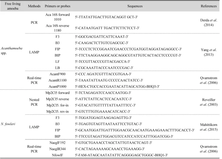
In order to select the most appropriate method to detect Acanthamoeba spp. and N. fowleri, the detection sensitivity of four published molecular methods (PCR, real-time PCR, nested PCR, and LAMP) and one commercially available real-time PCR kit (Genesig) were compared. The primers used, as well as the references to the original papers describing the methods, are listed in Table 1. Each method was performed following the published or the manufacturer’s protocol.
Results and Discussion
Comparison of the sensitivity of the molecular methods for detecting N. fowleri and Acanthamoeba spp. was conducted using ten-fold diluted (10
0~10
-6) solutions of genomic DNA (gDNA) extracted from N. fowleri and A. castellanii.
Table 1. The molecular methods for detecting N. fowleri and Acanthamoeba spp.
Free living
amoeba Methods Primers or probes Sequences References
Acanthamoeba spp.
PCR
Aca 16S forward
1010 5'-TTATATTGACTTGTACAGGT GCT-3'
Derda et al.
(2014) Aca 16S reverse
1180 5'-CATAATGATT TGACTTCTTCTCCT-3'
LAMP
F3 5'-GGCGACGATTCATTCAAAT-3'
Yang et al.
(2013) B3 5'-CAAGACTCTTGTCGAGCGC-3'
FIP 5'-TCCCTCTCCGGAATCGAACCCTCGATGGTAGGATAGAGGCC-3' BIP 5'-TTCTAAGGAAGGCAGCAGGCGTATTGTCACTACCTCCCCGT-3'
LF 5'-TCCGTTACCCGTTACGACCA-3' LB 5'-CGCAAATTACCCAATCCCGAC-3'
Real-time PCR
AcantF900 5'-CCC AGATCGTTTACCGTGAA-3'
Qvarnstrom et al. (2006) AcantR1100 5'-TAAATATTAATG CCCCCAACTATCC-3'
AcantP1000 5'-HEX-CTGCCACCGAATACATTAGCATGG-BHQ3-3'
N. fowleri
Nested PCR
Mp2Cl5 forward 5'-TCTAGAGATCCAACCAATGG-3'
Reveiller et al. (2003) Mp2Cl5 reverse 5'-ATTCTATTCACTCCACAATCC-3'
Mp2Cl5. for-in 5'-GTACATTGTTTTTATTAATTTCC-3' Mp2Cl5. rev-in 5'-GTCTTTGTGAAAACATCACC-3'
LAMP
F3 5'-TGGATGGAGTAAGAGAGTTG-3'
Mahittikorn et al. (2015) B3 5'-TGAGTGTAGTTAATAATTCCTGTAC-3'
FIP 5'-GCAATGGATTGATTTGGAACGCAACAATGAAAGAAACTTTGCACCT-3' BIP 5'-TTCCGTAGATTGGACGTCCATCCATCCATTTGGATCGG-3'
Real-time PCR
NaeglF192 5'-GTGCTGAAACCTAGCTATTGTAACTCAGT-3'
Qvarnstrom et al. (2006) NaeglR344 5'-CACTAGAAAAAGCAAACCTGAAAGG-3'
NfowlF 5'-FAM-ATAGCAATATATTCAGGGGAGCTGGGC-BHQ1-3'
The gDNAs of N. fowleri and A. castellanii were detected in 10
0~10
-5diluted samples by using the commercial real-time PCR kit (Table 2). The copy numbers of the diluted N. fowleri gDNA for the 10
0~10
-5dilutions were 7.7 × 10
6copies/ml, 7.7
× 10
5copies /ml, 8.2 × 10
4copies/ml, 8.8 × 10
3copies/ml, 1.0
× 10
3copies/ml, and 171 copies/ml. The copy numbers of the diluted A. castellanii gDNA for the 10
0~10
-5dilutions were 2.4
× 10
6copies/ml, 4.6 × 10
4copies/ml, 4.0 × 10
3copies/ml, 334 copies/ml, 25 copies/ml, and 3 copies/ml. No gDNA was detected at dilutions of 10
-6.
The duplex real-time PCR was conducted to detect gDNA of N. fowleri and A. castellanii. The results showed that the gDNA of N. fowleri and A. castellanii was detected in 10
0~10
-5diluted samples by duplex real-time PCR (Table 2).
Nested PCR was used to amplify gDNA from the type strain of N. fowleri. The nested PCR yielded clear bands for dilutions
of 10
0~10
-3. However, it was not possible to verify any bands for dilutions of 10
-4~10
-6(Table 2). PCR was done for A.
castellanii gDNA, using Aca 16S primers. As seen with N.
fowleri, the amplification resulted in clear bands for dilutions of 10
0~10
-3, and there were no bands for dilutions of 10
-4~10
-6(Table 2).
LAMP analysis was conducted using one pair of outer primers, one pair of inner primers, and one pair of loop primers. The results showed the typical ladder shapes for the LAMP products amplified from dilutions of 10
0~10
-3. No bands were seen for dilutions of 10
-4~10
-6. In the case of A. castellanii, as with N.
fowleri, the results showed typical ladder shapes for the LAMP products amplified from dilutions of 10
0~10
-3, and there were no ladder shapes for dilutions of 10
-4~10
-6(Table 2).
The results showed that the duplex real-time PCR and the commercial simplex real-time PCR were highly sensitive among
Table 2. Comparison of the three different molecular methods in terms of detection sensitivity
Detection methods Amoeba 10-fold dilution
100 10-1 10-2 10-3 10-4 10-5 10-6
Real-time PCR A. castellanii + + + + + + -
N. fowleri + + + + + + -
Duplex real-time PCR A. castellanii + + + + + + -
N. fowleri + + + + + + -
PCRa A. castellanii + + + + - - -
N. fowleri + + + + - - -
LAMP A. castellanii + + + + - - -
N. fowleri + + + + - - -
aA. castellanii, PCR; N. fowleri, nested PCR
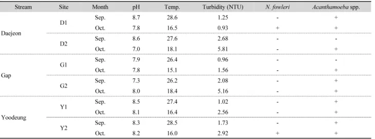
Table 3. Results of the raw water analysis in three streams in Daejeon
Stream Site Month pH Temp. Turbidity (NTU) N. fowleri Acanthamoeba spp.
Daejeon
D1 Sep. 8.7 28.6 1.25 - +
Oct. 7.8 16.5 0.93 + +
D2 Sep. 8.6 27.6 2.68 - -
Oct. 7.0 18.1 5.81 - +
Gap
G1 Sep. 7.9 26.4 0.96 - -
Oct. 7.8 15.1 1.56 - +
G2 Sep. 7.3 26.2 2.08 - +
Oct. 8.0 18.4 5.16 - +
Yoodeung
Y1 Sep. 8.5 27.4 1.02 - +
Oct. 8.1 16.4 2.56 - +
Y2 Sep. 8.3 28.5 1.73 - +
Oct. 8.2 16.0 2.92 + +
the compared molecular methods and the sensitivity of two methods was similar. However, we used the duplex real-time PCR for the surveillance of the FLAs in the aquatic environment of Daejeon city as the duplex real-time PCR could detect the two FLAs simultaneously in the single tube. In Table 3, Acanthamoeba spp. were detected in all samples except only two samples from the D2 and G1 sites, collected at September 2016. N. fowleri was detected in the samples from the D1 and Y2, collected at October 2016. Albeit the number of the surveyed sites and the samples was small, we firstly showed that the two harmful FLAs are distributed in the aquatic environment of the Republic of Korea.
According to De Jonckheere (2012), N. fowleri has been found on all continents except Antarctica. Also, it is known that Acanthamoeba spp. are also found throughout the world (da Rocha-Azevedo et al., 2009). They are classified as pathogenic protozoa in the contaminants candidate list of the U.S. Environ- mental Protection Agency. They are generally considered as organisms that need to be monitored and controlled to ensure drinking water safety, even though no legal regulations regarding amoebas specifically exist in a number of countries including Canada and Australia. In the Republic of Korea, there have been few environmental science studies on N. fowleri and Acanthamoeba spp., other than a few clinical studies (Jeong and Yu, 2005; Kim et al, 2008; Lee et al., 2011; Moon et al, 2016).
The high sterilization resistance of these FLAs becomes a risk factor in the drinking water treatment process. De Jonckheere et al.
(1976) and Sarkar and Gerba (2012) reported that Acanthamoeba spp., were highly resistant to chlorine and UV light, and UV resistance was even higher than that of Cryptosporidium oocysts.
De Jonckheere et al. (1976) found that N. fowleri showed high sterilization resistance. Cursons et al. (1980) reported that the inactivation of cysts required exposure to a residual chorine concentration of 0.74 mg/L for 30 min. Thus, it is necessary to investigate the presence and distribution of these FLAs in environmental water for the safety of drinking water. In addition to high sterilization resistance, these amoebas host other microorganisms such as bacteria, mold, and viruses and they can release these microorganisms into environments (Greub and Raoult, 2004; Thomas et al., 2004, 2010; Richards et al., 2013; Denoncourt et al., 2014). Also, it has been recognized
that bacteria inside the amoebas can show high resistance to various stresses and sanitizers. This is another impetus for investigating the ecology of amoebas; such knowledge is necessary to determine the nature and prevalence of dangerous microbiological factors in our drinking water and water sources and to develop water management plans.
Conclusion
We verified that real-time PCR was highly sensitive compared to PCR and LAMP as detection methods for two FLAs. Further, the duplex real-time PCR assay method could detect the two FLAs simultaneously. Among the twelve samples in three streams located in Daejeon city, N. fowleri and Acanthamoeba spp. were detected in two (16.6%) and ten (83.3%) samples, respectively. Although we tested only 12 samples, the results showed that the harmful FLAs were distributed in the Korean aquatic environment and Acanthamoeba was detected more frequently than N. fowleri. This implies that water resources should be managed and well treated to ensure the safety of public drinking water. In addition, it is necessary to survey the distributions of the two FLAs continuously in nationwide for maintaining the drinking water safety in Republic of Korea.
Acknowledgements
This work was supported by the research grant of K-water (KWCI2018-080-032).
적 요
가시아메바(Acanthamoeba spp.)와 파울러자유아메바
(Naegleria fowleri)는 자유생활아메바로 자연계에 널리 분포
하며 사람과 동물에게 치명적인 질병을 일으킨다. 본 연구에
서는 가사아메바와 파울러자유아메바를 물 환경에서 조사하
기 위해 기존에 보고된 네 종류의 분자생물학적 방법과 상용
real-time PCR 키트의 분석 민감도를 비교하였다. 그 결과
duplex real-time PCR 방법이 민감도가 가장 좋았으며, 동시에
두 종류의 자유생활아메바를 검출할 수 있었다. 따라서 이 방
법을 사용하여 한국의 대전시에 위치한 3개 하천, 6개 지점을
대상으로 그 분포를 2회 조사하였다. 가시아메바는 12개 시료 중 10개 시료에서 검출되었으며(83.3%), 파울러자유아메바 는 2개 시료에서 검출되었다(16.6%). 향후 이러한 유해 아메 바로부터 먹는 물의 안전성을 확보하기 위해 지속적인 분포 조사가 필요할 것이다.
References
Awwad, S.T., Petroll, W.M., McCulley, J.P., and Cavanagh, H.D.
2007. Updates in Acanthamoeba keratitis. Eye Contact Lens 33, 1–8.
Cursons, R.T., Brown, T.J., and Keys, E.A. 1980. Effect of disinfectants on pahtogenic free-living amoebae: in axenic conditions. Appl.
Environ. Microbiol. 40, 62–66.
da Rocha-Azevedo, B., Tanowitz, H.B., and Marciano-Cabral, F.
2009. Diagnosis of infections caused by pathogenic free-living amoebae. Interdiscip. Perspect. Infect. Dis. 2009, 251406.
De Jonckheere, J.F. 2004. Molecular definition and the ubiquity of species in the genus Naegleria. Protist 155, 89–103.
De Jonckheere, J.F. 2012. The impact of man on the occurrence of the pathogenic free-living amoeboflagellate Naegleria fowleri. Future Microbiol. 7, 5–7.
De Jonckheere, J. and van de Voorde, H. 1976. Differences in destruction of cysts of pathogenic and nonpathogenic Naegleria and Acanthamoeba by chlorine. Appl. Environ. Microbiol. 31, 294–297.
Denoncourt, A.M., Paquet, V.E., and Charette, S.J. 2014. Potential role of bacteria packaging by protozoa in the persistence and transmission of pathogenic bacteria. Front. Microbiol. 5, 240.
Derda, M., Wojtkowiak-Giera, A., and Hadas, E. 2014. Comparative analyses of different genetic markers for the detection of Acanthamoeba spp. isolates. Acta Parasitol. 59, 472–477.
Greub, G. and Raoult, D. 2004. Microorganisms resistant to free-living amoebae. Clin. Microbiol. Rev. 17, 413–433.
Im, K. and Kim, D.S. 1998. Acanthamoebiasis in Korea: two new cases with clinical cases review. Yonsei Med. J. 39, 478–484.
Jeong, H.J. and Yu, H.S. 2005. The role of domestic tap water in Acanthamoeba contamination in contact lens storage cases in Korea. Korean J. Parasitol. 43, 47–50.
Jung, E.Y., Jung, M.E., Park, H.G., Jung, J.M., Rho, J.S., and Ryu, P.J.
2008. Distribution of Acanthamoeba spp. in raw water and water treatment process. J. Environ. Sci. 17, 1121–1127.
Kim, J.H., Kim, D., and Shin, H.J. 2008. Contact-independent cell death of human microglial cells due to pathogenic Naegleria fowleri trophozoites. Korean J. Parasitol. 46, 217–221.
Lee, Y.J., Park, C.E., Kim, J.H., Sohn, H.J., Lee, J.L., Jung, S.Y., and Shin, H.J. 2011. Naegleria fowleri lysate induces strong cytopathic effects and pro-inflammatory cytokine release in rat microglial cells. Korean J. Parasitol. 49, 285–290.
Madarova, L., TrnKova, K., Feikova, S., Klement, C., and Obernauerova, M. 2010. A real-time PCR diagnostic method for detection of Naegleria fowleri. Exp. Parasitol. 126, 37–41.
Mahittikorn, A., Mori, H., Popruk, S., Roobthaisong, A., Sutthikornchai, C., Koompapong, K., Siri, S., Sukthana, Y., and Nacapunchai, D. 2015. Development of a rapid, simple method for detecting Naegleria fowleri visually in water samples by loop-mediated isothermal amplification (LAMP). PLoS One 10, e0120997.
Marciano-Cabral, F. and Cabral, G. 2003. Acanthamoeba spp. as agents of disease in humans. Clin. Microbiol. Rev. 16, 273–307.
Marciano-Cabral, F., MacLean, R., Mensah, A., and LaPat-Polasko, L.
2003. Identification of Naegleria fowleri in domestic water sources by nested PCR. Appl. Environ. Microbiol. 69, 5864– 5869.
Martinez, A.J. and Visvesvara, G.S. 1997. Free-living, amphizoic and opportunistic amebas. Brain Pathol. 7, 583–598.
Mathers, M.D., Nelson, S.E., Lane, J.L., Wilson, M.E., Allen, R.C., and Folberg, R. 2000. Confirmation of confocal microscopy diagnosis of Acanthamoeba keratitis using polymerase chain reaction analysis. Arch. Ophthalmol. 118, 178–183.
Moon, E.K., Park, H.R., Quan, F.S., and Kong, H.H. 2016. Efficacy of Korean multipurpose contact lens disinfecting solutions against Acanthamoeba castellanii. Korean J. Parasitol. 54, 697–702.
Qvarnstrom, Y., Visversvara, G.S., Sriram, R., and da Silva, A.J. 2006.
Multiplex real-time PCR assay for simultaneous detection of Acanthamoeba spp., Balamuthia mandrillaris, and Naegleria fowleri. J. Clin. Microbiol. 44, 3589–3595.
Reveiller, F.L., Cabanes, P.A., and Marciano-Cabral, F. 2003 Development of a nested PCR assay to detect the pathogenic free-living amoeba Naegleria fowleri. Parasitol. Res. 88, 443– 450.
Richards, A.M., Von Dwingelo, J.E., Price, C.T., and Kwaik, Y.A.
2013. Cellular microbiology and molecular ecology of Legionella- amoeba interaction. Virulence 4, 307–314.
Sarkar, P. and Gerba, C.P. 2012. Inactivation of Naegleria fowleri by chlorine and ultraviolet light. J. Am. Water Works Assoc. 104, E173–E180.
Schroeder, J.M., Booton, G.C., Hay, J., Niszl, I.A., Seal, D.V., Markus, M.B., Fuerst, P.A., and Byers, T.J. 2001. Use of subgenic 18S ribosomal DNA PCR and sequencing for genus and genotype identification of Acanthamoeba from humans with keratitis and from sewage sludge. J. Clin. Microbiol. 39, 1903–1911.
Schuster, F.L. and Visvesvara, G.S. 2004. Free-living amoebae as opportunistic and non-opportunistic pathogens of humans and animals. Int. J. Parasitol. 34, 1001–1027.
Seal, D.V. and Hay, J. 1994. Acanthamoeba keratitis. BMJ 309, 1019.
Siddiqui, R. and Khan, N.A. 2012. Biology and pathogenesis of Acanthamoeba. Parasit. Vectors 5, 6.
Thomas, V., Bouchez, T., Nicolas, V., Robert, S.l., Loret, J.F., and Levi, Y. 2004. Amoebae in domestic water systems: resistance to disinfection treatments and implication in Legionella persistence.
J. Appl. Microbiol. 97, 950–963.
Thomas, V., McDonnell, G., Denyer, S.P., and Maillard, J.Y. 2010.
Free-living amoebae and their intracellular pathogenic micro- organisms: risks for water quality. FEMS Microbiol. Rev. 34, 231–259.
Visvesvara, G.S., Moura, H., and Schuster, F.L. 2007. Pathogenic and opportunistic free-living amoebae: Acanthamoeba spp., Balamuthia Mandrillaris, Naegleria fowleri and Sappinia diploidea. FEMS
Immunol. Med. Microbiol. 50, 1–26.
Yang, H.W., Lee, Y.R., Inoue, N., Jha, B.K., Danne, D.B., Kim, H.K., Lee, J., Goo, Y.K., Kong, H.H., Chung, D.I., et al. 2013. Loop- mediated isothermal amplification targeting 18S ribosomal DNA for rapid detection of Acanthamoeba. Korean J. Parasitol. 51, 269–277.