47(1) : 24∼ 28 (2016)
24
인삼종자오일의 α-Glucosidase 및 α-Amylase 저해작용
안창호1·남윤민1,2·김신정2·양병욱2·김형춘3*·고성권1,2*
1세명대학교 한방식품영양학부, 2고연 인삼연구센터, 3강원대학교 약학대학
Inhibitory Effects of Ginseng Seed Oil on α-Glucosidase and α-Amylase Activity
Chang Ho Ahn1, Yun Min Nam1,2, Shin Jung Kim2, Byung Wook Yang2, Hyoung Chun Kim3*, and Sung Kwon Ko1,2*
1Department of Oriental Medical Food & nutrition, Semyung University, Jecheon 390-711, Korea
2Ginseng Research Center, Koyeon, Jecheon 390-711, Korea
3College of Pharmacy, Kangwon National University, Chunchon 200-701, Korea
Abstract − This study was to evaluate the effect of ginseng (Panax ginseng) seed oil on the α-glucosidase and α-amylase. Each ginseng seed oils (HE, SE, EE) exhibited a significant inhibitory effect (p<0.001) at all concentrations (10 and 20 mg/ml) on α-glucosidase activity. HE is the highest inhibitory activity (86.92%) at a concentration of 20 mg/ml, SE and EE showed an inhibitory effect of 77.13% and 65.83%, respectively. And also, Each ginseng seed oils (HE, SE, EE) exhibited a significant inhibitory effect (p<0.001) at all concentrations (1 and 2 mg/ml) on α-amylase activity. HE is the highest inhibitory activity (89.68%) at a concentration of 2 mg/ml, SE and EE showed an inhibitory effect of 76.99% and 65.70%, respectively.
Key words − α-Amylase, Ginseng, α-Glucosidase, Oil, Panax ginseng, Seed
인삼(Panax ginseng C. A. Meyer)의 뿌리는 주로 약으로 이용하며, 인삼의 주요한 생리활성물질은 인삼사포닌 (ginsenosides), 폴리아세틸렌(polyacetylenes), 인삼 단백질 (proteins), 다당체(polysaccharides), 페놀계 화합물(phenolic compounds)을 함유하고 있는 것으로 알려져 있다.1-4)
인삼사포닌(ginsenoside)은 생화학 및 약리작용을 나타내 는 인삼의 유효성분으로 주목 받아왔다. 그러므로 Shibata 등5-6)의 연구 시작으로 1960년대 후반부터 인삼에 대한 다 양한 연구들이 이루어지고 있다. 그 화학구조가 Shibata 그 룹5-6)의 연구에 의해 명확히 확인되었다. 또한 화학구조의 특성에 따라 protopanaxadiol(PD)계, protopanaxatriol(PT)계, oleanane계 사포닌으로 구분한다. 현재까지 각각 22종, 13종, 1종의 화합물의 화학구조가 규명되었다.
이러한 인삼사포닌(ginsenoside)의 약리 활성을 보면 항암 작용7)을 비롯하여 항당뇨 작용,8)중추신경 억제작용,9)동맥 경화 및 고혈압의 예방,10,11) 간기능 촉진 및 숙취제거효과,12)
항피로 및 항스트레스 작용,13,14) 항산화작용,15) 항염활성,16) 단백질 합성 촉진 작용,17)면역증강작용18)등이 보고되었다.
한편, 인삼의 지방산에 대한 연구로는 전 등19)이 홍삼으 로부터 myristic acid부터 nervonic acid까지 15종이 분리 확 인 되었고 불포화지방산인 linoleic acid가 80%, 포화지방산 인 palmitic acid가 10.5%로 두지방산이 유리지방산의 90.5%
를 차지한다고 발표하였고, Zhang 등20)은 인삼속 삼류 생약 (Panax ginseng, Panax notoginseng, Panax quinquefolium) 뿌리로부터 11종의 지방산(myristic acid, pentadecanoic acid, palmitic acid, palmitoleic acid, heptadecanoic acid, stearic acid, oleic acid, linoleic acid, α-linolenic acid, arachidic acid and eicosadienoic acid)을 GC-MS로 분석 보 고하였다. 또한, 고 등21)은 한국인삼 뿌리에 linoleic acid (42.62%), oleic acid(12.31%)를 함유한다고 보고하였고, Zhu 등22)은 한국, 중국, 미국산의 에테르 추출 인삼종자오일의 지방산 분석을 통하여 oleic acid가 미국삼(87.5%), 한국삼 (69.14%), 중국삼(61.19%) 함유한다고 보고하였다.
그러나, 인삼 지하부와 인삼종자오일에 대한 인삼사포닌 및 지방산 연구는 다수 이루어졌으나, 인삼종자오일에 대한
*교신저자(E-mail):[email protected], [email protected] (Tel): +82-33-250-6917, +82-43-649-1433
추출방법에 따른 성분 및 생리활성 차이에 대한 연구는 아 직 체계적으로 이루어지지 않고 있다. 이러한 점을 착안하 여, 본 연구에서는 인삼 및 홍삼에서 입증된 약리효능 중 항 당뇨작용8)에 대한 연구의 일환으로 국내산 인삼종자오일의 α-glucosidase 및 α-amylase에 대한 생리활성 연구를 수행 함으로서 우리 인삼종자오일의 당흡수저해를 통한 항당뇨 생리활성의 일부를 확인하고자 하였다.
실험재료 및 방법
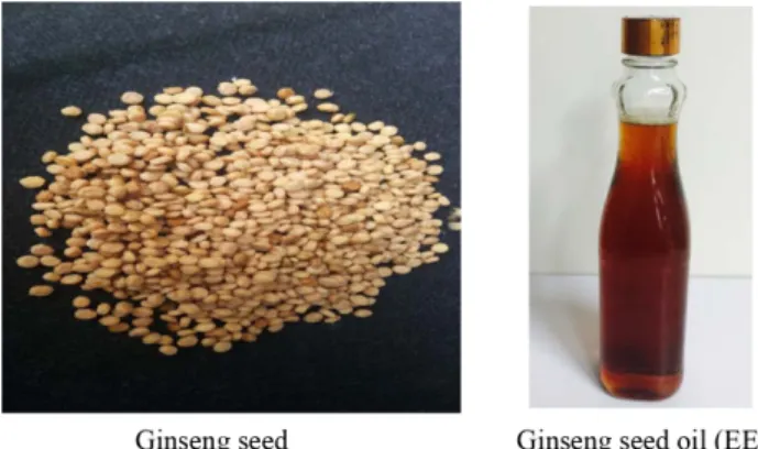
실험재료 − 본 연구에 사용한 고려인삼(Panax ginseng) 종 자는 한국 충북 음성에서 재배(재배자, 남성엽) 5년생 인삼 의 종자를 2014년 7월 14일에 채집하였고, 각각의 제품표 본은 세명대학교 한방식품연구실에 보관하고 있다(Fig. 1).
인삼종자오일 조제
1) 압출법에 의한 인삼종자오일(EE) 조제 − 인삼 종자 60 kg을 거피하여 7.35 kg의 거피 인삼종자를 얻었고, 얻은 거피 인삼종자를 압출하여 1.65 kg(약 2 l)을 얻었다(Fig. 1).
2) 초임계에 의한 인삼 종자 오일(SE) 조제 − 거피 인삼 종자 133 g을 Table I의 조건으로 초임계 추출하여 초임계 추출물 42.6 g을 얻었다.
3) 헥산 추출 인삼종자오일(HE) 조제 − 세말한 거피 인 삼종자 100 g에 hexane을 1 l를 넣고, 소니케이터(고도기업, 4020P, 한국)에서 2시간씩, 2회 추출하여 여과 후 여액을 합 하여 감압 농축하여 헥산(hexane)추출물을 얻었다.
인삼종자오일의 지방산 분석(GC법)23)− 인삼종자오일(EE, SE, HE)의 지방산 분석은 식품공전법에 따라서 GC법으로 분석하였다. 지방산 표준품은 SupelcoTM 37 Component FAME Mix, 100 mg Neat(Catalog No. 18919-1MP)를 사용 하였다. GC는 Agilent 7890A를 사용하였고, 컬럼은 SPTM- 2560, 100 m×0.25 mm ID, 0.20μm film를 사용하였다. 이 동상은 헬륨을 사용하였으며, 이동속도는 20 cm/sec이었다.
오븐의 온도는 140oC(5분)에서 240oC로 1분에 4oC씩 유지 하였다. 전개 크로마토그램 프로파일은 FID detector로 285oC 에서 검출하였다.
당 흡수저해 시험
1) α-Glucosidase 저해활성 측정24)− α-Glucosidase(0.35 U/ml)와 ρNPG(1.5 mM ρ-nitrophenyl-α-D-glucopyranoside) 는 0.1 M sodium phosphate buffer(pH 7.0)에 용해하여 사 용하며, 각각의 추출물 50 μl를 0.35 unit/ml α-glucosidase 효소액 100 μl와 혼합하여 37oC에서 10분간 전배양한 후 1.5 mM ρNPG 50 μl를 가하여 37oC에서 20분간 반응시킨 다. 1 M Na2CO3 1 ml을 가하여 반응을 정지시킨 후 ELISA 를 사용하여 405 nm에서 흡광도를 측정하고 저해율(%)을 계산하였으며, positive control로 acarbose(sigma, USA)를 사용하였다.
2) α-Amylase 저해활성 측정25)− α-Amylase 저해활성은 추출물 125 μl에 12 unit/ml pancreatin 기원의 α-amylase 효소액 62.5 μl, 200 mM potassium phosphate buffer(pH 6.8) 62.5μl와 혼합하여 37oC에서 10분간 전배양한 후 1%
starch를 125 μl 가하여 37oC에서 5분간 반응시킨다. 반응액 에 48 mM DNS(3,5-dinitrosalicylic acid and 30% sodium potassium tartrate in 0.5 M NaOH) 발색시약 125 μl를 넣고 100oC에서 15분간 끓여 발색을 시킨 후 충분히 냉각시키고, 이 반응액에 3배량의 증류수를 가한 후 ELISA를 사용하여 Fig. 1. Photos of ginseng seed and ginseng seed oil (EE).
Table I. Supercritical extraction conditions and amounts
Samples CO2 used per batch (l) Cumulative CO2 (l) Extraction amount of batch (g)
Cumulative extraction amount (g)
F1 537 537 10.686 10.686
F2 519 1056 9.32 20.006
F3 509 1565 9.522 29.528
F4 562 2127 9.105 38.633
F5 500 2627 3.135 41.768
F6 326 2953 0.835 42.603
540 nm에서 흡광도를 측정하고 저해율을 계산하였다.
결과 및 고찰
본 연구에서는 인삼종자오일의 추출방법에 따른 지방산 성분에 미치는 영향과 α-glucosidase 및 α-amylase에 대한 생리활성 연구를 수행함으로서 우리 인삼종자오일의 항당 뇨 생리활성의 일부를 확인하고자 하였다.
우리나라에서 재배되고 있는 5년생 인삼종자를 대상으로 추출 방법에 따른 개별 지방산의 함량 분포를 조사·비교함 으로써 지방산 함유패턴을 중심으로 하는 차이점을 검토하 였다. 분석한 지방산은 palmitic acid, palmitoleic acid, oleic acid, linoleic acid 이었으며 이들은 Fig. 1과 같이 GC를 통 하여 표품과 직접 비교·확인하였다.
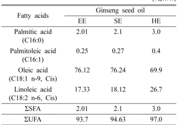
Table II에서 보는바와 같이 압출 인삼종자오일(EE)의 지 방산 함량은 oleic acid가 76.12%, linoleic acid가 17.33%, palmitic acid 2.01%, palmitoleic acid 0.25%이었으며, 초임 계추출 인삼종자오일(SE)의 지방산 함량은 oleic acid가 76.24%, linoleic acid가 18.12%, palmitic acid 2.10%, palmitoleic acid 0.27%를 나타내었고, 헥산추출 인삼종자오 일(HE)의 지방산 함량은 oleic acid가 69.9%, linoleic acid 가 26.7%, palmitic acid 3.0%, palmitoleic acid 0.40% 이 었다. 이와 같은 결과는 Zhu 등22)이 한국산 에테르 추출 인 삼종자오일의 지방산 분석을 통하여 oleic acid가 69.14%를 함유한다고 보고한 것과 비슷한 함량을 보여주었다.
콜레스테롤 저하작용26,27)이 알려져 있는 oleic acid와 linoleic acid 등의 불포화지방산 총함량은 HE가 97.0%로 가 장 높았고, SE(94.63%), EE(93.7%)의 순이었고, 주 함유 불
포화지방산인 oleic acid는 SE가 76.24%로 가장 높았고, EE(76.12%), HE(69.9%)의 순이었다.
한편, 홍삼에는 linoleic acid가 80%, 포화지방산인 palmitic acid가 10.5%로 두지방산이 90.5%를 차지한다고 보고19) 되 었으나, 반면에 본 연구를 통한 인삼종자오일의 지방산 성 분은 oleic acid와 linoleic acid가 EE(93.45%), HE(96.6%), SE(94.36%)로서 두지방산이 93% 이상을 함유함으로써 불 포화 지방산이 주요한 지방산 성분임을 확인할 수 있었다.
또한, 홍삼은 linoleic acid가 주지방산인 반면에 인삼종자오 일은 oleic acid가 주지방산임을 확인할 수 있었다.
α-Glucosidase 활성시험에서는 50% 이상의 저해활성을 나타내는 10 및 20 mg/ml의 농도에서 활성을 측정하였으 며, α-amylase 저해활성은 50%의 저해활성을 나타내는 1 및 2 mg/ml의 농도에서 활성평가를 실시하였다.
α-Glucosidase 활성시험에서는 Fig. 3에서 보는바와 같이 모든 시료가 유의적인 저해활성을 나타내었으며(p<0.001), 특히, HE는 10 mg/ml의 농도에서 64.39%의 가장 높은 저
Table II. Composition of fatty acids in the ginseng seed oil by various extracting conditions
(%,w/w) Fatty acids Ginseng seed oil
EE SE HE
Palmitic acid (C16:0)
2.01 2.1 3.0
Palmitoleic acid (C16:1)
0.25 0.27 0.4
Oleic acid (C18:1 n-9, Cis)
76.12 76.24 69.9 Linoleic acid
(C18:2 n-6, Cis)
17.33 18.12 26.7
ΣSFA 2.01 2.1 3.0
ΣUFA 93.7 94.63 97.0
*EE: extruded ginseng seed oil, SE: supercritical extracted ginseng seed oil, HE: hexene extracted ginseng seed oil, ΣSFA: total saturated fatty acid, ΣUFA: total unsaturated fatty acid
Fig. 2. GC chromatogram of fatty acids detected from the gin- seng seed oil by various extracting conditions.
해활성을 나타내었으며, SE와 EE는 각각 58.82% 및 52.25%
의 저해활성을 나타내었다. 20 mg/ml의 농도에서도 HE가 86.92%로 가장 우수한 저해활성을 나타내었으며, SE는 77.13%, EE는 65.83%의 저해활성을 나타내었다. 양성대조
물로 사용한 acarbose의 저해활성은 2.5 mg/ml의 농도에서 36.21%이었다.
α-Amylase 활성시험에서는 Fig. 4에서 보는바와 같이 모 든 시료가 유의적인 저해활성을 나타내었으며(p<0.001), 특 히, HE는 1 mg/ml의 농도에서 65.39%의 가장 우수한 저해 활성을 보여주었으며, SE가 57.25%, EE가 50.36%이었다.
2 mg/ml의 농도에서도 HE가 89.68%로 가장 우수한 활성을 보였으며, SE가 76.99%, EE가 65.70%이었다.
결 론
인삼종자오일의 지방산 성분은 불포화지방산 총함량에 있 어서 헥산 추출 인삼종자오일(HE)이 97.0%로 가장 높았고, 초임계 추출 인삼종자오일(SE, 94.63%), 압출 인삼종자오일 (EE, 93.7%)의 순이었고, 주 함유 불포화지방산인 oleic acid 는 SE가 76.24%로 가장 높았고, EE(76.12%), HE(69.9%) 의 순이었다.
α-Glucosidase 활성시험에서는 10과 20 mg/ml의 농도에 서 저해활성을 나타내었으며, HE는 20 mg/ml의 농도에서 가장 높은 저해활성(86.92%)을 나타내었다. α-amylase 활성 시험 결과, 1과 2 mg/ml의 농도에서 저해활성을 나타내었 고, HE는 2 mg/ml의 농도에서 89.68%의 가장 우수한 저해 활성을 보여주었다. 이와 같은 결과는 인삼종자오일이 α- glucosidase와 α-amylase 저해활성을 나타내므로 써, 장에서 당(sugar)의 흡수를 저해하고, 식후 혈당의 상승을 억제할 것으로 사료되며, 향후, 당흡수 저해 항당뇨 건강기능식품 의 개발이 기대된다.
사 사
“이 논문은 2014학년도 세명대학교 교내학술연구비 지원 에 의해 수행된 연구임”.
인용문헌
1. Namba, T. (1980) The Encyclopedia of Wakan-Yaku with Color Pictures (I), 1-3, Hoikusha, Osaka.
2. Park, J. D. (1996) Recent studies on the chemical constituents of Korean ginseng. Korean J. Ginseng Sci. 20: 389-415.
3. Sanata, S., Kondo, N., Shoji, J., Tanaka, O. and Shibata, S.
(1974) Studies on the saponins of ginseng. I. Structure of gin- seng-Ro, Rb1, Rb2, Rc and Rd. Chem. Pharm. Bull. 22: 421- 428.
4. Kitagawa, I., Taniyama, T., Shibuya, H., Nota, T. and Yoshi- kawa, M. (1987) Chemical studies on crude drug processing.
V. On the constituents of ginseng radix rubra (2): Comparison of the constituents of white ginseng and red ginseng prepared
*EE: extruded ginseng seed oil, SE; supercritical extracted ginseng seed oil, HE: hexene extracted ginseng seed oil, 1) Different alphabets depending on the concentration of the same sample that is significantly different (p<0.05), 2) Differ- ent alphabets between different samples of the same concen- tration that is significantly different (p<0.05), There is a significant difference between the concentration of the same sample (***: p<0.001).
Fig. 3. Inhibition effect of ginseng seed oil (EE, SE, HE) on α-glucosidase activity.
*EE: extruded ginseng seed oil, SE; supercritical extracted ginseng seed oil, HE: hexene extracted ginseng seed oil, 1) Different alphabets depending on the concentration of the same sample that is significantly different (p<0.05), 2) Differ- ent alphabets between different samples of the same concen- tration that is significantly different (p<0.05), There is a significant difference between the concentration of the same sample (***: p<0.001).
Fig. 4. Inhibition effect of ginseng seed oil (EE, SE, HE) on α-amylase activity.
from the same Panax ginseng root. Yakugaku Zasshi 107:
495-505.
5. Shibata, S., Tanaka, O., Ando, T., Sado, M., Tsushima, S. and Ohsawa, T. (1966) Chemical studies on oriental plant drugs (XIV). Protopanaxadiol, a genuine sapogenin of ginseng saponins. Chem. Pharm. Bull. 14: 595-600.
6. Tanaka, O., Nagai, M. and Shibata, S. (1966) Chemical stud- ies on the oriental plant drugs. XVI. The stereochemistry of protopanaxadiol, a genuine sapogenin of ginseng. Chem.
Pharm. Bull. 14: 1150-1156.
7. Mochizuki, M., Yoo, Y. C., Matsuzawa, K., Sato, K., Saiki, I., Tonooka, S., Samukawa, K. and Azuma, I. (1995) Inhibitory effect of tumor metastasis in mice by saponins, ginsenoside- Rb2, 20(R)- and (S)-ginsenoside-Rg3, of red ginseng. Biol.
Pharm. Bull. 18: 1197-1202.
8. Yokozawa, T., Kobayashi, T., Oura, H. and Kawashima, Y.
(1985) Studies on the mechanism of the hypoglycemic activ- ity of ginsenoside-Rb2 in streptozotocin-diabetic rats. Chem.
Pharm. Bull. 33: 869-872.
9. Takagi, K., Saito, H. and Nabata, H. (1972) Pharmacological studies of Panax ginseng root: estimation of pharmacological actions of Panax ginseng root. J. Pharmacol. 22: 245-249.
10. Jung, I. S. and Cho, Y. D. (1985) Effect of ginseng saponin fraction on absorption of cholesterol and serum lipid com- ponents. Korean J. Ginseng Sci. 9: 232-239.
11. Yoon, S. H. and Joo, C. N. (1993) Study on the preventive effect of ginsenosides against hypercholesterolemia and its mechanism. Korean J. Ginseng Sci. 17: 1-12.
12. Matsuda, H., Samukawa, K. and Kubo, M. (1991) Anti-hep- atitic activity of ginsenoside Ro. Planta Med. 57: 523-526.
13. Wang, B. X., Cui, J. C., Liu, A. J. and Wu, S. K. (1983) Stud- ies on the anti-fatigue effect of the saponins of stems and leaves of Panax ginseng(SSLG). J. Tradit. Chin. Med. 3: 89- 94.
14. Saito, H., Yoshida, Y. and Tagaki, K. (1974) Effects of Panax ginseng root on exhaustive exercise in mice. Jpn J. Phar- macol. 24: 119-126.
15. Jeong, C. S., Hyun, J. E. and Kim, Y. S. (2002) Anti-oxi- dative effect of ginsenoside Rb1 on the HCl ethanol-induced gastric tissue in rats. Korean J. Pharmacogn. 33: 252-256.
16. Matsuda, H., Samukawa, K. and Kubo, M. (1990) Anti- inflammatory activity of ginsenoside Ro. Planta Med. 56: 19- 23.
17. Yokozawa, T., Oura, H. (1990) Facilitation of protein bio- synthesis by ginsenoside-Rb2 administration in diabetic rats.
J. Nat. Prod. 53: 1514-1518.
18. Jie, Y. H., Cammisuli, S. and Baggiolini, M. (1984) Immu- nomodulatory effects of Panax ginseng C.A. Meyer in the mouse. Agents Actions 15: 386-391.
19. Jeon, B. S., Choi, K. J., Sung, H. S., Chang, K. S. and Koh, S. R. (1995) Effect of controlled atmosphere and modified atmosphere storage on the fatty acid of fresh and red ginseng.
Korean J. Ginseng Sci. 19: 260-266.
20. Zhang, X. J., Huang, L. L., Cai, X. J., Li, P., Wang, Y. T. and Wan, J. B. (2013) Fatty acid variability in three medicinal herbs of Panax species. Chem. Cent. J. 7: 7-12.
21. Ko, Y. S. and Chung, B. S. (1981) Studies on the oil soluble constituents of Korean ginseng. Korean J. Food Sci. Technol.
13: 15-19.
22. Zhu, X. M., Hu, J. N., Shin, J. A., Lee, J. H., Hong, S. T. and Lee, K. T. (2010) Comparison of seed oil characteristics from Korean ginseng, Chinese ginseng (Panax ginseng C.A.
Meyer) and American ginseng (Panax quinquefolium L.). J.
Food Sci. Nutr. 15: 275-281.
23. Korea Food and Drug Administration. (2001) Code of Korea Food Regulation 2001. 257-265, Korea Food and Drug Administration, Seoul.
24. Lee, K. Y., Hong, S. Y., Jeong, H. J., Lee, J. H., Lim, S. H., Heo, N. K., Kim, S. M. and Kim, H. Y. (2014) Isolation and identification of α-glucosidase inhibitory compounds, hyper- oside, and isoquercetin from Eleutherococcus senticosus Leaves. J. Korean Soc. Food Sci. Nutr. 43: 1858-1864.
25. Kim, D. Y., Jung, J. Y., Kim, K. B. W. R., Lee, C. J., Kwak, J. H., Kim, M. J., Woo, C. S., Kim, H. J., Jung, S. A., Kim, T. W., Cho, Y. J. and Ahn, D. H. (2011) Effects of heat and pH treatments on α-amylase inhibitory activity of Ecklonia cava ethanol extract. Kor. J. Fish Aquat. Sci. 44: 791-795.
26. Wolańska, D. and Kłosiewicz-Latoszek L. (2012) Fatty acids intake and serum lipids profile in overweighted and obese adults. Rocz Panstw Zakl Hig 63: 155-162.
27. Wanders, A. J., Brouwer, I. A., Siebelink, E. and Katan, M.
B. (2010) Effect of a high intake of conjugated linoleic acid on lipoprotein levels in healthy human subjects. PLoS 5:
e9000.
(2016. 1. 25 접수; 2016. 2. 22 심사; 2016. 3. 3 게재확정)