—1— ISSN: 1225-8598
서 론
어류는 해양생태계의 먹이사슬에서 최상위 집단에 속하 며, 현재 뿐 아니라 미래 식량자원으로서도 충분한 가치가 있다(Du Preez et al., 1990). 또한 특정지역의 환경상태를 유추할 수 있는 지표생물 또는 지표로서 이용될 수 있기 때문에 환경독성학적 연구에서 중요한 역할을 담당한다.
해양환경에 영향을 미치는 수온, 염분 변화 및 환경오염 등의 다양한 요인들은 서식생물들에게 여러 가지 문제를 일으킨다. 특히 연안에 서식하는 종들은 연안 및 내륙에서 발생하는 다양한 오염원들에게 직접적으로 노출된다. 그 중 유류오염은 연안에서 빈번하게 발생하지만, 내륙에서도 직
∙간접적으로 발생하기 때문에 대표적 환경오염의 하나로
인식되고 있다.
유류는 다양한 화합물로 구성되어 있으며(Wake, 2005), 해양환경에 유출되면 나프탈렌, 벤젠 등과 같은 다환방향족 탄화수소(ploycyclic aromatic hydrocarbons: PAHs)는 해수 에 용해되어 수용성의 독성물질로 전환된다. PAHs는 수서 생물들에게 빠르게 흡수되며, 정상적인 생리적 기능수행을 방해한다(Nagabhushanam et al., 1991). 그 중 나프탈렌은 PAHs 중에서도 가장 많이 함유되어 있으며, 높은 독성으로 인해 생물들에게 치명적인 영향을 미치는 것으로 알려져 있다(Vijayavel et al., 2004; Vijayavel and Balasubramanian, 2006; Pollino et al., 2009).
수중에서 다양한 형태의 영향을 직접적으로 받는 어류의 아가미는 삼투조절, 호흡, 배설을 위한 중요한 기관이며, 환 경상태를 유추할 수 있는 기관이다(Mallatt, 1985; Fanta et al., 2003). 아가미의 조직학적 지표는 작은 절편만으로도 분석이 가능하며, 환경모니터링 뿐 만 아니라 실내 생물학 적 검정 시험에서 자주 사용된다(Black et al., 1991; Spies et
나프탈렌에 노출된 조피볼락의 생존 및 아가미의 조직학적 변화
조재권∙김태익∙손맹현∙김경민∙진영국*
국립수산과학원 남서해수산연구소
Survival and Histological Changes in Gill of the Rockfish, Sebastes schlegeli Following Exposure to Naphthalene by Jae Kwon Cho, Tae Ik Kim, Maeng Hyun Son, Kyong Min Kim and Young Guk Jin*(Southwest Sea Fisheries Research Institute, NFRDI, Yeosu 556-823, Korea)
ABSTRACT Rockfish, Sebastes schlegeli (total length; 10.36±0.49 cm, total weight; 16.28±1.86 g, N; 290) were exposed to various concentrations of naphthalene for 28 days. Exposure concentrations of naphthalene established control, ethanol (solvent) control, 0.5, 1.0, 1.5 and 2.0 mg Nap L--1. After exposure, We observed survival rate, and degree of tissue change (DTC) in gill under optical microscopy.
Survival rate of the rockfish was more than 90% in control, ethanol control, 0.5 and 1.0 mg Nap L--1, whereas it decreased in 1.5 and 2.0 mg Nap L--1(respectively 80%, 62.2%). In histological observation of gill, hyperplasia of epithelial cells observed in all exposure groups. But no showed increase of DTC which was related to concentration. Whereas, DTC at fusion of gill lamellar, lamellar telangiectasia, stasis, aneurysm and necrosis showed dose dependent increase. Especially, fusion of gill lamellar, lamellar telangiectasia and stasis observed at more 1.0 mg Nap L--1, and aneurysm and necrosis at more 1.5 mg Nap L--1. These results showed naphthalene caused survival and severe change to the gill of the rockfish which was related to exposure concentration.
Key words : Sebastes schlegeli, naphthalene, survival, gill, DTC
*Corresponding author: Young Guk Jin Tel: 82-61-690-8976, Fax: 82-61-685-9073, E-mail: [email protected]
Accepted: March 20, 2013
http://www.fishkorea.or.kr
al., 1996). 또한 어류 아가미의 조직학적 분석을 통해 다양 한 환경오염 결과들이 보고되고 있으며, 유류에 오염된 종 들에서도 확인되고 있다(Prasad, 1991).
국내에서 발생한 유류오염은 2007년 허베이스피리트호 유류유출 사고를 비롯해 최근까지도 크고 작은 다수의 오 염사례들이 보고되고 있지만, 유류에 포함된 PAHs가 해양 생물들에게 미치는 영향에 대한 연구는 미미한 실정이다.
따라서 본 연구는 PAHs 중에서도 높은 독성을 가진 나프 탈렌이 연안성 어류인 조피볼락, Sebastes schlegeli의 생존과 아가미의 조직학적 구조에 미치는 영향을 조사하여, 해양생 태계의 보전 및 관리에 기초자료를 제공하고자 하였다.
재료 및 방법
1. 실험종 및 개체수
실험에 사용된 조피볼락은 전장 10.36±0.49 cm, 전중 16.28±1.86 g으로 실험 개체수는 270마리였다.
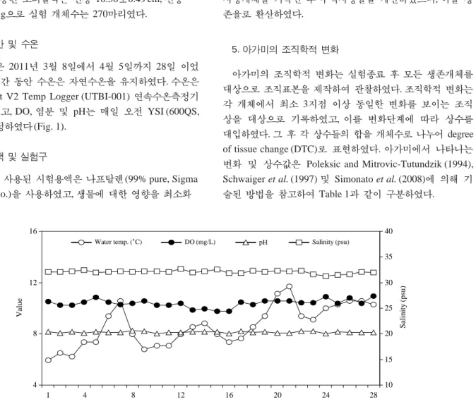
2. 실험기간 및 수온
실험기간은 2011년 3월 8일에서 4월 5일까지 28일 이었 으며, 실험기간 동안 수온은 자연수온을 유지하였다. 수온은 HOBO tidbit V2 Temp Logger (UTBI-001) 연속수온측정기 로 측정하였고, DO, 염분 및 pH는 매일 오전 YSI (600QS, USA)로 측정하였다(Fig. 1).
3. 시험용액 및 실험구
본 연구에 사용된 시험용액은 나프탈렌(99% pure, Sigma Chemicals Co.)을 사용하였고, 생물에 대한 영향을 최소화
하기 위해 에탄올에 녹인 후(DiMichele and Taylor, 1978) 증류수로 1 g Nap L-1표준용액을 만든 다음, 실험농도별로 희석하였다.
노출농도는 장기간 노출임을 고려해 96 h-LC5인 1.5 mg Nap L-1와 기존 연구(Nandini, 1988; Santos et al., 2006) 등 을 참고로 하여 control (0), ethanol (solvent) control, 0.5, 1.0, 1.5 및 2.0 mg Nap L-1을 설정하였다. 실험수조는 30 L 지수 형 순환여과식 유리수조를 사용하였으며, 각 실험 농도구 당 15개체씩 3반복으로 진행하였다. 매일 오전 10시에 5 L 의 사육수를 저면의 배설물을 제거함과 동시에 교환하였 고, 사육수의 증발을 최소화하기 위해 유리수조 윗면을 밀 폐하였다.
4. 생존율
생존율의 계산은 매일 오전 10시 사육수의 교환과 함께 사망개체를 기록한 후 누적사망률을 계산하였으며, 이를 생 존율로 환산하였다.
5. 아가미의 조직학적 변화
아가미의 조직학적 변화는 실험종료 후 모든 생존개체를 대상으로 조직표본을 제작하여 관찰하였다. 조직학적 변화는 각 개체에서 최소 3지점 이상 동일한 변화를 보이는 조직 상을 대상으로 기록하였고, 이를 변화단계에 따라 상수를 대입하였다. 그 후 각 상수들의 합을 개체수로 나누어 degree of tissue change (DTC)로 표현하였다. 아가미에서 나타나는 변화 및 상수값은 Poleksic and Mitrovic-Tutundzik (1994), Schwaiger et al. (1997) 및 Simonato et al. (2008)에 의해 기 술된 방법을 참고하여 Table 1과 같이 구분하였다.
4 8 12 16
1 4 8 12 16 20 24 28
Exposure time (day)
Value
10 15 20 25 30 35 40
Salinity(psu)
Water temp. (�C) DO (mg/L) pH Salinity (psu)
Fig. 1. Changes of water temperature, DO, pH and salinity during 28 days.
6. 조직학적 방법
28일 동안 노출 후 모든 실험구의 생존개체들은 계측형 질을 측정한 후 각 개체별로 외부로부터 두 번째 아가미를 절취하여 Bouin’s solution에 24시간 동안 고정하였다. 그 후 파라핀 절편법으로 연속절편(4μm) 한 다음 alcian blue- periodic acid and Schiff’s solution (AB-PAS, pH 2.5) 반응을 실시하여 광학현미경 관찰용 조직표본을 제작하였다. 제작
된 조직표본은 image measurement system (FOCUS techno- logy, 2005)이 부착된 광학현미경(Olympus, CX31)으로 아 가미의 조직학적 변화를 관찰하였다.
7. 통계분석
각 실험구간의 차이는 SPSS 10.0 (SPSS, Inc., Chicago, IL) 통계프로그램을 이용하여 그 유의성 여부를 검증하였다.
결 과
1. 생존율
28일 동안 나프탈렌에 노출된 조피볼락의 생존율은 Fig. 2 와 같다. 대조구의 경우 노출 16일까지 생존율에 변화가 없 었으며, 노출 16일 이후부터 노출 종료일인 28일까지 1개 체가 사망하여 97.8%의 생존율을 보였다. 에탄올(solvent) 대조구와 0.5 mg Nap L-1에 노출된 개체들의 생존율은 노 출 20일까지는 100%를 보였고, 그 이후 노출 종료일에 각 Table 1. Stage and scoring of histological changes in the gill of the
rockfish, Sebastes schlegeli following exposure to naphthalene Stage
List of severity of gill changes (gill change Scoring (Gc))
Hyperplasia of gill filament epithelial cells Gc 1 1 Hyperplasia of gill lamellar epithelial cells Gc 2 1 Rupture and lifting of gill lamellar epithelium Gc 3 1
Fusion of gill lamellar Gc 4 1
Telangiectasia of gill lamellar Gc 5 2
Stasis Gc 6 2
Aneurysm Gc 7 3
Necrosis Gc 8 3
40 60 80 100
1 4 8 12 16 20 24 28
Exposure time (day)
Survival(%)
0.5 mg Nap L-1 1.0 mg Nap L-1
Control Solvent control
1.5 mg Nap L-1 2.0 mg Nap L-1
Fig. 2. Survival (%) of the rockfish, Sebastes schlegeli following exposure to naphthalene.
c
b
ab ab
a a 0 2 4 6 8
Control Solvent 0.5 mg Nap L-1 1.0 mg Nap L-1 1.5 mg Nap L-1 2.0 mg Nap L-1
DTC
Fig. 3. Degree of tissue change (DTC) in gill tissues of the rockfish, Sebastes schlegeli following exposure to naphthalene.
각 95.6%와 97.8%를 나타냈다. 1.0 mg Nap L-1에 노출된 개체들은 노출 18일부터 사망개체가 관찰되기 시작하였으 며, 노출 종료일까지 총 3개체가 사망하여 93.3%의 생존율 을 보였다. 1.5 mg Nap L-1에 노출된 개체들은 노출 12일부 터 생존율의 감소를 보였으며, 노출 20일부터 꾸준히 감소 하여 80%의 생존율을 보였다. 2.0 mg Nap L-1에 노출된 개 체들의 경우 노출 11일부터 생존율이 감소하기 시작하였고, 노출 20일에서 24일, 노출 26일에서 28일 사이에 생존율의 급격한 감소를 보여 62.2%를 나타냈다.
2. 아가미의 조직학적 변화
나프탈렌에 노출된 조피볼락 아가미의 조직학적 손상정 도(DTC)는 Fig. 3과 같이 대조구의 경우 0.41로 에탄올 (solvent) 대조구 0.35와 별다른 차이를 보이지 않았다. 0.5 mg Nap L-1에 노출된 개체들의 DTC는 0.93, 1.0 mg Nap L-1 에 노출된 개체들의 DTC는 1.48로 대조구에 비해 높게 나 타났지만, 유의적인 차이는 관찰되지 않았다(P¤0.05). 하지 만, 1.5 mg Nap L-1과 2.0 mg Nap L-1에 노출된 개체들의 DTC는 대조구에 비해 각각 8배, 15배 이상으로 높게 나타 나 뚜렷한 차이를 보였으며, 이들 두 노출집단 사이에서도 유의한 차이를 보였다(P⁄0.05).
아가미에서 조직학적 변화를 보인 개체들은 대조구의 경 우 새엽 상피세포의 증식(Gc 1), 새판 상피세포의 증식(Gc 2) 및 새판 상피층의 파열 및 탈락(Gc 3) 증상이 주로 관찰 되었다(Fig. 4A). Gc 1은 생존개체 44마리 중 20.5%, DTC 0.20이었다(Tables 2 and 3). Gc 2는 9.1%, DTC는 0.09로 나 타났으며 Gc 3은 11.4%, DTC 0.11이었다(Tables 2 and 3).
에탄올(solvent) 대조구와 0.5 mg Nap L-1에 노출된 개체 들은 대조구와 동일한 변화를 보였다(Fig. 4B and C). 에탄 올 대조구의 경우 생존개체 43마리 중에서 Gc 1은 18.6%, DTC 0.19, Gc 2는 4.7%, DTC 0.05, Gc 3은 11.6%, DTC 0.12 로 대조구에 비해 차이가 나지 않거나 더 낮게 나타났다 (Tables 2 and 3). 0.5 mg Nap L-1에 노출된 개체들은 44마리 의 생존개체 중 Gc 1~3의 출현율이 각각 38.6%, 34.1%, 20.5%를 보였으며, DTC는 각각 0.39, 0.34, 0.20으로 대조 구와 에탄올 대조구에 비해 높게 나타났다(Tables 2 and 3).
1.0 mg Nap L-1에 노출된 개체들은 Gc 1~3의 변화 외에 도 새판의 융합(Gc 4), 새판 모세혈관확장(Gc 5) 및 울혈 (Gc 6) 증상을 보이고 있었다(Fig. 4D, E and F). 42마리의 생존개체 중 Gc 4의 경우 출현율 14.3%, DTC 0.14, Gc 5는 9.5%, DTC 0.19, Gc 6은 11.9%, DTC 0.24로 나타났다(Tables 2 and 3).
1.5 mg Nap L-1와 2.0 mg Nap L-1에 노출된 개체들은 Gc
Table 3. Degree of tissue change (DTC) in gill tissues of the rockfish, Sebastes schlegeli following exposure to naphthalene
Control Solvent 0.5 mg Nap L-1 1.0 mg Nap L-1 1.5 mg Nap L-1 2.0 mg Nap L-1
Gc 1 0.20 0.19 0.39 0.36 0.36 0.36
Gc 2 0.09 0.05 0.34 0.29 0.28 0.29
Gc 3 0.11 0.12 0.20 0.26 0.69 0.93
Gc 4 - - - 0.14 0.53 0.82
Gc 5 - - - 0.19 0.78 0.57
Gc 6 - - - 0.24 0.33 1.07
Gc 7 - - - - 0.25 1.50
Gc 8 - - - - 0.25 0.86
※Gc 1: Hyperplasia of gill filament epithelial cells, Gc 2: Hyperplasia of gill lamellar epithelial cells, Gc 3: Rupture and lifting of gill lamellar epithelium, Gc 4:
Fusion of gill lamellar, Gc 5: Gill lamellar telangiectasia, Gc 6: Stasis, Gc 7: Aneurysm, Gc 8: Necrosis.
Table 2. Frequency (F) of occurrence of histological changes in gill tissues of the rockfish, Sebastes schlegeli following exposure to naphthalene.
Number (N) of species with changes (number of examined species)
Control Solvent 0.5 mg Nap L-1 1.0 mg Nap L-1 1.5 mg Nap L-1 2.0 mg Nap L-1
N F (%) N F (%) N F (%) N F (%) N F (%) N F (%)
Gc 1 44 (9) 20.5 43 (8) 18.6 44 (17) 38.6 42 (15) 35.7 36 (13) 36.1 28 (10) 35.7
Gc 2 44 (4) 9.1 43 (2) 4.7 44 (15) 34.1 42 (12) 28.6 36 (10) 27.8 28 (8) 28.6
Gc 3 44 (5) 11.4 43 (5) 11.6 44 (9) 20.5 42 (11) 26.2 36 (25) 69.4 28 (26) 92.9
Gc 4 - - - - - - 42 (6) 14.3 36 (19) 52.8 28 (23) 82.1
Gc 5 - - - - - - 42 (4) 9.5 36 (14) 38.9 28 (8) 28.6
Gc 6 - - - - - - 42 (5) 11.9 36 (6) 16.7 28 (15) 53.6
Gc 7 - - - - - - - - 36 (3) 8.3 28 (14) 50.0
Gc 8 - - - - - - - - 36 (3) 8.3 28 (8) 28.6
※Gc 1: Hyperplasia of gill filament epithelial cells, Gc 2: Hyperplasia of gill lamellar epithelial cells, Gc 3: Rupture and lifting of gill lamellar epithelium, Gc 4:
Fusion of gill lamellar, Gc 5: Gill lamellar telangiectasia, Gc 6: Stasis, Gc 7: Aneurysm, Gc 8: Necrosis.
1~6 외에도 동맥류(Gc 7)와 괴사된 조직상(Gc 8)을 보이 고 있었다(Fig. 5). 두 농도구의 생존개체 36마리(1.5 mg Nap L-1)와 28마리(2.0 mg Nap L-1) 중 Gc 7의 출현율은 각각 8.3%, 50.5%, DTC 0.25, 1.50을 나타냈고, Gc 8의 경우 8.3%, 28.6%, DTC 0.25, 0.86을 보였다.
노출농도에 따라 변화된 조직상을 보이는 개체들은 Table
2에서 보는 바와 같이 Gc 1~3의 경우 대조구를 비롯해 전 노출 농도구에서 관찰되었다. Gc 1은 대조구에 비해 0.5 mg
Nap L-1농도구까지는 그 비율이 높아졌지만, 그 이상의 농
도로 갈수록 비슷하거나 감소하는 경향을 보였으며, 이 현 상은 에탄올 대조구에서도 관찰되었다. 하지만 Gc 3의 증상 을 보이는 개체들은 1.0 mg Nap L-1까지는 서서히 증가하 Fig. 4. Photomicrographs of gill tissue of the rockfish, Sebastes schlegeli following exposure to naphthalene. A: control, B: ethanol (solvent) control, C: 0.5 mg Nap L-1, D, E and F: 1.0 mg Nap L-1. 1; hyperplasia of gill filament epithelial cells. 2; hyperplasia of gill lamellar epithelial cells. 3; rupture and lifting of gill lamellar epithelium. 4; fusion of gill lamellar. 5; gill larmellar telangiectasia. Gf: gill filament, Gl: gill lamellae.
A B
C D
E F
다가 그 이후 급격한 증가폭을 나타냈다. Gc 4~6의 경우 1.0 mg Nap L-1이상의 노출 농도구에서만 나타났으며, Gc 5 의 일부 구간을 제외하고 노출 농도가 높을수록 출현개체 가 증가하는 현상을 보였다. Gc 7과 8의 변화를 보이는 개 체들은 1.5 mg Nap L-1이상의 노출 농도구에서만 관찰되었 으며, 그 출현율 역시 농도의존적 증가현상을 보였다.
고 찰
생물의 생존에 대한 오염물질들의 영향농도는 종, 생활 사, 서식환경, 노출기간 등 여러 가지 여건에 따라 매우 다 르기 때문에 특정 영향농도를 정확하게 언급하기란 매우 어렵다. 나프탈렌 역시 어류에 대한 영향농도가 매우 다양 하게 나타난다. Fathead minnow, Pimephales promelas의 경 우 4.38 mg Nap L-1이상에서 100%의 사망률을 보였으며 (DeGraeve et al., 1982), sheepshead minnow, Cyprinodon variegatus의 경우 96 h LC50은 2.0 mg Nap L-1로 보고되었 다(Heitmuller et al., 1981). 또한 rainbowfish, Melanotaenia
fluviatilis의 경우 72 h LC50은 1.21 mg/L로 나타났다(Pollino and Holdway, 2002).
본 연구에서 나타난 조피볼락의 생존율은 8주 동안 나프 탈렌에 노출된 speckled prawn, Metapenaeus monoceros (Nandini, 1988)의 생존 결과와 유사함을 보였다. 나프탈렌 에 노출된 speckled prawn은 대조구의 경우 노출 8주 후 92%, 0.75 mg Nap L-1의 경우 80%, 1.5 mg Nap L-1의 경우 72%, 3.0과 4.5 mg Nap L-1의 경우 노출 3주부터 생존율이 급격히 낮아지기 시작하여 노출 8주에 각각 56%와 20%를 보였다. 본 연구결과 역시 영향을 받는 농도는 차이가 있지 만, 1.5 mg Nap L-1이상의 농도에 노출된 개체들은 노출 후 약 3주부터 생존에 심한 영향을 받는 것으로 나타났다.
일반적으로 원유, 세척제, 암모니아, 페놀, 중금속 등의 오 염물질이나 다양한 형태의 스트레스에 노출된 어류 아가미 에서 나타나는 조직학적 변화는 상피세포의 증식 및 새판 상피층의 탈락 현상이 가장 먼저 관찰된다(Segers et al., 1984; Müller and Lloyd, 1994).
본 연구결과 역시 상피세포의 증식 현상은 기존 보고들 과 유사함을 보여주고 있었으며, 이 변화들이 대조구와 에 Fig. 5. Photomicrographs of gill tissue of the rockfish, Sebastes schlegeli following exposure to naphthalene. A and B: 1.5 mg Nap L-1. C and D:
2.0 mg Nap L-1. 6; stais. 7; aneurysm. 8; necrosis.
A B
C D
탄올대조구에서도 관찰됨을 볼 때 해양환경에서 빈번하게 나타나는 현상으로 보인다. 하지만 상피층 탈락의 경우 장 기간 노출 시 증가된 물과 혈액사이에 영향농도의 침투가 용이해져 호흡 및 이온조절에 영향을 받는다(Mallatt, 1985).
이는 본 결과에서 나타난 일정농도 이상에서 조직변화를 보인 개체의 출현율 증가, 높은 DTC 그리고 생존율의 감소 에서도 확인되었다.
새판의 융합은 PAHs, 구리 등의 오염물질에 노출된 어류 에서 보이는 주요한 변화이며(Prasad, 1991; Arellano et al., 1999), 높은 농도의 화학적 화합물이나 장기간 노출에 따른 증식에 의해 형성되어 가스 교환 등 정상적 기능수행을 방 해한다(Temmink et al., 1989).
새판 모세혈관확장, 울혈 및 동맥류 현상은 새엽과 새판 에서 혈액의 흐름을 제한하여 가스와 이온교환을 감소시키 며, 생존에 직접적인 영향을 미치기 때문에 회복이 불가능 한 심각한 변화에 속한다. 특히 울혈 및 동맥류는 혈구세포 의 밀집정도에 의해 결정되며, 본 결과에서 나타난 것처럼 높은 농도의 나프탈렌에 노출된 어류에서 관찰된다(Santos et al., 2006). 괴사는 생물의 생존여부를 판가름할 수 있는 가장 밀접한 변화 중 하나이며, 오염물질에 의한 영향에서 가장 마지막 변화에 속한다(Temmink et al., 1983). 본 연구 에서 괴사를 보이는 개체들은 생존율이 낮은 1.5 mg Nap L-1이상의 농도에서 관찰되었으며, 이는 기존 보고들에서 와 같이 생존에 직접적으로 영향을 미치는 한 원인으로 판 단된다.
DTC로 표현되는 정량적 조직변화는 관찰부위의 종류나 변화 정도의 등급 등에 따라 다르게 나타난다. 기존 보고들 에서는 0~10은 정상범위, 11~20은 경미한 변화, 21~50 은 보통의 변화, 51~100은 심각한 변화를 가르키며, 그 이 상은 회복되지 못하는 치명적인 변화를 나타낸다(Poleksic and Mitrovic-Tutundzik, 1994; Simonato et al., 2008). 하지만 본 연구에서 DTC는 1.0 미만은 정상적이거나 경미한 변화, 1.0에서 3.0 미만은 보통의 변화, 3.0에서 6.0 미만은 심각한 변화 그리고 그 이상은 치명적인 변화로 보여진다.
이상의 연구결과에서 나프탈렌에 노출된 조피볼락은 Rodrigues and Fanta (1998)가 언급한 것처럼 오염물질의 농 도 증가와 조직변화가 밀접한 관련을 보이고 있었다. 특히 일정 농도 이상에서 나타나는 생존율의 감소와 DTC는 나 프탈렌의 독성에 대한 영향농도를 결정하는데 매우 유용하 게 사용될 수 있을 것으로 보인다.
요 약
나프탈렌이 조피볼락의 생존과 아가미의 조직학적 변화 에 미치는 영향을 관찰하였다. 실험에 사용된 조피볼락은
전장 10.36±0.49 cm, 전중 16.28±1.86 g, 270개체이며, 실 험기간은 28일이었다. 실험농도는 6개 농도구를 설정하였 다(대조구, 에탄올(solvent) 대조구, 0.5, 1.0, 1.5, 2.0 mg Nap L-1). 실험기간 동안 사망개체를 매일 파악하여 생존율로 환산하였으며, 실험 종료 후 생존개체의 아가미는 조직표본 제작 후 조직손상의 정도(degree of tissue change (DTC))를 파악하였다. 생존율은 대조구, 에탄올 대조구, 0.5 및 1.0 mg Nap L-1에 노출된 개체들은 90% 이상을 나타냈다. 1.5 및 2.0 mg Nap L-1에 노출된 개체들은 노출 후 약 20일부터 급격히 감소하여 각각 80%와 62.2%를 나타냈다. 아가미의 조직학적 변화는 새엽 및 새판 상피세포의 증식이 대조구 를 포함해 전 노출 농도구에서 관찰되었으며, 농도 증가에 따른 DTC의 증가는 나타나지 않았다. 하지만, 새판 상피층 의 탈락 및 융합, 새판 모세혈관 확장, 울혈, 동맥류 및 괴사 는 농도의존적 증가를 보였다. 특히 새판의 융합, 모세혈관 확장 및 울혈은 1.0 mg Nap L-1이상의 농도에서 관찰되었 으며, 동맥류와 괴사는 1.5 mg Nap L-1이상의 농도에서만 관찰되었다. 이와 같은 결과는 생존율의 감소와 아가미의 조직학적 변화가 밀접한 관련이 있으며, 나프탈렌의 독성영 향을 결정하는데 있어 DTC의 유용성을 보여준다.
사 사
이 연구는 국립수산과학원(능성어 대량종묘생산 기술개 발, RP-2013-AQ-099)의 지원에 의해 수행되었습니다.
인 용 문 헌
Arellano, J.M., V. Storch and C. Sarasquete. 1999. Histological changes and copper accumulation in liver and gills of the senegales sole, Solea senegalensis. Ecotoxicol. Environ.
Saf., 44: 62-77.
Black, M.C., D.S. Millsap and J.F. McCarthy. 1991. Effects of acute temperature change on respiration and toxicant uptake by rainbow trout, Salmo gairdneri. Physiol. Zool., 64: 145- 168.
DeGraeve, G.M., R.G. Elder, D.C. Woods and H.L. Bergman. 1982.
Effects of naphthalene and benzene on fathead minnow and rainbow trout. Arch. Environ. Contam. Toxicol., 11: 487-490.
DiMichele, L. and M.H. Taylor. 1978. Histopathological and phy- siological responses of Fundulus heteroclitus to naphthalene exposure. J. Fish. Res. Board Canada, 35: 1060-1066.
Du Preez, H.H., A. McLachlan, J.F.K. Marais and A.C. Cockcroft.
1990. Bioenergetics of fishes in a high-energy surf-zone.
Mar. Biol., 106: 1-12.
Fanta, E., F.S. Rios, S. Romao, A.C.C. Vianna and S. Freiberger.
2003. Histopathology of the fish, Corydoras paleatus con- taminated with sublethal levels of organophosphorus in water and food. Ecotoxicol. Environ. Saf., 54: 119-130.
Heitmuller, P.T., T.A. Hollister and P.R. Parrish. 1981. Acute toxi- city of 54 industrial chemicals to sheepshead minnow, Cy- prinodon variegatus. Bull. Environ. Contam. Toxicol., 27:
596-604.
Mallatt, J. 1985. Fish gill structural changes induced by toxicants and other irritants: a statistical review. Can. J. Fish. Aquat.
Sci., 42: 630-648.
Müller, R. and R. Lloyd. 1994. Sublethal and chronic effects of pol- lutants on freshwater fish. Oxford. Oxford Blackwell Scen- tific.
Nagabhushanam, R., P.R. Machale, R.V. Kkatyayani, P.S. Peddy and R. Sarojini. 1991. Erythrophoretic responses induced by naphthalene in freshwater prawn, Caridina rajadhari. J.
Ecotoxicol. Environ. Monit., 1: 185-191.
Nandini, N.D. 1988. Effect of chronic exosure of speckled prawn, Metapenaeus monoceros to naphthalene. Indian J. Fish., 35:
226-228.
Poleksic, V. and V. Mitrovic-Tutundzic. 1994. Fish gills as monitor of sublethal and chronic effects of pollution. In: Müller, R.
and R. Lloyd. (eds.), Sublethal and chronic effects of pollu- tants on freshwater fish. Fishing News Books, Oxford, pp.
339-352.
Polino, C.A. and D.A. Holdway. 2002. Toxicity testing of crude oil and related comounds using early life stages of the crimson- spotted rainbowfish, Melanotaenia fluviatilis. Ecotoxicol.
Environ. Saf., 52: 180-189.
Polino, C.A., E. Georgiades and D.A. Haldway. 2009. Physiological changes in reproductively active rainbow fish, Melanotaenia fluviatilis following exposure to naphthalene. Ecotoxicol.
Environ. Saf., 72: 1265-1270.
Prasad, M.S. 1991. SEM study on the effects of crude oil on the gills and airbreathing organs of climbing perch, Anagas testudineus. Bull. Environ. Contam. Toxicol., 47: 882-889.
Rodrigues, E.L. and E. Fanta. 1998. Liver histopathlogy of the fish, Brachydanio rerio after acute exposure to sublethal levels of the organophosphate dimethoate 500. Rev. Bras. Zool., 15: 441-450.
Santos, T.C.A., V.N. Phan, M.J.A.C.R. Passos and V. Gomes. 2006.
Effects of naphthalene on metabolic rate and ammonia excre- tion of juvenile Florida Pompano, Trachinotus carolinus. J.
Exper. Mar. Biol. Ecol., 335: 82-90.
Schwaiger, J., R. Wanke, S. Adam, M. Pawert, W. Honnen and R.
Triebskorn. 1997. The use of histopathological indicators to evaluate contaminant related stress in fish. J. Aquat. Ecosyst.
Stress Recovery, 6: 75-86.
Segers, J.H.L., J.H.M. Temmink, J.H.J.Van den Berg and R.C.C.
Wegman. 1984. Morphological changes in gill of carp, Cyprinus carpio exposed to acutel toxic concentration of methyl bromide. Water Research, 18: 1437-1441.
Simonato, J.D., C.L.B. Guedes and C.B.R. Martinez. 2008. Bioche- mical, physiological, and histological changes in the neotro- pical fish, Prochilodus lineatus exposed to diesel oil. Eco- toxicol. Environ. Saf., 69: 112-120.
Spies, R.B., J.J. Stegeman, D.E. Hinton, B. Woodin, R. Smolowitz, M. Okihiro and D. Shea. 1996. Biomarkers of hydrocarbon exposure and sublethal effects in embiotocid fishes from a natural petroleum seep in the Santa Barbara Chanel. Aquat.
Toxicol., 34: 195-219.
Temmink, J., P. Bowmieister, P. Jong and J. Van der Berg. 1983.
An ultrastructural study of chromate-induced hyperplasia in the gill of rainbow trout, Salmo gairdneri. Aquatic Toxicol., 4: 165-179.
Temmink, J.H.M., J.A. Field, J.C. Van Haastrecht and R.C.M. Merkel- bach. 1989. Acute and subacute toxicity of bark tannings in carp, Cyprinus carpio. Water Res., 23: 341-344.
Vijayavel, K. and M.P. Balasubramanian. 2006. Changes in oxygen consumption and respiratory enzymes as stress indicators in an estuarine edible crab, Scylla serrata exposed to naphtha- lene. Chemosphere, 63: 1253-1531.
Vijayavel, K., R.D. Gomathi, K. Durgabhavani and M.P. Balasubra- manian. 2004. Sublethal effect of naphthalene on lipid per- oxidation and antioxidation status in the edible marine crab, Scylla serrata. Mar. Poll. Bull., 48: 429-433.
Wake, H. 2005. Oil refineries: a review of their ecological impacts on the aquatic environment. Estuarine Coastal She. Sci., 62:
131-140.