© Copyright
Keimyung University School of Medicine 2015
Department of Internal Medicine, Yonsei University Wonju College of Medicine, Wonju, Korea
Mucormycosis (formerly known as zygomycosis) is a life-threatening opportunistic mycosis that infects a broad range of hosts with qualitative or quantitative defects in innate immunity. The overall mortality rate of pulmonary mucormycosis is above 70%. The prognosis and outcome of pulmonary mucormycosis have not improved significantly over the last decade, mainly because of difficulty in early diagnosis and the limited activity of current antifungal agents against members of the order Mucorales. We report a case of pulmonary mucormycosis treated successfully with posaconazole as salvage therapy. We suggest that posaconazole may be considered as an alternative therapeutic approach in patients with invasive pulmonary mucormycosis who are unable to tolerate surgical treatment.
Key Words: Mucormycosis, Posaconazole, Salvage therapy
Introduction
Mucormycosis is a rare opportunistic infection caused by various fungi, such as Rhizopus, Rhizomucor, Mucor and Cunninghamella.
Mucormycosis is a life-threatening fungal infection that occurs primarily in patients with various immunocompromised states, such as individuals with diabetes mellitus or hematological malignancies, or renal transplant or stem cell transplant recipients [1].
Mucormycosis can be divided into the following categories on the basis of the site of infection: rhinocerebral, pulmonary, cutaneous, and disseminated. Pulmonary mucormycosis is the second most common clinical presentation among these, following rhinocerebral disease [2].
Received: September 4, 2015 Revised: October 12, 2015 Accepted: October 19, 2015
Corresponding Author: Won Yeon Lee, M.D., Department of Internal Medicine,
Yonsei University Wonju College of Medicine, 20 Ilsan-ro Wonju 26426, Korea
Tel: +82-33-741-1233, 0926 E-mail: [email protected]
* Author and contributions
Jiwon Choi designed this investigation and wrote the case report. Yeun Seoung Choi, Sang-Ha Kim, Suk Joong Yong, and Kye Chul Shin contributed to the case report design. Seok Jeong Lee and Ye-Ryung Jung collected the chest radiographic results and other results. Won-Yeon Lee designed this investigation and reviewed the case report.
Myoung Kyu Lee conducted statistical analysis and revision of the manuscript.
• The authors report no conflict of interest in this work.
Jiwon Choi, M.D., Yeun Seoung Choi, M.D., Myoung Kyu Lee, M.D., Sang Ha Kim, M.D., Suk Joong Yong, M.D., Kye Chul Shin, M.D., Seok Jeong Lee, M.D., Ye Ryung Jung, M.D.,
Won Yeon Lee, M.D.
Pulmonary Mucormycosis Treated Successfully with Posaconazole
as Salvage Therapy
Because of the rarity and heterogeneity of the infection, there is no consensus regarding treatment guidelines. However, the therapeutic a p p r o a c h t o m o s t c a s e s o f p u l m o n a r y mucormycosis involves antifungal treatment including amphotericin B combined with surgical resection [3]. Recently, there have been several case reports on the use of posaconazole, including as salvage therapy following the failure of amphotericin therapy [4,5].
Here, we report a case of invasive pulmonary m u c o r m y c o s i s t r e a t e d s u c c e s s f u l l y w i t h posaconazole as salvage therapy after the failure of liposomal amphotericin B without surgical debridment in a immunocompromiseel patient who could not tolerate surgical treatment.
Case Report
A 54-year-old man presented with a 3-week history of dyspnea, cough, and blood-tinged sputum. Although he had been treated in other hospitals prior to being admitted to our hospital, his symptoms worsened, and hypotension started to develop, leading to the referral to our institution.
He was a current smoker with a 34 pack-year smoking history and a chronic alcoholic with a drinking history of 1-2 bottles of beer daily. He had a 6-year history of diabetes mellitus controlled with insulin therapy, as well as alcoholic liver cirrhosis. In addition, he had been admitted to a tertiary hospital due to uncontrolled ascites 5 months earlier.
The patient’s vital sign on admission were as the following: blood pressure 81/37 mmHg, pulse rate 72/min, respiratory rate 22/min, and body temperature 36.0℃. Oxygen saturation was 94% on room air. On auscultation of the chest, coarse crackles were audible in both lung fields. There
was no palpable cervical lymphadenopathy or abdominal organomegaly. Laboratory tests on admission revealed a leukocyte count of 8,310/μL, hemoglobin 10.3 g/dL, platelets 70,000/μL, glucose 5 1 7 m g / d L , H b A 1 c 1 1 . 6 % , a s p a r t a t e a m i n o t r a n s f e r a s e (A S T) 20 I U/L, a l a n i n e transaminase (ALT) 20 IU/L, total bilirubin 2.92 mg/dL, and an elevated C-reactive protein of 16.23 mg/dL. Hepatitis B virus antigen and C virus a n t i b o d y t e s t s w e r e n e g a t i v e . H u m a n immunodeficiency virus antigen and antibody were negative. Serum Paragonimus antibody and Aspergillus antigen were negative. Chest radiography showed multifocal consolidation and cavities in both lung fields (Fig. 1A). Computed t o m o g r a p h y (C T) o f t h e c h e s t s h o w e d nonenhancing (68 to 70 Hounsfield unit [HU]) high-density multifocal cavities with the reversed halo sign in both lungs. Centrilobular nodules and interlobular septal thickening were also noted (Fig.
2A). Bronchoscopy revealed a necrotic obstructive mucosal lesion of the right middle lobe bronchus (Fig. 3). These radiographic findings were suspicious for invasive fungal infection. The patient was admitted to the intensive care unit with suspected invasive fungal infection, and amphotericin B was administered empirically. On hospital day 3, CT-guided needle aspiration biopsy was performed on the cavitary lesion of the left lower lobe for definitive diagnosis. Pathologic d i a g n o s i s w a s r e p o r t e d o n d a y 8. B o t h bronchoscopic biopsy on the right middle lobe and needle biopsy on the left lower lobe revealed irregularly wide fungal hyphae without septa showing broad angle branching (Fig. 4). These findings were consistent with mucormycosis.
Due to the presence of multiple bilateral lesions, as well as the patient’s unstable and chronic medical condition, surgical resection was not possible as a treatment option, so he was
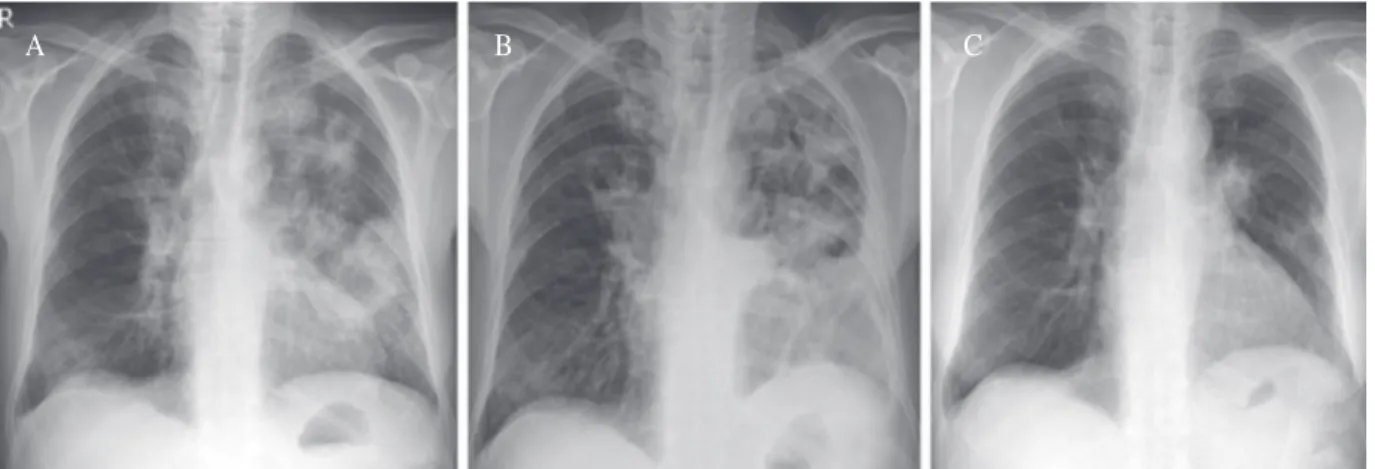
Fig. 1. (A) Initial chest radiography shows multifocal consolidation and cavities in both lungs. (B) Chest radiography shows a more aggravation of lung consolidation even after 4 weeks of amphotericin B treatment. (C) After 5 months of posaconazole treatment, it shows much improved multifocal consolidation and cavities.
A B C
Fig. 2. (A) High resolution computed tomography (CT) of chest shows non-enhanced, but high density multifocal cavities with reverse halo sign and consolidations in both lungs. (B) CT shows decreased size and thickness of multiple cavities with much improved consolidation after 5 months of posaconazole treatment.
A B
treated with a 4-week course of amphotericin B and liposomal amphotericin B. However, at the end of this course of treatment, his clinical status and chest radiograph had worsened (Fig. 1B).
Posaconazole (400 mg twice a day) was therefore chosen for the treatment of a refractory invasive fungal infection. After 2 weeks of posaconazole therapy, the patient’s respiratory condition and Fig. 4. (A) This slide reveals ulcerative lesion of bronchial mucosa that consisted with inflammatory cells and numerous fungal hyphae (H&E stain, × 200). (B) There are a lot of irregular fungal hyphae invading tissues with broad angular branching without septa (H&E stain, × 400).
A B
Fig. 3. (A) Fiberoptic bronchoscopy shows obstructive mucosal lesion with necrotic materials on medial segment of right middle lobe bronchus (black arrow). (B) The bronchoscopy after posaconazole treatment shows a complete improvement of a necrotic obstructive lesion on right middle lobe bronchus (white arrow).
A B
chest radiograph showed dramatic improvement.
The patient was discharged in stable condition on hospital day 43. Posaconazole therapy was continued for an additional 19 weeks on an outpatient basis. A follow-up chest radiograph (Fig. 1C) and CT (Fig. 2B) performed after 5 months revealed decreased size and thickness of multiple cavities and multifocal ill-defined consolidation in both lungs, and posaconazole therapy was discontinued. Since that time, the patient has reported intermittent blood-tinged sputum, but the chest radiographic findings have been stable, and bronchoscopy demonstrated complete improvement of the necrotic obstructive lesion on the right middle lobe bronchus (Fig. 3B) at the last evaluation 3 years after the completion of posaconazole treatment.
Discussion
Pulmonary mucormycosis is an uncommon but life-threatening infection caused by fungi belonging to the order Mucorales [1]. The infection tends to be rapidly progressive with prominent angioinvasion, resulting in tissue necrosis [6].
Pulmonary manifestations include cavitary lesions, pneumonia, solitary nodules, and disseminated lesions. The overall mortality rate of invasive pulmonary mucormycosis is greater than 70% [7].
Given the high morbidity and mortality of mucormycosis, establishing a timely diagnosis, followed by immediate treatment, is of major importance [8]. The most common finding on chest r a d i o g r a p h y a n d h i g h-r e s o l u t i o n C T i s consolidation, which is usually unilobar, although with disease progression, multilobar involvement may develop. Cavitation, especially producing an air crescent or halo (or reversed halo) sign, is highly suggestive of fungal infection [9], but this
does not distinguish it from aspergillosis. The diagnosis of mucormycosis is established by obtaining a biopsy specimen of the involved tissue, and a histologic assessment of the tissue sample with hematoxylin and eosin (H&E) or specialized fungal stains should be performed [6,8].
Based on clinical experience and small case studies, the mainstay of treatment requires a combination of prolonged effective antifungal therapy and extensive surgical debridement to remove the necrotic tissue, along with correction of the predisposing underlying condition [3]. The first-line antifungal agent is lipid amphotericin B [3,10]. Posaconazole, a second-generation triazole with broad-spectrum activity, may be a promising alternative [3,4]. In the case described here, posaconazole was used successfully for salvage therapy after the failure of amphotericin B as first- line treatment in an immunocompromised patient who was unable to tolerate surgical treatment.
Posaconazole is still used as second-line therapy and additionally has 3 main indications: salvage therapy, step-down to an oral agent after initial treatment with amphotericin B, and alternative antifungal agent in the case of side effects of amphotericin B [3]. However, clinical experience with posaconazole in pulmonary mucormycosis, although it has been documented as effective in in vivo studies against mucormycosis, including in rhino-orbital infections [4,11], has been limited.
Especially in Korea posaconazole is expensive and not covered by medical insurance, which results in increased medical costs and places limitations on the amount of experience and clinical data available regarding the medicine. There have been several cases of pulmonary mucormycosis that were treated with posaconazole in Korea. Lee et al. [12] reported on a total of 11 patients who received posaconazole over the last 5 years, and among t h e m w e r e t w o p a t i e n t s w i t h p u l m o n a r y
mucormycosis who received posaconazole due to intolerance of amphotericin B and lipophilic amphotericin B and due to maintenance of oral antifungal treatment. Surgical debridement was also performed in these two cases. Partial response was achieved in one case, and stable response was reported in the other. In addition, one case of i s o l a t e d p u l m o n a r y m u c o r m y c o s i s i n a n immunocompetent host was reported [13]. In that case, posaconazole oral suspension was used for home maintenance therapy after 1 week of amphotericin B treatment and surgical resection was not performed due to poor lung function. After 3 months of posaconazole treatment, the patient’s pulmonary mucormycosis was improved. In this case, the purpose for posaconazole use was not a first line therapy, which was amphotericin B, but just a step- down.
In our case discussed here, salvage therapy with posaconazole without surgical resection resulted in prompt clinical improvement in a patient with pulmonary mucormycosis. We suggest that posaconazole may be considered as an alternative treatment for invasive pulmonary mucormycosis in patients who are unable to tolerate surgical treatment.
References
1. Hamilos G, Samonis G, Kontoyiannis DP. Pulmonary mucormycosis. Semin Respir Crit Care Med 2011;32:693-702.
2. Kim MJ, Park PW, Ahn JY, Kim KH, Seo JY, Jeong JH, et al. Fatal pulmonary mucormycosis caused by Rhizopus microsporus in a patient with diabetes. Ann Lab Med 2014;34:76-9.
3. Spellberg B, Ibrahim AS. Recent advances in the treatment of mucormycosis. Curr Infect Dis Rep 2010;12:423-9.
4. Gelston CD, Durairaj VD, Simoes EA. Rhino-orbital mucormycosis causing cavernous sinus and internal carotid thrombosis treated with posaconazole. Arch Ophthalmol 2007;125:848-9.
5. Sedlacek M, Cotter JG, Suriawinata AA, Kaneko TM, Zuckerman RA, Parsonnet J, et al. Mucormycosis peritonitis: more than 2 years of disease-free follow-up after posaconazole salvage therapy after failure of liposomal amphotericin B. Am J Kidney Dis 2008;51:302-6.
6. Smith JA, Kauffman CA. Pulmonary fungal infections.
Respirology 2012;17:913-26.
7. Roden MM, Zaoutis TE, Buchanan WL, Knudsen TA, Sarkisova TA, Schaufele RL, et al. Epidemiology and outcome of zygomycosis: a review of 929 reported cases. Clin Infect Dis 2005;41:634-53.
8. Walsh TJ, Gamaletsou MN, McGinnis MR, Hayden RT, Kontoyiannis DP. Early clinical and laboratory diagnosis of invasive pulmonary, extrapulmonary, and disseminated mucormycosis (zygomycosis). Clin Infect Dis 2012;54 Suppl 1:S55-60.
9. Wahba H, Truong MT, Lei X, Kontoyiannis DP, Marom EM. Reversed halo sign in invasive pulmonary fungal infections. Clin Infect Dis 2008;46:1733-7.
10. Petrikkos GL. Lipid formulations of amphotericin B as first-line treatment of zygomycosis. Clin Microbiol Infect 2009;15 Suppl 5:87-92.
11. Vehreschild JJ, Birtel A, Vehreschild MJ, Liss B, Farowski F, Kochanek M, et al. Mucormycosis treated with posaconazole: review of 96 case reports. Crit Rev Microbiol 2013;39:310-24.
12. Lee HJ, Kwon JC, Kim SH, Choi SM, Lee DG, Park SH, et al. Posaconazole treatment in Korea: single- center experience over 5 years. Yonsei Med J 2013;54:1234-40.
13. Lee JS, Kim HC, Park SW, So HS, Woo CY, Choi JH, et al. A case of isolated pulmonary mucormycosis in an immunocompetent host. Tuberc Respir Dis (Seoul) 2013;74:269-73.