Review of the Current Status of Intra- Arterial Thrombolysis for Treating Acute Cerebral Infarction: a Retrospective
Analysis of the Data from Multiple Centers in Korea
Objective: The purpose of the study was to review the current status of intra- arterial (IA) thrombolysis in Korea by conducting a retrospective analysis of the data from multiple domestic centers.
Materials and Methods: The radiologists at each participating institution were asked to fill out case report forms on all patients who had undergone IA recanal- ization due to acute anterior circulation ischemia. These forms included clinical, imaging and procedure-related information. A central reader analyzed the CT/MR and angiographic results. The rates of successful recanalization, hemorrhagic transformation and functional outcome were obtained. The univariate analyses were performed together with the multivariate analysis.
Results: We analyzed the data from 163 patients, and they had been treated at seven institutes. The initial imaging modalities were CT for 46 patients (28%), MR for 63 (39%), and both for 54 (33%). Various mechanical treatment methods were applied together in 50% of the patients. Radiologically significant hemorrhage was noted in 20/155 patients (13%). We found various factors that influenced the recanalization rate and the occurrence of significant hemorrhagic transforma- tions. The favorable outcome rate, reported as modified Rankin Scale 2, was 40%, and the mortality rate was 11%. The factors that predicted a poor functional outcome were old age (p = 0.01), initially severe neurological symptoms (p <
0.0001), MR findings of a wide distribution of lesions (p = 0.001), involvement of the basal ganglia (p = 0.01), performance of procedures after working hours (p = 0.01), failure of recanalization (p = 0.003), contrast extravasation after the proce- dure (p = 0.007) and significant hemorrhagic transformation (p = 0.002). The sub- sequent multivariate analysis failed to show any statistically significant variable.
Conclusion: There was a trend toward increased dependency on MR imaging during the initial evaluation and increased usage of combined
pharmacologic/mechanical thrombolysis. The imaging and clinical outcome results of this study were comparable to those of the previous major thrombolytic trials.
ith the first successful use of local intra-arterial (IA) thrombolysis for vertebrobasilar disease (1), many studies have shown that IA application of thrombolytics can successfully treat hyperacute ischemic strokes (2 8). However, in Korea, this procedure has yielded variable results (9 11). A combina- tion of microcatheter techniques with their improved performance has increased the effectiveness and safety of IA thrombolysis. In addition to the local infusion of Deok Hee Lee, MD1
Dong Gyu Na, MD2 Yon Kwon Ihn, MD3 Dong Joon Kim, MD4 Eung Yeop Kim, MD5 Yong Sun Kim, MD6 Soo Mee Lim, MD7 Hong Gee Roh, MD5, 8 Chul-Ho Sohn, MD9 Stroke Study Group10
Index terms : Brain ischemia Brain infarction Thrombolytic therapy
Korean J Radiol 2007 ; 8 : 87-93 Received February 28, 2006; accepted after revision May 29, 2006.
Department of Radiology, 1University of Ulsan College of Medicine, Seoul 138-736, Korea; 2Seoul National University College of Medicine, Seoul 110-744, Korea; 3The Catholic University of Korea, Seoul 137-701, Korea; 4Yonsei University College of Medicine, Seoul 120-752, Korea;
5Sungkyunkwan University School of Medicine, Seoul 135-710, Korea; 6College of Medicine, Kyungpook National University, Daegu 702-701, Korea; 7College of Medicine, Ewha Womans University, Seoul 120-750, Korea; 8Konkuk University Hospital, Seoul 143-701, Korea; 9Keimyung University School of Medicine, Daugu 704- 701, Korea; 10Korean Society of
Neuroradiology and Head & Neck Radiology This study was supported by a grant from Guerbet Korea, Seoul, Korea.
Address reprint requests to:
Deok Hee Lee, MD, Department of Radiology and Research Institute of Radiology, Asan Medical Center, University of Ulsan College of Medicine, 388-1 Poongnap 2-dong, Songpa-gu, Seoul 138- 736, Korea.
Tel. (822) 3010-5944 Fax. (822) 476-4719 e-mail: [email protected]
W
thrombolytics, balloon angioplasty catheters, various metallic stents and other mechanical measures have also been used to increase the recanalization rate (12 14).
In the single successful randomized controlled trial of IA thrombolysis that has been conducted to date, the PROACT-II Trial (7), thrombolytic pro-urokinase was infused over two hours without manipulation of the clot.
Yet the techniques used for IA thrombolysis differ from one center to another, according to the endovascular technique, and the outcomes of this procedure, as measured by the recanalization rate and the functional outcome, have not been consistent (15). This lack of standardization, along with the inconsistent outcomes, has prompted us to investigate the current IA thrombolysis techniques and their radiological and clinical results in Korea. Therefore, in conjunction with the Stroke Study Group of the Korean Society of Neuroradiology and Head
& Neck Radiology (KSNHNR), we retrospectively analyzed the current status of IA thrombolytic therapy for the treatment of acute cerebral ischemic lesions. We determined what modalities are used for the radiological evaluation and the findings, the treatment patterns and the short-term clinical results, including the rate of
hemorrhagic transformation, the mortality rate and the clinical outcome, and we determined some of factors that are prognostic for a poor outcome.
MATERIALS AND METHODS
The study was designed as a multicenter, retrospective collection of cases with using a case reporting form. Only patients who underwent an IA recanalization procedure due to acute anterior circulation ischemia in the internal carotid artery (ICA) and the middle cerebral artery (MCA), and this was performed within the past five years, were included in this review.
The case reporting form was provided to each enrolled institute. This form included the basic clinical information, the initial imaging results, the angiography and procedure- related information, the follow-up imaging results and the clinical outcome of each patient together with the actual imaging data. We requested that the clinical and procedure-related parts of the form be filled out by the radiologist at each center.
The basic clinical information included age, gender, National Institute of Health Stroke Scale (NIHSS) score at the time of presentation and the presence of underlying diabetes, hypertension and/or heart disease. Selection of the initial imaging modality, the time from symptom onset to the time of imaging, the time from symptom onset to the initiation of angiography (the first angiographic run), and
the time of day of the procedure (i.e., during or after working hours) were analyzed.
The pre- and post-procedure CT/MR images and angiographic findings were evaluated by the board- certified radiologist who had more than five years of experience with IA thrombolysis. The CT results were analyzed for the presence of early CT signs, involvement of basal ganglia and the presence of hyperdense MCA signs. The MR findings were analyzed to determine the extent of the lesion on the diffusion-weighted images (DWI), involvement of the basal ganglia, the presence of perfusion/diffusion mismatch and presence of microbleeds.
The angiographic results were analyzed for the anatomic location of the culprit steno-occlusive lesions and the initial and post-procedure Thrombolysis in Myocardial Infarction (TIMI) grades. We regarded a post-procedure TIMI grade of II or III as indicative of successful recanalization.
The immediate post-procedure CT was reviewed for the presence of hemorrhages and parenchymal hyperdensity that was observed due to the contrast media used during the thrombolytic procedure. The follow-up CT or MR findings were analyzed for occurrence of hemorrhagic transformation, which was evaluated by the European Cooperative Acute Stroke Study (ECASS) classification method. We calculated the rates of occurrence of any hemorrhage and any radiologically significant hemorrhage, with the latter regarded as hemorrhage with a mass effect, such as parenchymal hematoma type 2 or some cases of hemorrhagic infarction type 2. These rates were surrogate indicators of symptomatic hemorrhages (16, 17).
The clinical outcome analysis was performed using the modified Rankin Scale (mRS), which was recorded at various time points after the procedure (at 1 12 months, except for the patients who died in the interim). If the clinical outcome data were not available, the case was dropped from the clinical outcome analysis. We calculated
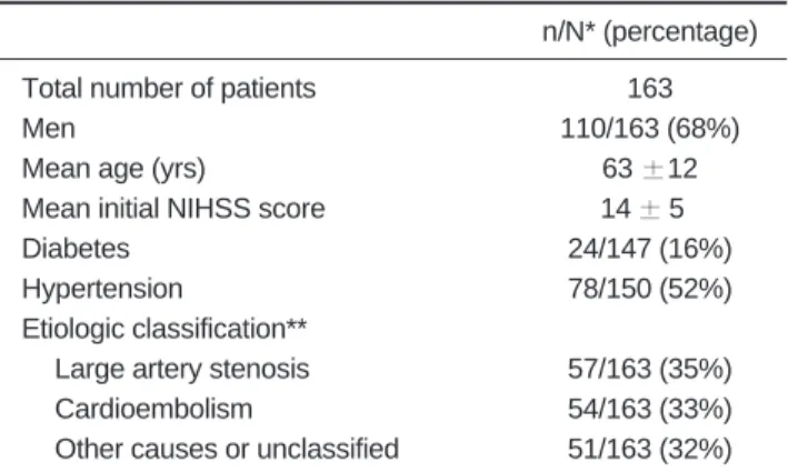
Table 1. Demographic Characteristics at the Baseline
n/N* (percentage)
Total number of patients 163
Men 110/163 (68%)
Mean age (yrs) 63 12
Mean initial NIHSS score 14 50
Diabetes 24/147 (16%)
Hypertension 78/150 (52%)
Etiologic classification**
Large artery stenosis 57/163 (35%)
Cardioembolism 54/163 (33%)
Other causes or unclassified 51/163 (32%) Note. n = number, * N = the number of patients with available data.
**According to the Trial of Org 10172 in Acute Stroke Treatment (TOAST).
the percentage of patients with each mRS score, as well as the mortality rate, which was defined as the rate of procedure-related deaths within one month.
Univariate logistic regression methods were used to analyze the factors that had an influence on successful recanalization, significant hemorrhagic transformation and poor functional outcome results. The proportion of patients with a mRS of more than 3 was used for the latter.
Variables with p values less than 0.05 on the univariate analyses were chosen as the variables for the multivariate logistic regression analysis. In both analyses, p values less than 0.05 were considered statistically significant.
RESULTS
Patient Demographics
The basic demographic characteristic of the 163 patients from seven domestic institutes are summarized in Table 1.
The mean time interval from the symptom onset to the initial CT scanning was 139 145 minutes and the mean time interval from the symptom onset to the first angiogra- phy was 280 178 minutes (Table 2). Out of the 149 patients for whom we were able to obtain the time of day of their angiography, 92 (62%) had the procedures
performed during normal working hours (09:00 18:00).
Initial Imaging Results
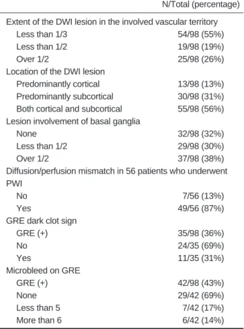
The initial imaging modalities were CT in 46 patients (28%), MR in 63 (39%), and both CT and MR in 54 (33%). We were able to review the CT images of 69 of the 100 (69%) patients who initially underwent CT. The basic initial CT and MR findings are summarized in Tables 3 and 4, respectively.
Angiography and Procedure
The site of arterial stenosis (TIMI grade 1, n = 18) or occlusion (TIMI grade 0, n = 145) was the ICA, including the carotid ‘T’ occlusion, in 62 patients (38%), the MCA, including M2 occlusion, in 99 (61%) and the anterior cerebral artery in two patients (1.2%) (Table 5). Before the initiation of IA thrombolysis, 73 patients (45%) were administered intravenous tissue plasminogen activator (t- PA, 0.6 or 0.9 mg/kg). Urokinase at a median dosage of 400,000 IU (range 40,000 1,000,000 IU) was the IA fibrinolytic agent in 139 patients (85%), t-PA at a median dosage of 10 mg (range 5 20 mg) was used in 11 patients
Table 2. Time from Symptom Onset to the Initial Imaging and Treatment (the First Angiography)
Time to Imaging Minutes
From symptom onset to CT (n = 71) 139 145 From symptom onset to MR (n = 61) 206 207
From CT to MR (n = 47) 60 52
Time to Treatment
From symptom onset (n = 144) 280 178
From CT (n = 71) 128 680
From MR (n = 61) 107 530
Table 3. Initial CT Findings in 69 Patients
N (percentage) Early CT sign
None 31 (45%)
Less than 1/3 of the MCA territory 31 (45%) Over 1/3 of the MCA territory 7 (10%) Low density involving the basal ganglia
No 52 (75%)
Less than 1/2 8 (12%)
Over 1/2 9 (13%)
Positive hyperdense MCA sign
No 49 (71%)
Yes 20 (29%)
Note. N = number, MCA = middle cerebral artery.
Table 4. Initial MR Findings in 98 Patients
N/Total (percentage) Extent of the DWI lesion in the involved vascular territory
Less than 1/3 54/98 (55%)
Less than 1/2 19/98 (19%)
Over 1/2 25/98 (26%)
Location of the DWI lesion
Predominantly cortical 13/98 (13%) Predominantly subcortical 30/98 (31%) Both cortical and subcortical 55/98 (56%) Lesion involvement of basal ganglia
None 32/98 (32%)
Less than 1/2 29/98 (30%)
Over 1/2 37/98 (38%)
Diffusion/perfusion mismatch in 56 patients who underwent PWI
No 07/56 (13%)
Yes 49/56 (87%)
GRE dark clot sign
GRE (+) 35/98 (36%)
No 24/35 (69%)
Yes 11/35 (31%)
Microbleed on GRE
GRE (+) 42/98 (43%)
None 29/42 (69%)
Less than 5 07/42 (17%)
More than 6 06/42 (14%)
Note. N = number, DWI = diffusion-weighted image, PWI = perfusion- weighted image, GRE = gradient echo image.
(7%), and the procedure was performed without IA fibrinolytics in 13 patients (8%). In the latter case, mechan- ical methods were primarily applied, except for one patient, who received an IA infusion of abciximab. Various mechanical methods were applied, along with local infusion of fibrinolytics, in 82 patients (50%), including those patients who were treated with primary mechanical thrombolysis. The methods employed included mechanical disruption of the clot using a micro-guidewire (n = 49, 60%), stenting of the steno-occlusive lesion using various kinds of metallic stents (n = 17, 21%), balloon catheter angioplasty or clot disruption (n = 12, 14%) and aspiration of the clot using a large-bore guiding catheter (n = 4, 5%).
A combination of these methods was used in patients for whom the primary mechanical method was ineffective in recanalization.
Postprocedure angiography was available for 152 patients. The final TIMI grades were zero in 21 patients (14%), one in 23 (15%), two in 31 (20%) and three in 77 (51%). The successful recanalization (TIMI grade 2 or 3) rate was 71%.
Postprocedure Imaging Results
Immediate postprocedure CT was available for 117 patients. Various patterns of parenchymal high densities were observed in 54 patients (47%), and 24 (21%) of them showed obvious hematoma formation and/or high density with a mass effect.
Hemorrhagic transformation of the lesion was evaluated in 155 patients whose follow-up CT and/or MR images were available for the review, and 55 (35%) of these patients showed variable amounts of hemorrhage. The hemorrhage pattern was hemorrhagic infarction (HI) type
1 in 15 patients (27%), HI type 2 in six (11%), parenchy- mal hematoma (PH) type 1 in 17 (31%) and PH type 2 in 17 (31%). Radiologically significant hemorrhage was noted in 20 of these 155 patients (13%).
Clinical Outcome
The clinical follow-up data were available for 158 patients. The follow-up period varied from one to 12 months, except for those patients who died in the interim.
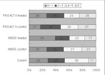
We noted mRS scores of zero in 17 patients (11%), one in 27 (17%), two in 19 (12%), three in 20 (13%), four in 33 (21%) and five in 24 (15%); these findings were compara- ble with those of other major thrombolysis trials (Fig. 1).
Out of the 158 patients, 18 died, yielding a mortality rate of 11%.
Results of the Univariate and Multivariate Analyses Among the various factors considered, an initial NIHSS score of less than 15 was the only factor that positively influenced successful recanalization (p = 0.001). Table 6 shows the univariate analysis of the factors that influenced significant hemorrhage after the procedure, and Table 7 shows the factors that influenced the poor functional outcome. Subsequent multivariate analyses failed to show any statistically significant variables both for significant hemorrhage and for a poor functional outcome.
DISCUSSION
The results of our analysis provide an overview on the current practice status of IA thrombolysis in Korea.
Although more than 30 centers in Korea actively perform neurointerventional procedures (see the 2005 member list of the Korean Society of Interventional Neuroradiology),
Table 5. Anatomic Sites of Stenosis or Occlusion on the Initial Angiography
Anatomic Site N (percentage)
ICA 62 (38%)
Proximal ICA 13 (8%)
Proximal ICA and tandem
MCA occlusion 7 (4%)
Distal ICA 6 (4%)
Carotid T lesion 36 (22%)
MCA 99 (61%)
Proximal MCA trunk 35 (22%)
Mid MCA trunk 16 (10%)
Distal MCA trunk 35 (22%)
M2 13 (8%)
ACA 2 (1%)
Note. N = number, ICA = internal carotid artery, MCA = middle cerebral artery, ACA = anterior cerebral artery.
Fig. 1. Functional outcome represented as modified Rankin Scale and comparison of the current results with those of the control and treated groups of the major thrombolysis trials.
only seven of these centers participated in this study. It is likely that many of the other centers have been reluctant to perform IA thrombolysis, primarily due to a shortage of trained personnel. In Western countries also, IA thrombol- ysis is performed less frequently than the IV method due to a shortage of interventional specialists (15).
We found that from the symptom onset, the mean time before the imaging was 2.3 hours, and the mean time before the treatment was 4.7 hours; these results are comparable with those reported in the PROACT II Trial (7). The mean delay between the CT and MR imaging was about 60 min, which was one of the in-hospital delay factors for IA thrombolysis (18).
About 40% of these procedures were performed before or after normal working hours, and the functional outcome results of these patients were generally poorer than those of the patients who were treated during normal working hours. One of the primary reasons for the poorer outcome in the former group of patients may be the in-hospital
delay. A well-organized stroke team and an on-call system may improve the results for these patients.
One notable finding of our survey was the preference for MR as the initial imaging modality. MR was the sole initial imaging modality in 39% of patients, and it was combined with the CT imaging in 33%. Although MR has the potential disadvantage of time delay, it has shown an advantage for the initial evaluation of acute ischemic stroke (19 22). However, a delay in the initiation of the optimal treatment due to logistic limitations of MR imaging cannot be justified. Many centers use multi-detector CT imaging to overcome the inevitable logistic limitation of MR imaging (21, 23).
It was notable that about 10% of the cases in the study were in violation of the ‘one-third rule’, which is one of the most important exclusion criteria of the European
thrombolysis trial (24) and the PROACT II trial (7). Out of all the patients evaluated by the MR imaging, 26% showed
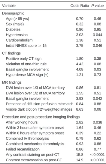
Table 6. Univariate Relationships of the Significant Hemorrhage after Thrombolysis
Variable Odds Ratio P value
Demographic
Age (> 65 yrs) 0.70 0.46
Sex (male) 0.32 0.08
Diabetes 0.96 0.95
Hypertension 3.03 0.044
Cardioembolism 1.38 0.51
Initial NIHSS score 15 3.75 0.046
CT findings
Positive early CT sign 1.80 0.38
Violation of one-third rule 4.42 0.08
Basal ganglia involvement 4.08 0.03
Hyperdense MCA sign (+) 1.21 0.73
MR findings
DWI lesion over 1/3 of MCA territory 0.86 0.81 DWI lesion over 1/2 of MCA territory 1.55 0.51 Basal ganglia involvement 3.93 0.042 Presence of diffusion-perfusion mismatch 0.84 0.88 Visible dark clot on T2*-weighted images 8.63 0.08 Procedure and post-procedure imaging findings
After working hours 2.82 0.038
Within 3 hours after symptom onset 1.64 0.46 Within 6 hours after symptom onset 0.39 0.22
Combined IV thrombolysis 0.79 0.62
Combined mechanical thrombolysis 0.93 0.88
Failed recanalization 0.86 0.77
Any contrast staining on post-CT 15.8 < 0.0001 Contrast extravasation on post-CT 14.9 < 0.0001 Note. DWI = diffusion-weighted image, MCA = middle cerebral artery.
Table 7. Univariate Relationships with a Poor Functional Outcome (mRS > 3)
Variable Odds Ratio P value
Demographic
Age (> 65 yrs) 02.27 0.010
Sex (male) 00.87 0.680
Diabetes 01.52 0.350
Hypertension 01.12 0.730
Cardioembolism 00.88 0.700
Initial NIHSS score 15 04.88 < 0.0001 CT findings
Positive early CT sign 01.57 0.370
Violation of the one-third rule 05.80 0.120 Basal ganglia involvement 01.26 0.680
Hyperdense MCA sign (+) 01.21 0.730
MR findings
DWI lesion over 1/3 of MCA territory 02.35 0.040 DWI lesion over 1/2 of MCA territory 06.14 0.001 Basal ganglia involvement 03.22 0.010 Presence of diffusion-perfusion mismatch 0.003 Visible dark clot on T2*-weighted images 02.10 0.330 Procedure and post-procedure imaging findings
After working hours 02.41 0.010
Within 3 hours after symptom onset 00.98 0.970 Within 6 hours after symptom onset 00.93 0.870 Combined IV thrombolysis 01.08 0.800 Combined mechanical thrombolysis 00.96 0.890
Failed recanalization 00.31 0.003
Any contrast staining on post-CT 02.33 0.030 Contrast extravasation on post-CT 04.00 0.007
Significant hemorrhage 26.70 0.002
Note. DWI = diffusion-weighted image, MCA = middle cerebral artery.
DWI lesions covering more than half of the involved vascular territory. This was probably due to a lack of certain inclusion or exclusion criteria for MR imaging in respect to the patient selection for IA thrombolysis.
Although we limited our study to patients with anterior circulation infarction and so increasing the homogeneity of the study group, we found a wide variation in the
occlusion site, from the proximal ICA to the proximal portion of the MCA.
Our observed recanalization rate, 71%, was slightly higher than that reported in the PROACT II trial (7), which may have been due, at least in part, to the liberal use of mechanical methods (14). We were disappointed to find that only one factor, the lower initial NIHSS score, had a significant effect on the successful recanalization rate. In contrast to reports on the combined or sequential
thrombolytic treatments (25 27), we found that the use of IV thrombolytics prior to the IA procedure did not affect the recanalization rate. However, in many of these patients, however, the initial IV administration of thrombolytics was ineffective. About half of the patients received a combination of IA and mechanical thromboly- sis, although the use of mechanical methods failed to improve the recanalization rate (p = 0.083).
One of the major concerns after the IA thrombolytic treatment is hemorrhagic transformation of the affected lesion (28, 29). We found that the rate of any amount hemorrhagic transformation was 35%, and the rate of the significant hemorrhagic transformation was 13%. The univariate analysis showed that several factors that had influence on significant hemorrhagic transformation, including the presence of hypertension, severe initial neurological symptoms, involvement of the basal ganglia on CT/MR and the performance of procedures after normal working hours. In addition, the risk of significant hemorrhage was increased by the presence of any contrast density on the CT obtained immediately after the
thrombolysis procedure. Violation of the one-third rule on the initial CT tended to increase the occurrence of signifi- cant hemorrhage (p = 0.08), whereas extensive involve- ment of the affected vascular territory on the DWI (p = 0.51) and the combined use of IV thrombolytics and mechanical methods had no effect.
Although direct comparison is not possible because of the differences in the patient groups, the functional outcome in our series of patients was similar to that of other major thrombolysis trials (7, 30). Among the factors that were found to influence the poor functional outcome were the old age, severe initial neurological symptoms, more extensive initial DWI lesions and involvement of basal ganglia on MR, failed recanalization, any contrast
staining on CT immediately after the procedure and the presence of significant hemorrhagic transformation. Other studies have found that these same factors were predictive of poor prognosis (8, 31, 32).
The primary limitations of this analysis were its
retrospective design and the collection of data from several centers, all of which had different clinical and imaging indications for the procedure and different clinical settings.
In addition, the data we collected were not representative of practice throughout Korea because only a few active centers contributed to the study. Due to our inability to collect some data from the participating centers, we were unable to perform appropriate multivariate analyses. This could be one of the possible reasons for our failure to determine any significant variables that affected the results of significant hemorrhagic transformation and poor functional outcomes. Furthermore, our analysis of the imaging data was limited due to the between-center differ- ences in the imaging protocols, including the use of contrast enhancement, MR pulse sequences and the angiographic filming techniques. Nevertheless, the results presented here may be representative of the nation-wide data on current IA thrombolytic treatment, and they could serve as a basis for clinical practice guidelines, as well as for designing a prospective study on the basis of this protocol.
In conclusion, our multicenter, retrospective analysis of IA thrombolysis showed an increased dependency on MR imaging during the initial patient evaluation and increased application of combined pharmacologic/mechanical thrombolysis in Korea. Both our imaging and clinical outcome results were comparable with those of the other major thrombolytic trials. Our results could be used as the basis for clinical practice and for future prospective studies.
References
1. Zeumer H, Hacke W, Ringelstein EB. Local intraarterial thrombolysis in vertebrobasilar thromboembolic disease. AJNR Am J Neuroradiol 1983;4:401-404
2. Mori E, Tabuchi M, Yoshida T, Yamadori A. Intracarotid urokinase with thromboembolic occlusion of the middle cerebral artery. Stroke 1988;19:802-812
3. Theron J, Courtheoux P, Casasco A, Alachkar F, Notari F, Ganem F, et al. Local intraarterial fibrinolysis in the carotid territory. AJNR Am J Neuroradiol 1989;10:753-765
4. Barr JD, Mathis JM, Wildenhain SL, Wechsler L, Jungreis CA, Horton JA. Acute stroke intervention with intraarterial urokinase infusion. J Vasc Interv Radiol 1994;5:705-713 5. Freitag HJ, Becker VU, Thie A, Tilsner V, Philapitsch A,
Schwarz HP, et al. Lys-plasminogen as an adjunct to local intra- arterial fibrinolysis for carotid territory stroke: laboratory and clinical findings. Neuroradiology 1996;38:181-185
6. del Zoppo GJ, Higashida RT, Furlan AJ, Pessin MS, Rowley HA, Gent M. PROACT: a phase II randomized trial of recombi-
nant pro-urokinase by direct arterial delivery in acute middle cerebral artery stroke. PROACT Investigators. Prolyse in Acute Cerebral Thromboembolism. Stroke 1998;29:4-11
7. Furlan A, Higashida R, Wechsler L, Gent M, Rowley H, Kase C, et al. Intra-arterial prourokinase for acute ischemic stroke. The PROACT II study: a randomized controlled trial. Prolyse in Acute Cerebral Thromboembolism. JAMA 1999;282:2003-2011 8. Arnold M, Schroth G, Nedeltchev K, Loher T, Remonda L,
Stepper F, et al. Intra-arterial thrombolysis in 100 patients with acute stroke due to middle cerebral artery occlusion. Stroke 2002;33:1828-1833
9. Lee JH, Kim JS, Lee MC, Suh DC, Lee MS. A comparison study on therapeutic efficacy of urokinase vs. Heparin in acute ischemic stroke. J Korean Neurol Soc 1994;12:225-236 10. Kim SY, Suh JH. Local intracranial intraarterial thrombolytic
therapy in acute cerebral infarction. J Korean Radiol Soc 1996;34:703-710 (Korean)
11. Shi HB, Suh DC, Lim SM, Lee JH, Kim JK, Jeong AK, et al.
Outcome evaluation of intra-arterial infusion of urokinase for acute ischemic stroke. J Korean Radiol Soc 2000;42:897-904 12. Nesbit GM, Clark WM, O’Neill OR, Barnwell SL. Intracranial
intraarterial thrombolysis facilitated by microcatheter naviga- tion through an occluded cervical internal carotid artery. J Neurosurg 1996;84:387-392
13. Nakano S, Iseda T, Yoneyama T, Kawano H, Wakisaka S.
Direct percutaneous transluminal angioplasty for acute middle cerebral artery trunk occlusion: an alternative option to intra- arterial thrombolysis. Stroke 2002;33:2872-2876
14. Nesbit GM, Luh G, Tien R, Barnwell SL. New and future endovascular treatment strategies for acute ischemic stroke. J Vasc Interv Radiol 2004;15:S103-110
15. Saver JL. Intra-arterial thrombolysis. Neurology 2001;57:S58- 60
16. Fiorelli M, Bastianello S, von Kummer R, del Zoppo GJ, Larrue V, Lesaffre E, et al. Hemorrhagic transformation within 36 hours of a cerebral infarct: relationships with early clinical deterioration and 3-month outcome in the European Cooperative Acute Stroke Study I (ECASS I) cohort. Stroke 1999;30:2280-2284
17. Berger C, Fiorelli M, Steiner T, Schabitz WR, Bozzao L, Bluhmki E, et al. Hemorrhagic transformation of ischemic brain tissue: asymptomatic or symptomatic? Stroke 2001;32:1330- 1335
18. Nedeltchev K, Arnold M, Brekenfeld C, Isenegger J, Remonda L, Schroth G, et al. Pre- and in-hospital delays from stroke onset to intra-arterial thrombolysis. Stroke 2003;34:1230-1234 19. Schellinger PD, Jansen O, Fiebach JB, Pohlers O, Ryssel H,
Heiland S, et al. Feasibility and practicality of MR imaging of stroke in the management of hyperacute cerebral ischemia.
AJNR Am J Neuroradiol 2000;21:1184-1189
20. Lee DH, Lee JH. MR imaging of hyperacute ischemic stroke J Korean Radiol Soc 2004;50:1-17 (Korean)
21. Hjort N, Butcher K, Davis SM, Kidwell CS, Koroshetz WJ, Rother J, et al. Magnetic resonance imaging criteria for thrombolysis in acute cerebral infarct. Stroke 2005;36:388-397 22. Lee DH, Kang DW, Ahn JS, Choi CG, Kim SJ, Suh DC.
Imaging of the ischemic penumbra in acute stroke. Korean J Radiol 2005;6:64-74
23. Yi CA, Na DG, Ryoo JW, Moon CH, Byun HS, Roh HG, et al.
Multiphasic perfusion CT in acute middle cerebral artery ischemic stroke: prediction of final infarct volume and correla- tion with clinical outcome. Korean J Radiol 2002;3:163-170 24. Hacke W, Kaste M, Fieschi C, von Kummer R, Davalos A,
Meier D, et al. Randomised double-blind placebo-controlled trial of thrombolytic therapy with intravenous alteplase in acute ischaemic stroke (ECASS II). Second European-Australasian Acute Stroke Study Investigators. Lancet 1998;352:1245-1251 25. Lewandowski CA, Frankel M, Tomsick TA, Broderick J, Frey J,
Clark W, et al. Combined intravenous and intra-arterial r-TPA versus intra-arterial therapy of acute ischemic stroke:
Emergency Management of Stroke (EMS) Bridging Trial. Stroke 1999;30:2598-2605
26. IMS Study Investigators. Combined intravenous and intra- arterial recanalization for acute ischemic stroke: the Interventional Management of Stroke Study. Stroke 2004;35:904-911
27. Lee KY, Kim DI, Kim SH, Lee SI, Chung HW, Shim YW, et al.
Sequential combination of intravenous recombinant tissue plasminogen activator and intra-arterial urokinase in acute ischemic stroke. AJNR Am J Neuroradiol 2004;25:1470-1475 28. Suarez JI, Sunshine JL, Tarr R, Zaidat O, Selman WR, Kernich
C, et al. Predictors of clinical improvement, angiographic recanalization, and intracranial hemorrhage after intra-arterial thrombolysis for acute ischemic stroke. Stroke 1999;30:2094- 2100
29. Chalela JA, Katzan I, Liebeskind DS, Rasmussen P, Zaidat O, Suarez JI, et al. Safety of intra-arterial thrombolysis in the postoperative period. Stroke 2001;32:1365-1369
30. Tissue plasminogen activator for acute ischemic stroke. The National Institute of Neurological Disorders and Stroke rt-PA Stroke Study Group. N Engl J Med 1995;333:1581-1587 31. Wechsler LR, Roberts R, Furlan AJ, Higashida RT, Dillon W,
Roberts H, et al. Factors influencing outcome and treatment effect in PROACT II. Stroke 2003;34:1224-1229
32. Lisboa RC, Jovanovic BD, Alberts MJ. Analysis of the safety and efficacy of intra-arterial thrombolytic therapy in ischemic stroke. Stroke 2002;33:2866-2871