pISSN 2466-1384 eISSN 2466-1392 Korean J Vet Res (2019) 59(2):105~108 https://doi.org/10.14405/kjvr.2019.59.2.105
105 CASE REPORT
Remnant parietal serosa detection in a cat with true diaphragmatic hernia using computed tomography
Sang-Kwon Lee, Wooram Jeong, Jihye Choi*
College of Veterinary Medicine and BK21 Plus project team, Chonnam National University, Gwangju 61186, Korea
Abstract: A 4-year-old cat was referred for a suspected pulmonary mass. True diaphragmatic hernia presence was diagnosed via computed tomography (CT). There was a thin membrane covering the diaphragmatic defect. The membrane was thinner than the diaphragm. After contrast injection, the membrane was less enhanced than that of the normal diaphragm. The membrane was identified as a remnant of the parietal pleura. In addition, contrast-enhanced CT images provided clarity in viewing the herniated liver and falciform fat. A thinner membrane, covering the diaphragmatic defect, and attached to the thicker normal diaphragm, is considered a unique CT feature of true diaphragmatic hernia.
Keywords: cat, computed tomography, intact membrane, parietal serosa, true diaphragmatic hernia
Introduction
True diaphragmatic hernia, a rare congenital diaphragmatic defect, is an incom- plete tear of the diaphragm in which communication between peritoneal and pleural cavities is prevented by the remnant intact serosa on the thoracic surface.
Thus, true diaphragmatic hernia is characterized as a hernia sac surrounded by serosa, and these unique features are different from those of other types of dia- phragmatic hernia. Most previous cases with true diaphragmatic hernia were con- fused with pulmonary mass or peritoneopericardial diaphragmatic hernia on radiography and confirmed as true diaphragmatic hernia through surgery or nec- ropsy [1-5]. In four cases, positive contrast peritoneography diagnosed true diaphragmatic hernia by revealing incomplete diaphragmatic defects between peritoneal and pleural cavities [6-8]. Recently, a single case report described com- puted tomography (CT) features of true diaphragmatic hernia in a cat [8]. How- ever, there are no previous reports focusing on the imaging characteristics of the remnant intact serosa. This case report describes a thin membrane connected with the diaphragm and creating a hernia sac on CT images in a cat with true diaphragmatic hernia. This membrane was indicated as a remnant serosa, and this CT features may be a specific sign of true diaphragmatic hernia.
A 4-year-old male neutered Persian cat was referred to Chonnam National University Veterinary Teaching Hospital. The cat was referred for evaluation of a suspected pulmonary mass, identified on thoracic radiography during a health inspection. On presentation, the cat was asymptomatic, and its com- plete blood count and serum biochemistry results were unremarkable.
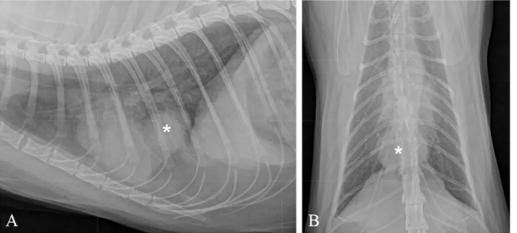
On thoracic radiographs, a round, soft tissue mass was observed between the caudal border of the heart and the diaphragm (Fig. 1). In contrast to the dis- tinct cranial margin, the mass had an indistinct caudal margin, and a silhou- ette sign was present with the diaphragm. Differential diagnoses for the lesion included a mass originating from the accessory lung lobe, diaphragm, or cau- dal mediastinum; diaphragmatic rupture; and true diaphragmatic hernia.
The intrathoracic mass was identified on ultrasonography. A hyperechoic diaphragm was not observed between the mass and the liver, and the mass appeared to be connected to the liver. The mass was more hypoechoic than the liver, and vascular structures were not clearly observed. Based on the ultraso- nographic observations, diaphragmatic hernia of the liver was suspected.
*Corresponding author Jihye Choi
College of Veterinary Medicine, Chonnam National University, Yongbong-ro, Buk-gu, Gwangju 61186, Korea
Tel: +82-62-530-2821 Fax: +82-62-530-2809 E-mail: [email protected] ORCID:
Sang-Kwon Lee
https://orcid.org/0000-0002-3097-0345 Wooram Jeong
https://orcid.org/0000-0002-7711-524X Jihye Choi
https://orcid.org/0000-0002-1258-7158 Conflict of Interest
The authors declare no conflicts of interest.
Received: May 5, 2019 Revised : May 13, 2019 Accepted: May 23, 2019
106 Sang-Kwon Lee, Wooram Jeong, Jihye Choi
The CT was performed by using a 16-row multi-detector CT scanner (Siemens Emotion 16, Siemens, Germany). Gen- eral anesthesia was induced by ketamine hydrochloride (Ket- amine, Yuhan, Seoul, Korea; 5 mg/kg, IM), and maintained with isoflurane (Terrell, Piramal Critical Care, USA; 1-2%) and oxygen (1 L/min). The cat was positioned in sternal recumbency and pre- and post-contrast CT images of the tho- rax and abdomen were acquired at the following settings:
slice thickness, 1 mm; pitch, 0.7; rotation duration, 500 msec;
tube voltage, 120 kV, and tube current; 120 mA. To reduce motion artifact, the cat was hyperventilated before CT scan- ning. Post-contrast CT study was performed after a bolus injection of 600 mg I/kg iohexol (Omnipaque 300, GE Healthcare, Oslo, Norway). Soft and sharp algorithms were used to generate images, and the transverse, dorsal, and sag- ittal planes were reconstructed. On CT images, the diaphragm had a defect at the center (Fig. 2). The thoracic mass pro-
truded through the defect in the diaphragm, from the abdo- men caudal into the thorax. The mass consisted of liver and falciform fat, and was confined within a thin membrane that created a pouch-like hernia sac. The membrane (less than 1 mm) was thinner than the adjacent diaphragm (4.1 mm);
however, it was connected to the diaphragm and prevented communication between the hernia sac and the pleural cav- ity. Mediastinal fat, adjacent to the hernia sac, enabled visu- alization of the pleural side of the membrane. After contrast injection, the hepatic vein and hepatic portal veins were observed in the herniated liver. Vascular obstruction or thrombi were not observed. Based on the distribution of the intrahe- patic vessels, the herniated liver portion was determined to be the left medial or quadrate liver lobe. Contrast enhance- ment of the herniated liver lobe (177 Hounsfield units [HU]) was higher than that of the abdominal liver lobes (149 HU).
A small volume of fluid was observed within the hernia sac, Fig. 1. Thoracic radiographs of a cat; right lateral (A) and ventrodorsal (B) views. A round soft tissue mass (asterisk) is located between heart and diaphragm. The caudal margin of the mass effaced the cranial border of the diaphragm.
Fig. 2. Transverse (A) and dorsally reformatted (B) soft tissue window images before contrast injection. The diaphragmatic defect is observed between the intrathoracic mass (asterisk) and the peritoneal cavity (B). The herniated mass is composed of soft tissue struc- tures and falciform fat, and a thin membrane (arrows) surrounds the herniated mass. This membrane is connected with the outer layer of the normal diaphragm (dashed arrows), and is thinner than the diaphragm (B).
CT features of true diaphragmatic hernia 107
and mild congestion of the herniated liver was suspected.
The thin membrane enhanced to a lesser degree than that of the normal diaphragm, although the membrane was too thin to be numerically measured via CT (Fig. 3). There were no other abnormal findings in the thorax and abdomen. The cat was diagnosed with true diaphragmatic hernia, along with a mildly congested liver and falciform fat. The cat was asymp- tomatic, and there was no pulmonary collapse identified on CT images; therefore, monitoring of the herniated liver lobes was recommended, without additional treatment.
Because true diaphragmatic hernia is a rare diaphragmatic hernia and most of them may be mistaken for a mass origi- nating from the lung, diaphragm, or caudal mediastinum; dia- phragmatic rupture and peritoneopericardial diaphragmatic hernia on radiography and ultrasonography, reports on the diagnostic features of true diaphragmatic hernia are limited.
Positive contrast peritoneography in four cases confirmed true diaphragmatic hernia diagnoses by revealing round pouch-like structures protruding into the thoracic cavity and no commu- nication between the structures and the pleural cavities [3,6,7].
However, this contrast technique is limited in its ability to characterize the herniated organ and the diaphragmatic layer.
In humans, CT is preferred for evaluation of diaphragmatic hernia because the technique can confirm the presence of a hernia and assess the herniated organs and the involved dia- phragm [9,10]. A diaphragmatic eventration, one of the dia- phragmatic diseases, is commonly reported in humans [2]. A congenital eventration is characterized by muscular aplasia of the diaphragm. In acquired eventration, diaphragmatic paral- ysis and cranial displacement of the entire diaphragm due to trauma or surgical injury are observed. A diaphragmatic even- tration is referred to as true diaphragmatic hernia in veteri- nary medicine and is defined as a subtotal diaphragmatic tear in which the serosa on the thoracic surface of the diaphragm remains intact [4,6]. On CT images of diaphragmatic even- tration, focal bulging of the affected diaphragm forms a her- nia sac containing herniated abdominal structures [10]. In
particular, a thin layer of the diaphragm can be identified between the hernia and the pleural cavity, and the layer forms a sharp transitional edge between the normal diaphragm and itself. This characteristic CT finding is dissimilar from those for congenital diaphragmatic hernia or diaphragmatic rupture.
On CT images of the present case, a hernia sac, consisting of a thin membrane, was observed instead of a complete dia- phragmatic defect, and the membrane enclosed the herniated liver lobe and falciform fat. The thin membrane prevented communication between the peritoneal and pleural cavities and was connected to the normal diaphragm. These CT observa- tions correspond with previous surgical examinations reveal- ing that the encapsulating layer surrounding the herniated organ was confluent with the diaphragm, and that the mem- brane was considered as a remnant of the parietal serosa of the diaphragm [1,4,5].
The remaining diaphragmatic membrane is not considered pathognomonic of true diaphragmatic hernia, since it may be only identified when it borders fat. In this case, the pleural side of the membrane adjoined the mediastinal fat, and the perito- neal side adjoined the falciform fat. In a previous case diag- nosed via CT [8], the remaining diaphragmatic layer was silhouetted against the herniated liver and not identified sep- arately from it.
Most cases of true diaphragmatic hernia were associated with a lack of clinical signs except for two neonatal cats with severe dyspnea due to compression of the lung lobes by cranial displacement of abdominal organs [1-8]. Exploratory laparot- omy was performed in most previous cases in order to diag- nose true diaphragmatic hernia and correct it. The present cat had CT-confirmed true diaphragmatic hernia but was asymp- tomatic; thus, clinical monitoring was recommended without surgical correction.
In conclusion, CT evaluation was useful for determination of the type of diaphragmatic hernia and for assessment of the herniated abdominal structures. A thin membrane, covering the diaphragmatic defect and connecting with the diaphragm Fig. 3. Transverse (A) and sagittal (B) images after contrast injection. Hepatic vasculature is evident in the herniated soft tissue struc- ture. Observation of hepatic vessels confirms the liver herniation; in this case, the left medial or quadrate lobe. Attenuation of the her- niated liver lobe (asterisk) is higher than that of the remaining lobes (l), and a small volume of fluid (arrow) is observed adjacent to the herniated liver.
108 Sang-Kwon Lee, Wooram Jeong, Jihye Choi
indicated a remnant of the parietal serosa; such a membrane can be used as a specific CT feature of true diaphragmatic hernia.
Acknowledgment
This research was supported by Basic Science Research Program through the National Research Foundation of Korea (NRF) funded by the Ministry of Science, ICT and Future Planning (NRF-2018R1A2B6006775).
References
1. Cariou MP, Shihab N, Kenny P, Baines SJ. Surgical management of an incidentally diagnosed true pleuroperitoneal hernia in a cat. J Feline Med Surg 2009;11:873-877.
2. Gombač M, Vrecl M, Svara T. Congenital diaphragmatic eventration in two closely related British Shorthair cats. J Feline Med Surg 2011;13:276-279.
3. Parry A. Positive contrast peritoneography in the diagnosis of a pleuroperitoneal diaphragmatic hernia in a cat. J Feline Med Surg 2010;12:141-143.
4. Voges AK, Bertrand S, Hill RC, Neuwirth L, Schaer M. True diaphragmatic hernia in a cat. Vet Radiol Ultrasound 1997;
38:116-119.
5. White JD, Tisdall PL, Norris JM, Malik R. Diaphragmatic hernia in a cat mimicking a pulmonary mass. J Feline Med Surg 2003;5:197-201.
6. Choi J, Kim H, Kim M, Yoon J. Imaging diagnosis--positive contrast peritoneographic features of true diaphragmatic hernia.
Vet Radiol Ultrasound 2009;50:185-187.
7. Green EM, Thamm DH. What is your diagnosis? A soft- tissue mass in the thoracic cavity between the heart and the right crus of the diaphragm. J Am Vet Med Assoc 2000;
216:23-24.
8. Rose AM, Ryan SD, Johnstone T, Beck C. Imaging diagnosis:
the computed tomography features of a pleuroperitoneal hernia in a cat. Vet Radiol Ultrasound 2017;58:E55-E59.
9. Kaur R, Prabhakar A, Kochhar S, Dalal U. Blunt traumatic diaphragmatic hernia: Pictorial review of CT signs. Indian J Radiol Imaging 2015;25:226-232.
10. Nason LK, Walker CM, McNeeley MF, Burivong W, Fligner CL, Godwin JD. Imaging of the diaphragm: anatomy and function. Radiographics 2012;32:E51-E70.