서 론
포유류1)및 조류2)의 신경계에 가장 많이 분포하고 있 는 신경전달물질인 Neuropeptidy Y(이하 NPY)는 tyrosine 이 풍부한 신경펩타이드로 이 tyrosine은 혈류로 유입된 후 뇌로 이동, 흡수, 농축되고 신경세포절(ganglion)을 이 루는 세포들에서 TH (tyrosin hydroxylase)에 의해 dehy- droxyphenyl alanine (DOPA)로 전환되며, 조류에서는 카 테콜라민, 카테콜라민과 관련있는 신경원, 부신의 chromaffin
cell과 공존하고 있는 것으로 알려져 있다.3).
사람을 비롯한 포유류의 사이뇌에 분포하는 NPY의 정확한 기능에 대해서는 많은 것이 밝혀져 있지 않으나 신경자극전달 및 내분비 작용 등에 의해 식욕조절, 주야 리듬조절, 심혈관조절, 체온조절, 성성숙 등 많은 기능 이 있는 것으로 알려져 있다. 식욕조절 작용에 대한 연 구로는 셋째 뇌실 내에 NPY를 주입하였을 때에 음식물 과 물의 섭취가 증가하는 것을 확인한 바 있으며4,5,6)시 상하부 뇌실옆핵(paraventricular nucleus, PVN)에 주입할
발생중인 병아리의 사이뇌에서 NPY 양성반응세포의 분포
이미영, 박일권, 이경열, 권효정, 송치원, 박미선, 이강이1, 하권수2, 김원식3, 김무강* 충남대학교 수의과대학, 1대전대학교 한의과대학, 2기초과학지원연구소, 3충남대학교 의과대학
(게재승인 : 2002년 2월 21일)
NPY Distribution in Diencephalon of Developing Chick
Mee-Young Lee, Il-Kwon Park, Kyoung-Youl Lee, Hyo-Jung Kwon, Mee-Sun Park, Kang-Iee Lee
1, Kwon-Soo Ha
2, Won-Sik Kim
3, Moo-Kang Kim
*College of Veterinary Medicine, Chungnam National University,
1
College of Oriental Medicine, Taejon University,
2
Korea Basic Science Institute,
3College of Medicine, Chungnam National University
(Accepted : February 21, 2002)ABSTRACT This study deals with the distribution and morphological study of the neurons with the
neuropeptide Y (NPY) in developing chick brain. The developing brains of Korean native chicks at embryonic days 8 (E8), E10, E12, E14, E16, E18, and E20 were used. The chicks were perfused with 4%paraformaldehyde through the left ventricle and aorta. The brains were removed and transferred into 30%
sucrose, and then cut in a cryostat into 60㎛ in thickness. The sections were immunostained with free-floating and avidin-biotin complex (ABC) methods.
The numerous neurons with NPY were first observed in nucleus rotundus of diencephalon at E8. In particular, neurons in nucleus rotundus had the well-developed processes. At E12, the neurons with NPY were distributed widely in diencephalon; nucleus septalis lateralis, medialis, nucleus magnocelluaris preopticus dorsalis, medialis, ventralis, nucleus preopticus medialis, nucleus dorsolateralis anterior thalami pars magnocellularis, and nucleus paraventricularis magnocellularis (paraventricular nucleus) except nucleus rotundus. From E12 to E20, positive neurons were oval-shaped, changed gradually into the spherical- and multipolar-shaped. The shapes of processes were also omnidirectional and the number of those were less than in telencephalon. As the chicks developed, the morphology of neurons with NPY showed the tendency to increase in their sizes and numbers.
Key words : NPY, diencephalon, chick, development
*Corresponding author : +82-42-821-6752 (phone), [email protected] (email) 7
of hypothalamus)에 고농도로 분포하는 NPY는 noreadrenalin 전달 활성의 조절에 관여하는 신경내분비(neuroendocrine) 의 기능이 있고10, 11, 12)
황체형성호르몬분비인자(luteinizing hormone releasing factor, LHRF)를 자극하여 황체형성호 르몬(luteinizing hormone, LH)의 분비를 증진시킬 뿐 아 니라 성장호르몬의 분비에도 영향을 주는 것으로 알려 져 있다.13, 14, 15)
또 혈관에도 영향을 미쳐 중추신경계의 심혈관 조절과16, 17) 대뇌동맥의 수축에도 관여하는 것으 로 알려져 있으며18) 부신에서는 tyrosine hydroxylase gene expression을 조절함으로써 catecholamine의 생합성에 관 여한다는 보고가 있다.19) 그외 NPY는 인지기능에도 관 여하는 것으로 알려져 있다.20)
1972년 Ziger21)등은 닭의 종뇌와 사이뇌에 병변이 발 생하면 무섭식증(aphagia)이 초래되는데 이와같은 현상 은 섭식에 관련된 forebrain의 신경계조절과 관련이 있다 고 하였고, 1985년 Denbow22, 23) 등은 조류와 포유류에 있어서 NPY의 섭식조절 메카니즘은 매우 유사하며 그 작용은 lateral hypothalamus, ventromedial hypothalamus, olfatory bulb 등이 관여한다고 하였다. 또한 1989년 Kuenzel24) 은 조류의 섭식에는 삼차감각운동신경계(tri- germinal sensorimotor system), 시각계/기저핵통로(visual system/basal ganglia pathway), 미각계(gustatory system), 후각통로(olfactory pathway), 자율신경계/부교감신경통로 (autonomic nervous system/rasympathetic pathway) 등의 5 가지의 신경계경로가 관여하는데 삼차감각운동신경계 에는 앞새줄무늬체(neostriatum frontale), 뒤새줄무늬체 (neostriatum caudale)와 중간원줄무늬체(archistriatum in- termedium) 부위가 해당되며 이 부위들은 섭식시 하악의 움직임에 관여한다고 하였다. 두번째 경로인 visual system/basal ganglia pathway는 중요한 ascending pathway 인 tectofugal pathway와 thalamofugal pathway의 두가지 경로가 있으며 전자는 moving stimuli에 직접 반응하는 경로이고 후자는 먹이의 크기와 위치자극에 반응하는 경로라고 하였다. 세번째 경로인 gustatory system에 속하
NPY면역반응신경세포의 또 다른 기능으로는 조류의 성성숙에 관여하는 것으로 설파메타진을 수컷병아리에 처리하면 시상하부 정중융기에서 NPY의 농도가 증가되 어 성성숙25)을 유발하고 있음을 보고한 바 있으며, 체온 조절26)에도 NPY면역반응신경세포가 관여하고 있는 것 으로 알려져 있다.
이처럼 많은 연구자들에 의해 조류의 섭식 및 성행위 에 관련한 연구가 이루어져 왔고 뇌의 각 부위에서의 NPY면역반응신경세포의 기능에 대한 연구도 이루어졌 지만 발생중인 병아리 사이뇌에서의 각 일령별 NPY면 역반응신경세포의 시기별 분포상태, 세포형태, 돌기형 태, 크기 및 수의 변화 등에 관한 연구는 아직까지 이루 어지지 않아 이 분야연구의 기초자료를 마련하고자 본 실험을 시행하였다.
재료 및 방법
1. 실험동물 및 조직처리
본 실험에 사용된 실험동물은 대전 축산기술 연구소 로부터 제공받은 한국고유재래종 닭의 유정란을 부화기 (대명과학, 온도 38℃, 습도 70%)에서 부화시킨 후 부화 8일령부터 20일령까지 2일 간격으로 발생중인 병아리 (embryo chick) 각 10마리씩을 사용하였다.
부화중인 유정란의 양막과 요막을 제거한 후 흉강을 열고 좌심실을 통하여 오름대동맥으로 26게이지(gage) 주사바늘이 부착된 1cc용 주사기를 삽입하여 제정맥과 제동맥을 결찰한 후 우심실을 약간 절개하여 배출구를 만들고 좌심실을 통해 PBS(phosphate buffer saline)로 충 분히 관류세척한 다음, 4% paraformaldehyde(in 0.1M PBS pH7.4)로 관류고정 하였다. 그 후 두개(skull)와 경막 (duramater)을 제거하고 뇌를 적출한 다음 4℃의 동일고 정액에 12시간 후고정 시켰다. 후고정된 뇌는 빙결 방지 를 위하여 30% sucrose(in 0.1M PBS)액에 뇌가 완전히 가라앉을 때(평균 48hr)까지 침적시킨 다음 절편 할 때
까지 뇌가 변질되지 않도록 하기 위하여 냉동(-70℃) 보 관하였다. 절편제작은 냉동절편기(Reichert-Jung)를 이용 하여 사이뇌를 60μm두께로 후각망울쪽에서부터 시작하 여 연수부위쪽으로 관상절편 하였다.
2. 항체 및 시약
본 면역조직화학염색에 사용한 1차 항체인 rabbit anti- NPY(Calbiochem)항체를 1% Normal goat serum(NGS, Vector)과 1% bovine serum albumin(BSA, Sigma) 그리고 0.3% Triton X-100이 함유된 0.1M PBS(PH 7.4)에 1: 2000 의 비율로 희석하여 사용하였고, 2차항체인 biotinylated swine anti-rabbit Ig G(Vector)도 역시 1% BSA 및 0.3%
Triton X-100 이 첨가된 0.1M PBS(pH 7.4)로 1: 200이 되 도록 희석하여 사용하였으며 발색에 필요한 peroxidase 를 2차항체에 결합시키기 위해 streptavidin을 이용하였 다. Peroxidase에 대한 발색은 3,3’- diaminobenzidine- 4HCl(DAB, Sigma)을 0.1M PBS(pH 7.4)에 용해하여 (40mg/100ml) 2번 여과한 후 과산화수소수(H2O2)를 0.0045% 첨가시켜(DAB용액) 사용하였다.
3. 면역조직화학염색
모든 면역염색과정은 항원성의 손실을 최대한 방지하 기 위하여 조직을 slide에 부착시키는 대신 24well plate 를 이용한 부유법(free-floating methods)을 시행하였으며 모든 반응 과정은 4℃에서 시행하였다. 조직절편은 먼 저 조직내에 존재하는 내재성 페록시다제(endogenous peroxidase)를 비활성화시키기 위해 0.1M PBS(pH 7.4)에 희석한 1% H2O2용액에 30분간 반응시킨 다음 과산화수 소용액을 제거하기 위하여 0.1M PBS(pH 7.4)에 10분씩 3회 세척하였다.
이어서 비특이성 항원에 대한 면역반응을 방지하기 위하여 PBS용액에 0.3% Triton X-100(Sigma), 1% NGS 및 1% BSA를 첨가한 Preincubation용액에 24시간 반응 시켰다. 그 후 1차항체를 4℃에서 72시간 반응시킨 후 PBS용액으로 10분씩 3회 세척하였고, 이어 2차 항체인 biotinylated swine anti-rabbit immuno-globulins(Incstar)을 4
℃에서 48시간 반응시킨 후 PBS용액으로 10분씩 3회 세 척하였다. 이어서 peroxidase- conjugated streptavidin을 4
℃에서 24시간 반응시킨 후 PBS용액으로 10분씩 3회 세척한 후 발색제인 DAB용액으로 면역반응정도에 따라 5-10분간 발색시켰다. 발색된 절편은 5% gelatin액 속에 서 붓을 이용하여 슬라이드 위에 부착시켰으며 50℃ 항 온기에서 10분간 수분을 제거하여 조직을 슬라이드에 완전히 밀착시킨 후 실온에서 3일간 건조하였다. 건조 가 끝난 슬라이드표본은 50%, 60%, 70%, 80%, 90%,
95%, 100% Ⅰ, Ⅱ알콜용액에 각각 5분간씩 통과시켜 탈수 시켰으며, xylene투명과정을 거쳐 canadian balsam으로 봉 입하여 영구표본을 제작한후 검경하고 사진촬영 하였다.
결 과
1. 중격영역(septal area)
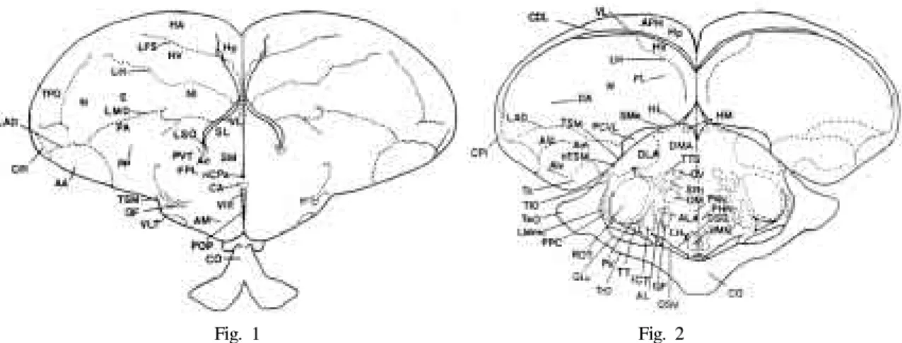
사이뇌의 중격을 중심으로 외측중격핵(nucleus septalis lateralis, SL)과 내측중격핵(nucleus septalis medialis, SM) 으로 나뉘어져 있었으며 외측중격핵은 뇌실과 인접하게 위치하고 있었다(Fig. 1, 2). 본 실험결과 NPY면역반응 신경세포는 외측 및 내측중격핵에 고농도로 분포하고 있었으며 발생 12일령부터 관찰되기 시작하여 발생 20 일령까지 관찰되었다(Fig. 3). 세포의 형태는 최초로 관 찰된 발생 12일령에는 대부분이 난원형과 구형이었으며 (Fig. 3) 이러한 세포의 형태는 발생 20일령까지 큰 변화 를 보이지 않았고 모든 실험 일령에서 난원형의 세포형 태를 취하고 있는 것이 가장 많이 관찰되었다. 본실험에 서 관찰한 모든 일령에서 내측중격핵보다 외측중격핵에 많은 수의 NPY면역반응신경세포가 밀집되어 있었으며 발생 12일령에서 발생 16일령까지는 세포수의 증가만 관찰되고 세포체의 크기는 큰 변화가 없었으나 발생 18 일령에는 세포체의 크기가 눈에 띄게 증가하였으며(Fig.
3-D) 발생 20일령에는 세포체의 크기가 발생 18일령에 비해 더욱더 커졌음이 뚜렷이 관찰되었다(Fig. 3-E). 또 한 세포수 역시 발생 18일령부터 눈에 띄게 증가하였고 내측중격핵 에서보다 외측중격핵에 더 많은 수의 NPY 면역반응신경세포가 밀집되어 있음이 관찰되었다(Fig.
3-F). 세포돌기는 세포체에 비해 발달되어 있지 않았으 며 대부분이 다방향성 돌기를 가지고 있었고 간혹 홑극 형 또는 두극형 돌기를 가지고 있는 것도 관찰 되었으 며 세포형태와 마찬가지로 세포돌기도 발생 12일령에서 20일령까지 큰 변화를 보이지 않았다(Fig. 3)
2. 시각영역(Optic area)
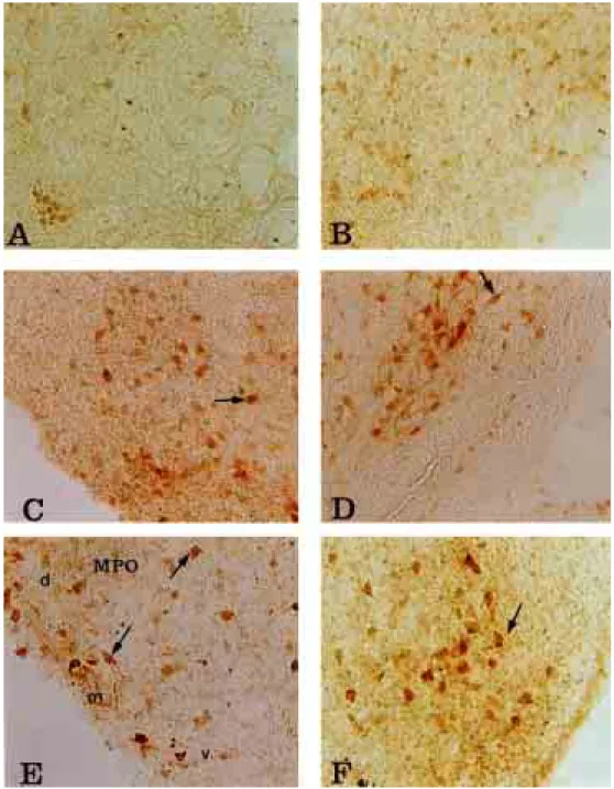
사이뇌의 시각영역은 크게 preoptic과 supraoptic으로 구분되어 있었으며 시각상위배측핵(nucleus supraopticus pars ventralis, SOv), 시각상위외측핵(nucleus supraopticus pars externs, SOe), 시신경교차전방 내측핵(nucleus preopticus medialis, POM), 시신경교차전방 뇌실주위핵(nucleus preopticus periventricularis, POP), 시신경교차전방 거대세포 핵(nucleus magnocelluaris preopticus, MPO), 외측무릎체 핵(nucleus geniculatus lateralis, GLv) 등으로 세분되어져 있었다.(Fig. 1, 2). 본 실험결과 사이뇌의 시각영역에서 NPY면역반응신경세포는 시신경교차전방 거대세포핵과
시신경교차전방 내측핵에서 발생 12일령부터 관찰되기 시작하여 발생 20일령까지 관찰되었다(Fig. 4)시신경교 차전방 거대세포핵의 등쪽, 내쪽, 배쪽과 시신경교차전 방 내측핵은 각각의 영역이 관찰 전기간동안 뚜렷이 구 분되어 있지 않았으며(Fig. 4-E) NPY면역반응신경세포 도 각각의 영역에 고루 분산되어 있었다. 세포의 형태는 발생 12일령에는 매우 적은 수의 세포가 난원형의 형태 를 취하고 있었고 발생일령이 증가할수록 구형, 난원형,
뭇각형의 다양한 세포형태가 관찰되었으나 대부분이 난 원형이었으며 이와 같은 양상은 발생 20일령까지 계속 되었다(Fig. 4). 세포수에 있어서는 발생 초에는 매우 적 은 수의 세포가 관찰되었으나(Fig. 4-A) 사이뇌의 다른 부위에서와 마찬가지로 발생 일령이 증가할수록 세포수 도 증가하였으며 세포체의 크기 또한 조금씩 커지고 있 음을 관찰 할 수 있었다(Fig. 4). 세포돌기는 발생초에는 난원형의 세포에 홑극형 또는 두극형 돌기를 취하고 있
Fig. 1-2. Schematic coronal plane drawing Gallus domesticus base on the
atlas of Kuenzel and Wayne J(1953), E:Ectostriatum, N: Neostriatum, HA: Hyperstriatum accessorium, HP: Hippocampus, HV: Hyperstriatum ventrale, HD:
Hyperstriatum dorsale, LFS: Lamina frontalis superior, LH: Lamina hyperstriatica, NI: Neostriatum intermedium, INP:
Nucleus intrapeduncularis, PA: Paleostriatum augmentatum(Caudate putamen), PP: Paleostriatum (Globus pallidus), SL:
Nucleus septalis lateralis, Va: Vallecula telencephali, AA: Archistriatum anterior [rostrale] DMA: Nucleus dorsomedialis anterior [rostralis], DA: Tractus dorso-archistriaticus, FA: tractus fronto-archistriaticus, AL: Ansa lenticularis, Ald:
Archistriatum intermedium, pars dorsalis (Zeier and Karten), Alv: Archistriatum intermedium, pars ventralis, FDB: Fasciculus diagonalis Brocae, AC: Nucleus accumbens, LMmc: Nucleus lentiformis mesencephali.pars magnocellularis, CO: Chiasma opticum, nCPa: Nucleus commissurae pallii (Bed nucleus pallial commissure), CPi: Cortex piriformis, DSD: Decussatio supraoptica dorsalis, LAD: Lamina archistriatalis dorsalis, LMD: Lamina medullaris dorsalis, CDL: Area corticoidea dorsolateralis, Sme: Stria medullaris, QE: Tractus quintofrontalis, HIS: Hyperstriatum intercalatum supremum, FPL: Fasciculus prosencephali lateralis (Lateral forebrain bundle), HL: Nucleus habenularis lateralis, PCVL: Plexus choroideus ventriculi lateralis (Choroid plexus within lateral ventricle), VL: Ventriculus lateralis, ALA: Nucleus ansae lenticularis anterior [rostralis], DLA: Nucleus dorsolateralis anterior [rostralis], LFS: Lamina frontalis superior, VLT: Nucleus ventrolateralis thalami, GLv: Nucleus geniculatus lateralis, pars ventralis, nTSM: Nucleus tractus septomesenceph alicus, Am:
Archistriatum mediale (Zeier and Karten), LFSM: Lamina frontalis suprema, AM: Nucleus anterior[rostralis] (Ananteior commissure), HM: Nucleus habenularis medialis, OM : Tractus occipitomesence
phalicus, VMN: Nucleus ventromedialis hypothalami, PVN: Nucleus paraventricularis magnocellularis, PHN: Nucleus periventrcularis hypothalami, Tn: Nucleus taeniae, NI: neostriatum intermedium, SCNm: Nucleus suprachiasmaticus, pars medialis, BAS: Nucleus basalis, CO: Chiasma opticum, LPO: Lobus parolfactorius, LSO: Organum septi laterale (Lateral septal organ), TeO: Tectum opticum, TIO: Tractus isthmo-opticus, TrO: Tractus opticus, POM: Nucleus preopticus medialis, SOv: Nucleus supraopticus (Ralph), pars ventralis, Aph: Area parahippocampalis, CPi: Cortex piriformis, TPO:
Area temporo-parieto-occipitalis, POP: Nucleus preopticus periventricularis, QE: Tractus quintofroontalis, SRt: Nucleus subnucleus rotundus, SM: Nucleus septalis medialis, ICT: Nucleus intercalatus thalami, ROT: Nucleus rotundus, T:
Nucleus triangularis, TT: Tractus tectothalamicus, TTS: Tractus thalamostriaticus, DSV: Decussatio supraoptica ventralis, V:
Nucleus ovoidalis, PV: Nucleus posteroventralis thalami PVT: Paleostriatum ventrale (Kitt and Brauth), VⅢ: Ventriculus tertius (Third ventricle), LHy: Regio lateralis hypothalami (Lateral hypothalamic area)
Fig. 1 Fig. 2
Fig. 3-A. NPY-IR neurons in nucleus septalis medialis(SM) and nucleus septalis lateralis(SL) at E12 day of chick (×200) Fig. 3-B. NPY-IR neurons in nucleus septalis medialis(SM) and nucleus septalis lateralis(SL) at E14 day of chick (×200) Fig. 3-C. NPY-IR neurons in nucleus septalis medialis(SM) and nucleus septalis lateralis(SL) at E16 day of chick (×200) Fig. 3-D. NPY-IR neurons in nucleus septalis medialis(SM) and nucleus septalis lateralis(SL) at E18 day of chick (×200),
st : septumFig. 3-E. NPY-IR neurons in nucleus septalis medialis(SM) at E20 day of chick (×200)
Fig. 4-A. NPY-IR neurons in nucleus preopticus medialis(POM) at E12 day of chick (×200)
Fig. 4-B. NPY-IR neurons in nucleus preopticus medialis(POM) at E14 day of chick (×200)
Fig. 4-C. NPY-IR neurons in nucleus preopticus medialis(POM) at E16 day of chick (×200)
Fig. 4-D. NPY-IR neurons in nucleus preopticus medialis(POM) at E18 day of chick (×200)
Fig. 4-E. NPY-IR neurons in nucleus magnocelluaris preopticus(MPO) at E20 day of chick (×200)
Fig. 4-F. NPY-IR neurons in nucleus preopticus medialis(POM) at E20 day of chick (×200)
Fig. 5-A. NPY-IR neurons in nucleus dorolateralis anterior[rostralis] thalami at E12 day of chick (×200), DLAm: neurons
in Nucleus dorolateralis anterior[rostralis] thalami, DLAI: neurons in nucleus dorolateralis anterior[rostralis] thalamiFig. 5-B. NPY-IR neurons in nucleus dorolateralis anterior[rostralis] thalami at E14 day of chick (×200)
Fig. 5-C. NPY-IR neurons in nucleus dorolateralis anterior[rostralis] thalami at E16 day of chick (×200), TSM : Tractus
septomesencephalicusFig. 5-D. NPY-IR neurons in nucleus dorolateralis anterior[rostralis] thalami at E18 day of chick (×200)
Fig. 5-E. NPY-IR neurons in nucleus dorolateralis anterior[rostralis] thalami at E20 day of chick (×200)
Fig. 5-F. NPY-IR neurons in nucleus dorolateralis anterior[rostralis] thalami at E20 day of chick (×200)
Fig. 6-A. NPY-IR neurons in nucleus paraventricularis magnocellularis(PVN) at E12 day of chick (×200) Fig. 6-B. NPY-IR neurons in nucleus paraventricularis magnocellularis(PVN) at E14 day of chick (×200)
Fig. 6-C. NPY-IR neurons in nucleus paraventricularis magnocellularis paraventricular nucleus, PVN) at E16 day of chick
(×400)Fig. 6-D. NPY-IR neurons in nucleus paraventricularis magnocellularis paraventricular nucleus, PVN) at E18 day of chick
(×200)Fig. 6-E. NPY-IR neurons in nucleus paraventricularis magnocellularis (paraventricular nucleus, PVN) at E20 day of chick
Fig. 7-A. NPY-IR neurons in nucleus rotundus at E8 day of chick (×40)
Fig. 7-B. NPY-IR neurons in nucleus rotundus at E12 day of chick (×40)
Fig. 7-C. NPY-IR neurons in nucleus rotundus at E16 day of chick (×40)
Fig. 7-D. NPY-IR neurons in nucleus rotundus at E20 day of chick (×40)
Fig. 7-E. NPY-IR neurons in nucleus rotundus at E18 day of chick (×400)
Fig. 7-F. NPY-IR neurons in nucleus rotundus at E20 day of chick (×400)
상 앞쪽 등내측핵(nucleus doromedialis anterior thalami, DMA), 시상 뒤쪽 등외측핵(nucleus dorolateralis posterior thalami, DLP), 시상 뒤배측핵(nucleus posteoventralis thalami), 시상 뒤쪽 등중간내측핵(nucleus dorsointermedialis posterial thalami, DIP), 시상 뒤쪽 등내측핵(nucleus doromedialis posterior thalami, DMP), 시상 중간사이핵(nucleus inter- calatus thalami, ICT)으로 세분화되어 있었다(Fig. 1, 2).
본 실험결과 NPY면역반응신경세포는 시상영역중 시상 앞쪽 등외측핵에 고농도로 분포하고 있었으며 시상 등 외측 거대세포핵 에서도 소수의 NPY면역반응신경세포 를 관찰할 수 있었다. 시상 앞쪽 등외측핵과 등외측 거 대세포핵 에서의 NPY면역반응신경세포는 발생 12일령 에 처음으로 관찰되기 시작하여 발생 20일령까지 관찰 되었다(Fig. 5). 세포의 형태는 발생 12일령에 난원형과 뭇각형의 세포가 많이 관찰되었고(Fig. 5-A) 이후 일령 에서는 대부분이 구형과 난원형의 세포형태를 취하고 있었다(Fig. 5-B, C, D, E, F). 또한 발생 20일령에는 중뇌 중격로(tractus septomesencephalicus, TSM) 주변부위에 존 재하는 세포의 돌기가 매우 발달되어 있었다(Fig. 5-F).
발생초 12일령에 많은 수의 세포가 관찰되었으며 세포 돌기는 잘 발달되어 있지 않았고(Fig. 5-A) 발생일령이 증가할수록 세포의 수 및 세포체의 크기가 증가하였으 며 세포돌기는 발생 20일령에 매우 잘 발달되어 있음을 관찰할 수 있었다(Fig. 5-F). 관찰한 모든 일령에서 세포 돌기는 대부분이 난원형과 구형의 세포에 다방향성 돌 기(Fig. 5-F)를 가지고 있는 것이 많이 관찰되었으며 홑 극형(Fig. 5-E)과 두극형 돌기(Fig. 5-C)를 가지고 있는 것도 관찰되었고 이와 같은 양상은 발생일령에 따라 큰 차이를 보이지 않았다(Fig. 5).
4. 시상하부(hypothalamus)
시상하부 각각의 영역은 외측시상하부영역(lateral hypothalamic area, LHY), 시상하부 배쪽 내측핵(nucleus ventromedialis hypothalami, VMN), 시상하부 뇌실주위핵
옆핵에는 고농도로 분포하고 있었다(Fig. 6). 시상하부 배쪽 내측핵, 앞쪽 내측핵과 뇌실옆핵에서의 NPY면역 반응신경세포는 발생 12일령에 관찰되기 시작하여 발생 20일령까지 관찰되었다(Fig. 6).
시상하부 배쪽 내측핵, 시상하부 앞쪽 내측핵과 뇌실 옆핵에서의 NPY면역반응신경세포는 세포의 형태, 돌기, 크기에 큰 차이가 없었으며 발생 12일령에는 적은 수의 세포가 관찰되었고 대부분이 뭇각형의 세포형태를 취하 고 있었으나(Fig. 6-A) 이후 일령에서는 난원형의 세포 가 많이 관찰되었고 간혹 구형과 뭇각형의 세포도 관찰 되었으나 대부분이 난원형의 세포였다(Fig 6). 세포체의 크기는 발생 12일령 이후 조금씩 커짐을 관찰할 수 있 었으며 발생 18일령에는 세포체의 크기에는 큰 변화가 없었고(Fig. 6-E) 세포수만이 눈에 띄게 증가함을 관찰할 수 있었다(Fig. 6-E). 세포돌기는 실험 전일령 에서 난원 형의 세포에 다방향성 돌기(Fig. 6-F)를 가지고 있는 것 이 대부분 이었으며 때로는 두극형 돌기(Fig. 6-C)를 가 지고 있는 것도 관찰되었다.
5. 원형핵(Nucleus rotundus, ROT)
조류사이뇌에서 시각을 담당하는 부위로 원형핵과 아 래원형핵(nucleus subrotundus, SRt)으로 구분되어 있었으 며(Fig. 1, 2) 원형핵의 등쪽 내측에 위치하고 있는 아래 원형핵에서는 실험 전일령에서 NPY면역반응신경세포 를 관찰 할 수 없었으나 원형핵에서는 발생 8일령에 관 찰되기 시작하여 발생 20일령까지 관찰되었다(Fig. 7).
발생 8일령에서 20일령까지의 전일령에서 NPY면역반응 신경세포는 원형핵 주변부위(Fig. 7)를 뚜렷이 둘러싸고 있는 특이한 형태를 취하고 있었으며 발생일령이 증가 할수록 둘러싸고 있는 양상이 더욱 뚜렷이 관찰되었다 (Fig. 7-A, B, C, D).
세포의 형태는 발생 8일령에는 대부분 난원형의 세포 가 관찰되었고(Fig. 7-E,F) 12일령에는 구형과 뭇각형 세 포가 다수 관찰되었으며 소수의 난원형 세포도 관찰되
었다. 16일령 이후에는 대부분이 뭇각형으로 관찰되었 으며(Fig. 7-E) 발생일령이 증가될수록 구형과 뭇각형의 세포가 난원형의 세포보다 많은 수를 차지하였다(Fig.
7-F). 원형핵 부위에서 전체적인 세포의 크기는 발생 8 일령 이후 조금씩 커지고 있음을 관찰할 수 있었고, 세 포의 수 또한 발생 일령이 증가함에 따라 조금씩 증가 하고 있음을 확인할 수 있었다. 세포돌기는 일령의 증가 에 따른 큰 변화가 없었으며 전 일령에서 다방향성 세 포돌기가 가장 많이 관찰되었다(Fig. 7-E, F). 원형핵은 시각이 매우 발달한 조류에서 시각기능을 담당하는 부 위로 사이뇌의 다른 출현부위에 비해 세포체와 세포돌 기가 매우 발달되어 있었다(Fig. 7-E, F). 또한 원형핵에 서의 세포돌기는 다른 출현부위에서 일정한 방향 없이 사방으로 뻗어 있었던 것과는 달리 원형핵 외측방향으 로 향하고 있었다(Fig. 7-E, F).
고 찰
NPY를 1982년 Tatemoto34)가 돼지의 뇌에서 분리 추출 하여 보고한 이후 많은 연구가 실시되었으며 최근에는 이에 대한 면역조직화학적 연구도 시작되었다. 과거 연 구자들이 신경계통에서 Pancreatic Polypeptide(이하 PP) 라고 보고했던 연구결과들이 계속된 연구결과에서 NPY 에 대한 연구결과와 일치하였으며 이것은 신경계통에서 의 결과들은 PP가 아니라 NPY의 형태로 존재함이 밝혀 졌다.1)
발생중인 병아리 사이뇌에서 NPY면역반응신경세포 는 외측 및 내측중격핵과 시신경교차전방 거대세포핵, 시신경교차전방 내측핵, 시상의 앞쪽 등외측핵과 등외 측 거대세포핵, 시상하부의 배쪽 내측핵, 앞쪽 내측핵과 뇌실옆핵에서 발생 12일령에 최초로 관찰되기 시작하여 발생 20일령까지 관찰되었다. 그러나 조류와28, 31) 파충 류32)의 시각기능을 담당하는 것으로 알려진 원형핵 에 서는 발생 8일령부터 관찰되었다. 발생중인 병아리의 사이뇌에서는 발생 12일령에 처음으로 NPY면역반응신 경세포가 관찰 되었지만, 흰쥐29)의 경우는 발생 14일령 에 사이뇌에서 처음 관찰된다고 하여 발생기간이 비슷 한 흰쥐에서보다 조류에서 좀더 일찍 관찰되는 것을 알 수 있었다. 성숙한 포유동물에서의 NPY면역반응신경세 포의 분포는 흰쥐27)에서 시상하부 뇌실주위핵과 preoptic 부위에서 종뇌에서 보다 고농도로 분포한다고 보고하였 고, 돼지30)에서는 사이뇌 대부분의 부위에서 NPY면역반 응신경세포가 고농도로 관찰된다고 보고하였는데, 이러 한 결과는 발생중인 병아리의 사이뇌에서 많은 수의 세 포가 관찰된 본 실험에서의 결과와 유사하였다.
세포형태는 원형핵부위를 제외한 부위에서 발생초에 난원형과 구형을 취하고 있는 것이 대부분 이었으며 이 같은 형태는 발생일령이 증가함에 따른 큰 변화없이 발 생 20일령까지 같은 형태로 관찰되었다. 이는 발생중인 병아리의 종뇌에서 발생초에는 난원형으로 관찰되고 발 생후기에 대부분이 뭇각형으로 관찰되었던 결과와 차이 를 보였다. 세포돌기는 종뇌에 비해 발달되어 있지 않았 으며 종뇌에서는 발생초부터 발생후기까지 원형, 난원 형, 뭇각형의 세포에 다방형성돌기를 가지고 있는 것이 많이 관찰되었으나 사이뇌에서는 원형핵을 제외한 부위 에서 난원형의 세포에 홑극형, 두극형 그리고 많은 수의 다방향성 돌기를 가지고 있는 것을 관찰할 수 있었다.
세포체의 크기는 발생초에 비해 일령이 증가할수록 크 기가 조금씩 커지는 것이 관찰되었고, 발생후기에는 눈 에 띄는 크기의 변화는 관찰되지 않았다. 세포수는 발생 16일령까지는 점차적으로 증가하다가 그 이후 일령에서 는 큰 변화가 관찰되지 않았다.
본 실험에서는 포유류에서 관찰할 수 없었던 원형핵 에서 NPY면역반응신경세포를 발생 8일령부터 관찰할 수 있었는데, 원형핵은 시각이 매우 발달한 조류에서 중 요한 시각기능을 담당하고 있는 부위이기 때문인 것으 로 사료된다. 조류에서의 원형핵33)은 포유류와는 달리 4 개의 층으로 이루어져 있는데 표면으로부터 내부로 향 한 층의 구조가 작은 세포층(small cell layer), 그물모양 층(plexiform layer), 큰 세포층(large cell layer), 가장자리 층(marginal layer)으로 이루어져 있다.32) 조류에서 망막 으로부터 종뇌를 통하는 시각정보에는 원심성시상경로 (thalamofugal pathway)와 원심성피개경로(tectofugal path- way)의 두가지 경로가 있는데 원심성피개경로에는 optic tectum, 원형핵(nucleus rotundus), 삼각형핵(nucleus trian- gularis), 시상하부 뒤쪽 등외측핵(nucleus dorsolateralis posterior thalami), 외줄무늬체(ectostriatum), 중간부신줄 무늬체(neostriatum intermedium), 배쪽상줄무늬체(hyper- striatum ventrale), 중간부상줄무늬체(hyperstriatum inter- medium) 부위가 포함되며 원심성시상경로에는 시상하 부 앞쪽 외측핵(nucleus lateralis anterior thalami), 시상하 부 앞쪽등외측 및 외측핵(nucleus dorsolateralis anterior thalami pars lateralis), 시상하부 앞쪽 등외측핵(nucleus dorsolateralis anterior thalami), 부상줄무늬체(hyperstriatum accessorium), 등쪽상줄무늬체(hyperstriatum dorsale)가 포 함되는데 조류의 시각계에 관여하는 대부분의 부위에서 NPY면역반응신경세포가 고농도로 분포하고 있는 것이 관찰되었으며 특히 시각계에서 중요한 역할을 하는 원 형핵에서는 원형핵 주변부를 뚜렷히 둘러싸고 NPY면역 반응신경세포를 관찰할수 있었다. 원형핵에서의 NPY면
20일령까지 눈에 띄는 변화는 관찰되지 않았으나 다른 출현부위에서는 세포돌기가 일정한 방향성을 갖지 않았 던 것과는 달리 원형핵 외측방향으로 향하고 있는 돌기 형태를 관찰 할 수 있었다.
발생중인 병아리의 사이뇌에서의 NPY면역반응신경 세포는 종뇌에 비해 세포의 크기와 면역성이 약했으며 세포의 수는 많이 관찰되었다. 발생일령이 비슷한 흰쥐 의 사이뇌에서는 발생 14일령에 처음 NPY면역반응신경 세포가 관찰된 것과는 달리 본 실험에서는 발생 8일령 에 원형핵에서 처음으로 관찰되었고 그 분포에 있어서 도 차이가 있을 것으로 보아 포유류에서와 발생중인 병 아리에서의 부위별 NPY면역반응신경세포에 의한 기능 에도 차이가 있을 것으로 사료된다. 또한 포유동물의 사 이뇌에서 발생에 따른 NPY면역반응신경세포의 변화를 관찰한 연구는 많지 않았으며 조류의 사이뇌부위에 대 한 연구는 아직까지 거의 이루어지지 않았다. 그러므로 앞으로 이 분야에 대한 연구를 더 많이 해서 우수한 결 론들을 이끌어낼 필요가 있을 것으로 사료된다.
결 론
본 연구에서는 한국재래종 닭의 유정란을 부화시켜 발생 8일령에서 20일령까지의 병아리 사이뇌에서 NPY 면역반응신경세포의 출현일 및 발생일령에 따른 세포의 형태, 돌기, 세포수 및 크기의 변화를 관찰하고자 면역 조직화학염색을 실시하여 관찰하였던 바 다음과 같은 결론을 얻었다.
발생일령별 NPY면역반응신경세포 관찰시기는 발생 8 일령에는 원형핵부위에서 관찰되었고 발생 12일령에는 외측 및 내측중격핵과 시각영역의 시신경교차전방 거대 세포핵과 시신경교차전방 내측핵, 시상의 앞쪽 등외측 핵과 등외측 거대세포핵, 시상하부영역의 배쪽내측핵, 앞쪽내측핵과 뇌실옆핵등 사이뇌 대부분의 영역에서 관 찰되었다. 세포의 형태는 발생 8일째부터 난원형으로
로 원형핵 외측방향으로 향하고 있는 것이 관찰되어 뇌 의 다른 출현부위에서 사방으로 뻗어있는 것과는 차이 가 있었다. 세포의 분포 또한 다른 출현부위에서 산재해 있던 것과는 달리 원형핵 주변부위를 둘러싸고 있었으 며 발생일령이 증가할수록 원형핵 주변부위를 둘러싸고 있는 양상이 더욱 뚜렷하게 관찰되었다. 세포체의 크기 및 수는 사이뇌의 다른 출현부위에서와 마찬가지로 발 생일령에 비례하여 증가하는 것을 관찰할 수 있었다. 원 형핵부위에서의 세포형태는 발생초에는 난원형의 세포 가 많았고 일령이 증가할수록 구형, 난원형, 뭇각형의 다양한 세포형태가 관찰되었으나 대부분이 뭇각형의 세 포형태였다. 이는 사이뇌의 다른 부위에서 세포형태가 발생일령이 증가함에도 불구하고 큰 변화 없이 대부분 이 난원형의 세포형태를 취하고 있었던 것과는 차이를 보였다.
이상과 같은 결과로 볼 때 발생중인 병아리 사이뇌의 발생단계에 따른 NPY면역반응신경세포의 출현빈도, 세 포모양, 돌기모양, 세포의 크기 등이 발생초기부터 뇌의 부위에 따라 차이가 생김으로 뇌의 부위에 따르는 뇌기 능에도 차이가 생길 뿐 아니라 포유류와도 형태적 차이 에 의한 기능적 차이가 있을 것으로 사료된다.
참고문헌
1. Allen YS, Andrian TE, Allen JM, Tatemoto, K, Crow TJ, Bloom SR and Polk JM. Neuropeptide Y distribution in the rat brain. Science 221, 877-879.
1983.
2. Kuenzel WJ and McMurtry JM. Neuropeptide Y: brain localization and central effects on plasma insulin levels in chicks. Physiol. Behav. 44, 669-678. 1988.
3. Lundberg JM, Hokfelt T, Anggard A, Terenius L, Elde R, Markey K, Goldstein M and Kimmel J (1982).
Organizationl principle in the peripheral sympathetic
nervous system : subdivision by coexisting peptides(somatostain-, avian pancreatic polypeptide-, and vasoactive intestinal polypeptide-like immunoreactive materials). Proc. Natl. Acad. Sci. USA 79, 303-1307.
4. Clark JJ, Kalra PS, Crawley WR, Kalra SP. Neu- ropeptide Y and human pancreatic polypeptide stimulate feeding behavior in rats. Endocrinology 115 : 427-431,
1984.
5. B. Glen Stanley, Sarah F. Leibowitz. Neuropeptide Y injected in the paraventricular hypothalamus : A powerful stimulant of feeding behavior. Neurobiology. 82 :
3940-3943. 1985.
6. Gray TS, Morley JE. Neuropeptide Y : Anatomical distri- bution and possible function in mammalian nervous system. Life Sci 38 : 389-401, 1986.
7. Stanley BG, Daniel DR., Chin AS and Leibowits SF Paraventricular nucleus injections of peptide YY and neuropeptide Y preferentially enhance carbohydrate ingestion. Peptides 6:1205-1211, 1985.
8. Yvan Dumont, Alain Fournier. Expression and Charac- terization of the Neuropeptide Y Y5 Receptor subtype in the Rat Brain. The Journal of Neuroscience, August
1, 18(15) : 5565-5574, 1998..
9. Albers HE, Ferris CF. Neuropeptide Y : Role in light- dark cycle entrainment of hamster circadian rhythms.
Neurosci Lett 50 : 163-168, 1984.
10. Kalra SP, Crowley WR. Norepinephrine-like effects of neuropeptide Y on LH release in the rat. Life Sci 35 :
1173-1176, 1984.
11. Lundberg JM, Terenius L, Hökfelt T, Marthing CR, Tatemoto K, Mutt V et al. Neuropeptide Y(NPY)-like immunoreactivity in periperal noradrenergic neurons and effects of NPY on sympathetic function. Acta
physiol Scand 116 : 477-480, 1982.
12. Allen JM, Adrian K, Tatemoto K, Polak JM, Hughes J, Bloom SR. Two novel related peptides, neuropeptide Y(NPY) and peptide YY(PYY), inhibit the contraction of the electrically stimulated mouse vas deferens.
Neuropeptides 3 : 71-77, 1982.
13. Allen LG, Kalra PS, Crowley WR, Kalra SP. Comparison of the effect of neuropeptide Y and adrenergic trans- mitters on LH release and food intake in male rats.
Life Sci 37 : 617-623, 1985.
14. Mcdonald JK, Lumpkin MD, Samson WK. Mclann SM. Neuropeptide Y affects secretion of lutenizing
hormone and growth hormone in ovariectomized rats.
Proc Natl Acad Sci 82 : 561-564, 1985.
15. Crowley WR, Hassid A, Kalra SP. Neuropeptide Y enhances the release of lutenizing hormone induced by lutenizing hormone-releasing hormone. Endocrinology
120 : 941-945, 1987.
16. Fuxe K, Agnati LF, Harfstrand A, Zini I, Tatemoto K, Pich, Hokfelt T, Mutt V, Terenius L. Central admini- stration of neuropeptide Y induces hypotension bradypenea and EEG synchronization in the rat. Acta physiol Scand
118(2) : 189-192. 1984.
17. Aguirre JA, Fuxe K, Hedlund P, Narvaez JA, Cintra A, Rosen L, Agrati LF. Neuropeptide Y /angiotensin Ⅱ interaction in central cardiovascular regulation of the rat. Brain Res 566 : 61-69, 1991.
18. Edvinsson L, Copeland JR, Emson P, Mculloch J, Uddman R. Nerve fibers containing neuropeptide Y in the cerebrovascular immunocytochemistry, radioimmu- noassay and vasomotor effects. J Cerebr Bool Flow
Metabol 7 : 45-57, 1987.
19. Hong M, Lis Fournier A, St-Pierre and Pellejer G.
Role of neuropeptid Y in the regulation of tyrosine hydroxylase gene expression in rat adrenal glands.
Neuroendocrinology 61 : 85-88, 1995.
20. Hlguchi H. Neuropeptide Y functions and biosynthesis as a peptidergic neurotransmitter and the regulation of neuron-specific expression of NPY gene. Folia phamacol
Jpn 93 : 203-218, 1989.
21. Zeigler HP and Karten HJ. Brain mechanism and feeding behavior in the pigeon (Columba livia). I.
Quinto-frontal structures. J. Comp. Neurol. 152, 59-82.
1972.
22. Denbow DM. Food intake control in birds. Neurosci.
Biobehav. Rev. 9, 223-232. 1985.
23. Kuenzel WJ. Neuroanatomical substrates involved in the control of food intake. Poultry Sci. 68, 926-937.
1989.
24. Shimjzu T, Cox K and Karten HJ. Intratelencephalic projections of the visual wulst in pigeons (Columba livia). J Comp. neurol. 359, 551-572. 1995.
25. Walsh KM, Kuenzel WJ. Effect of sulfamethazine on sexual precocity and neuropeptide Y neurons within the tuberoinfundibular region of the chick brain. Brain Res
Bull, 1997, 44:6, 707-13.
26. Fraley GS and Kuenzel WJ. Precocious puberity in
Bloom SR. Ontogeny of a novel peptide, neuropeptide Y (NPY) in rat brain. Brain Res, Jun, 303:1, 197-200.
1984.
30. Chiba A. Immunohistochemical distribution of neuropeptide Y-related substance in the brain and hypophysis of the
Comp.Neurol. 223, 592-610. 1984.
34. Tatemoto YK, Cartquist M and Mun V. Neuropeptide Y a novel brain peptide with structural simmilaritics to peptide YY and pancreatic polypeptide. Nature 296-670,

![Fig. 5-A. NPY-IR neurons in nucleus dorolateralis anterior[rostralis] thalami at E12 day of chick (×200), DLAm: neurons in Nucleus dorolateralis anterior[rostralis] thalami, DLAI: neurons in nucleus dorolateralis anterior[rostralis] thalami Fig](https://thumb-ap.123doks.com/thumbv2/123dokinfo/5268751.369296/7.786.119.671.131.834/dorolateralis-anterior-rostralis-nucleus-dorolateralis-rostralis-dorolateralis-rostralis.webp)


