• 검색 결과가 없습니다.

Cardiac Sarcoidosis Presenting With Complete Atrioventricular Block and Sustained Monomorphic Ventricular Tachycardia

N/A
N/A
Protected

Academic year: 2022

Share "Cardiac Sarcoidosis Presenting With Complete Atrioventricular Block and Sustained Monomorphic Ventricular Tachycardia"

Copied!
4
0
0

로드 중.... (전체 텍스트 보기)

전체 글

(1)

571 Copyright © 2012 The Korean Society of Cardiology

Korean Circulation Journal

Introduction

Sarcoidosis is a multisystem granulomatous disease of unknown etiology. Cardiac manifestations, seen in approximately 5% of pa- tients,

1)2)

typically include congestive heart failure with left ventric- ular dysfunction, conduction abnormalities, and ventricular arrhyth- mias. Moreover, sudden death has been reported in up to 67% of instances where cardiac sarcoidosis is diagnosed postmortem.
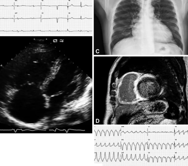
3)

Herein, we describe a patient whose cardiac sarcoidosis presented as symptomatic complete atrioventricular (AV) block with conges- tive heart failure and ventricular tachycardia. Tissue diagnosis was achieved via paratracheal node sampling, obtained by endobron- chial ultrasonography guided transbronchial lymph node aspira-

Case Report

http://dx.doi.org/10.4070/kcj.2012.42.8.571 Print ISSN 1738-5520 • On-line ISSN 1738-5555

Cardiac Sarcoidosis Presenting With Complete Atrioventricular Block and Sustained Monomorphic Ventricular Tachycardia

Joo Myung Lee, MD 1 , Il-Young Oh, MD 2 , and Dong-Ju Choi, MD 2

1

Cardiovascular Center and Department of Internal Medicine, Seoul National University Hospital, Seoul,

2

Division of Cardiology, Department of Internal Medicine, College of Medicine, Seoul National University and Cardiovascular Center, Seoul National University Bundang Hospital, Seongnam, Korea

Sarcoidosis is a rare but potentially fatal multisystem granulomatous disease of unknown etiology. While a number of clinical manifesta- tions may develop, cardiac involvement (prior to or coincident with sarcoidosis of other organs) is an important prognostic factor. Re- cently, we encountered a patient with cardiac sarcoidosis who presented with complete atrioventricular (AV) block and sustained ventricu- lar tachycardia. An implantable cardioverter-defibrillator was inserted as a precautionary measure for ventricular tachycardia and symp- tomatic complete AV block.

18

F-fluoro-2-deoxyglucose positron emission tomography confirmed a dramatic response to high-dose steroid at four weeks, as demonstrated by a marked decrease in cardiac sarcoid activity from baseline status. (Korean Circ J 2012;42:571-574) KEY WORDS: Sarcoidosis; Heart failure; Magentic resonance imaging; Tachycardia, ventricular.

Received: November 10, 2011 Revision Received: December 21, 2011 Accepted: January 12, 2012

Correspondence: Dong-Ju Choi, MD, Division of Cardiology, Department of Internal Medicine, College of Medicine, Seoul National University and Cardiovascular Center, Seoul National University Bundang Hospital, 82 Gumi-ro 173beon-gil, Bundang-gu, Seongnam 463-707, Korea

Tel: 82-31-787-7031, Fax: 82-31-787-4051 E-mail: [email protected]

• The authors have no financial conflicts of interest.

This is an Open Access article distributed under the terms of the Creative Commons Attribution Non-Commercial License (http://creativecommons.

org/licenses/by-nc/3.0) which permits unrestricted non-commercial use, distribution, and reproduction in any medium, provided the original work is properly cited.

tion (EBUS-TBNA). The patient then received an implantable cardio- verter-defibrillator (ICD) and systemic steroid therapy. Follow-up

18

F-fluoro-2-deoxyglucose positron emission tomography (

18

F-FDG- PET) showed a marked decrease in cardiac sarcoid activity compared to the baseline.

Case

A 55-year-old male was admitted to another hospital six months previously for dyspnea on exertion (DOE) and dizziness. At that time, there was evidence of severe global hypokinesia of the left ventricle (LV), with systolic dysfunction {left ventricular ejection fraction (LVEF) 33% per M-mode} on echocardiogram, as well as enlargement of the right atrium and right ventricle (RV), and severe RV dysfunction.

A corresponding electrocardiogram documented complete right

bundle branch block with sinus bradycardia, while 24-hour Holter mo-

nitoring recorded a maximum RR interval of 2.76 seconds. No high-

level AV block or arrhythmia was apparent, and by coronary angio-

graphy, epicardial coronary arteries were normal. On this basis, a

diagnosis of idiopathic dilated cardiomyopathy was rendered and

thereafter treated with ramipril 2.5 mg, furosemide 20 mg, and

spironolactone 25 mg daily. Upon admission to our center, the pa-

tient again presented with DOE (NYHA class III) and intermittent diz-

ziness, which were both aggravated in the preceding 15 days. His

electrocardiogram showed complete AV block (heart rate, 27 beats

per minute) (Fig. 1A) as did a 24-hour Holter monitor, with a maxi-

(2)

572 Cardiac Sarcoidosis With Conduction Disturbance and Ventricular Tachycardia

http://dx.doi.org/10.4070/kcj.2012.42.8.571 www.e-kcj.org

mum RR interval of 4.7 seconds. An echocardiogram demonstrated severe LV dysfunction (LVEF 34.4% per modified Simpson method), a dyskinetic basal septum, basilar cardiac akinesia, and an enlarg- ed, severely dysfunctional RV, without pulmonary hypertension {pul- monary artery systolic pressure (RVSP) 27.64 mm Hg} (Fig. 1B). Mild cardiomegaly was noted on chest X-ray (cardiothoracic ratio 55%), but hilar lymph nodes were not prominent (Fig. 1C). Gadolinium-di- etylene triamine penta-acetic acid (DTPA)-enhanced cardiac MRI sh- owed delayed hyper-enhancement of the entire RV wall and subepi- cardial (apical) or transmural (apical, basal) portions of the LV (Fig. 1D).

While cardiac sarcoidosis was strongly suspected, endomyocar- dial biopsy findings were nonspecific, and angiotensin-converting enzyme (ACE) was within normal reference range (27.5 IU/L; normal range: 8.0-55.0 IU/L). During endomyocaridal biopsy, sustained ven- tricular tachycardia developed repetitively (Fig. 1E). Enlarged right paratracheal (17.27×8.35 mm) and subcarinal (18.63×11.97 mm) lymph nodes, found incidentally with coronary CT angiography six

months prior (Fig. 2A and B, respectively), were instead sampled via EBUS-TBNA to histologically confirm the presence of noncaseating epitheloid granulomata with giant cells (Fig. 2C and D). In accord- ance with the Japanese Ministry of Health and Welfare guideline,

4)

cardiac sarcoidosis was diagnosed and a systemic glucocorticoid (prednisolone, 60 mg daily) was initiated. An ICD was also inserted as a precautionary measure for ventricular tachycardia and symp- tomatic complete AV block. Because the ACE level was within the normal reference range, we used

18

F-FDG-PET to monitor therapy.

Baseline results at >18 hours prolonged fasting showed inhomo- geneous

18

F-FDG uptake, accentuated in LV myocardium {maxi- mum standardized uptake value (SUV

MAX

)=7.9 g/mL} (Fig. 2E and F).

On discharge, the patient was asymptomatic. One month later, fol- low-up

18

F-FDG-PET showed a marked decrease in myocardial hy- permetabolism (SUV

MAX

=3.1 g/mL) (Fig. 2G and H), and the right pa- ratracheal lymph node had decreased in size upon chest CT. The ACE level also dropped to 14.1 U/L. The patient is now doing well, with- A  

E   C  

B  

D  

Fig. 1. Initial presentation of the patients. A: admission electrocardiogram: complete atrioventricular block (heart rate, 27 beats per minute). B: echocar- diogram: RV and right atrium enlargement and severe RV dysfunction, without pulmonary hypertension (pulmonary artery systolic pressure, 27.64 mm Hg). C: chest X-ray: mild cardiomegaly (cardiothoracic ratio 55%); no hilar lymph node enlargement. D: Gadolinium-dietylene triamine penta-acetic acid-en- hanced cardiac MRI: delayed hyper-enhancement of the entire RV wall and subepicardial (apical) or transmural (apical, basal) portions of the left ventricle.

E: sustained ventricular tachycardia during endomyocardial biopsy. RV: right ventricle.

(3)

573 Joo Myung Lee, et al.

http://dx.doi.org/10.4070/kcj.2012.42.8.571 www.e-kcj.org

out recurrent symptoms, including ventricular tachycardia.

Discussion

Sarcoidosis is a multisystem disease that has a worldwide distri- bution, characterized by the presence of noncaseating granulomata in involved organs. Its etiology is currently unknown. The prevalence of this disease varies from 10 per 100000 in white populations to 35

per 100000 in African Americans.

5-7)

In the US, around 20-27% of patients with sarcoidosis have myocardial involvement,

8)

while in Ja- pan, up to 58% of patients have cardiac lesions, accounting for as many as 85% of recorded deaths from sarcoidosis.

9)

Reports also indicate that cardiac involvement is largely unrecog- nized antemortem. In only about 5% of patients with autopsy evi- dence of cardiac sarcoid (i.e., noncaseating granulomata) is cardiac disease apparent prior to death.

10)

Common presentations of cardi- Fig. 2. Pathologic finding of lymph nodes and results of initial and follow-up

18

F-FDG-PET. A and B: right paratracheal (17.27×8.35 mm) and subcarinal (18.63×11.97 mm) lymph node enlargement discovered incidentally in coronary CT angiography six months prior. C and D: lymph node aspirate: noncase- ating epitheloid granulomata with giant cells. E and F: baseline

18

F-FDG-PET: inhomogeneous uptake, accentuated in left ventricular myocardium {maxi- mum standardized uptake value (SUV

MAX

)=7.9 g/mL}. G and H:

18

F-FDG-PET at one-month follow-up; marked decrease in myocardial hypermetabolism (SU- V

MAX

=3.1 g/mL).

18

F-FDG-PET:

18

F-fluoro-2-deoxyglucose positron emission tomography.

A  

C  

E  

G  

B  

D  

F  

H  

(4)

574 Cardiac Sarcoidosis With Conduction Disturbance and Ventricular Tachycardia

http://dx.doi.org/10.4070/kcj.2012.42.8.571 www.e-kcj.org

ac sarcoidosis include congestive heart failure with left ventricular dysfunction, cardiac rhythm disturbance, and most importantly sud- den death. The latter has been cited in up to 67% of patients where cardiac sarcoidosis was diagnosed postmortem.

3)

Among the available noninvasive diagnostic modalities, gadolini- um-DTPA-enhanced cardiac MRI offers greater utility for early diag- nosis and follow-up, compared with the low-sensitivity of endo- myocardial biopsy (approaching 20%).

11-13)

Recent studies have similarly demonstrated the potential of

18

F-FDG-PET as a diagnos- tic and follow-up tool.

14-16)

Because the ultimate goal for these pa- tients is to preserve cardiac function and prevent fatal arrhythmias, early detection, even in absence of symptoms, is critical. In this re- spect, gadolinium-DTPA-enhanced cardiac MRI and

18

F-FDG-PET may more reliably facilitate early diagnosis and thus result in better treatment outcomes.

14)

In our patient, delayed myocardial enhance- ment by cardiac MRI was a key diagnostic clue. Despite procurement of an adequate endomyocardial biopsy, a tissue diagnosis could not be established with this approach. It was through EBUS-TBNA of right paratracheal and subcarinal lymph nodes that the diagnosis of car- diac sarcoidosis was eventually made.

Systemic steroids have shown survival benefits with cardiac sar- coidosis and have been generally accepted as the treatment of ch- oice

17)18)

However, the effect of steroids on ventricular arrhythmias is less certain, and some recommend an ICD as primary therapy in patients presenting with ventricular tachycardia.

10)19)

In addition to a systemic steroid, our patient received an ICD to deal with symp- tomatic complete AV block and ventricular tachycardia.

Serum ACE level typically reflects sarcoid activity and can be used as a biomarker, although our patient’s levels were normal. Fur- thermore, cardiac MRI was precluded for therapeutic monitoring, due to the precautionary ICD, so

18

F-FDG-PET was used. At the one- month interval, myocardial hypermetabolism had markedly de- creased by

18

F-FDG-PET, which correlated well with the patient’s cli- nical response and a corresponding decline in his serum ACE level.

In conclusion, cardiac sarcoidosis presenting with symptomatic complete AV block and sustained ventricular tachycardia, was suc- cessfully treated in this instance with a systemic steroid and an ICD, while serum levels of ACE and

18

F-FDG-PET aided in therapeutic monitoring.

References

1. Hagemann GJ WK. The clinical, electrocardiographic and pathological features of cardiac sarcoid. In: Jones Williams W, Davies BH, editors.

Sarcoidosis and Other Granulomatous Disorders: Proceedings of the 8th International Conference Cardiff. Cardiff: Alpha & Omega Press;

1980. p.601-6.

2. Johns CJ, Michele TM. The clinical management of sarcoidosis: a 50-

year experience at the Johns Hopkins Hospital. Medicine (Baltimore) 1999;78:65-111.

3. Roberts WC, McAllister HA Jr, Ferrans VJ. Sarcoidosis of the heart: a clinicopathologic study of 35 necropsy patients (group 1) and review of 78 previously described necropsy patients (group 11). Am J Med 1977;63:86-108.

4. Hiraga H, Yuwai K, Hiroe M. Guidelines for the Diagnosis of Cardiac Sarcoidosis: Study Report of Diffuse Pulmonary Diseases. Tokyo: Japa- nese Ministry of Health and Welfare;1993. p.23-4.

5. Chapelon-Abric C, de Zuttere D, Duhaut P, et al. Cardiac sarcoidosis: a retrospective study of 41 cases. Medicine (Baltimore) 2004;83:315-34.

6. Hillerdal G, Nöu E, Osterman K, Schmekel B. Sarcoidosis: epidemiology and prognosis: a 15-year European study. Am Rev Respir Dis 1984;130:

29-32.

7. Rybicki BA, Major M, Popovich J Jr, Maliarik MJ, Iannuzzi MC. Racial differences in sarcoidosis incidence: a 5-year study in a health main- tenance organization. Am J Epidemiol 1997;145:234-41.

8. Silverman KJ, Hutchins GM, Bulkley BH. Cardiac sarcoid: a clinicopath- ologic study of 84 unselected patients with systemic sarcoidosis. Cir- culation 1978;58:1204-11.

9. Matsui Y, Iwai K, Tachibana T, et al. Clinicopathological study of fatal myocardial sarcoidosis. Ann N Y Acad Sci 1976;278:455-69.

10. Winters SL, Cohen M, Greenberg S, et al. Sustained ventricular tachy- cardia associated with sarcoidosis: assessment of the underlying car- diac anatomy and the prospective utility of programmed ventricular stimulation, drug therapy and an implantable antitachycardia device.

J Am Coll Cardiol 1991;18:937-43.

11. Shimada T, Shimada K, Sakane T, et al. Diagnosis of cardiac sarcoidosis and evaluation of the effects of steroid therapy by gadolinium-DTPA- enhanced magnetic resonance imaging. Am J Med 2001;110: 520-7.

12. Vignaux O, Dhote R, Duboc D, et al. Clinical significance of myocardial magnetic resonance abnormalities in patients with sarcoidosis: a 1-year follow-up study. Chest 2002;122:1895-901.

13. Uemura A, Morimoto S, Hiramitsu S, Kato Y, Ito T, Hishida H. Histologic diagnostic rate of cardiac sarcoidosis: evaluation of endomyocardial biopsies. Am Heart J 1999;138(2 Pt 1):299-302.

14. Ohira H, Tsujino I, Yoshinaga K.

18

F-Fluoro-2-deoxyglucose positron emission tomography in cardiac sarcoidosis. Eur J Nucl Med Mol Imag- ing 2011;38:1773-83.

15. Braun JJ, Kessler R, Constantinesco A, Imperiale A. 18F-FDG PET/CT in sarcoidosis management: review and report of 20 cases. Eur J Nucl Med Mol Imaging 2008;35:1537-43.

16. Kim JS, Judson MA, Donnino R, et al. Cardiac sarcoidosis. Am Heart J 2009;157:9-21.

17. Reuhl J, Schneider M, Sievert H, Lutz FU, Zieger G. Myocardial sarcoid- osis as a rare cause of sudden cardiac death. Forensic Sci Int 1997;89:

145-53.

18. Takada K, Ina Y, Yamamoto M, Satoh T, Morishita M. Prognosis after pacemaker implantation in cardiac sarcoidosis in Japan: clinical eval- uation of corticosteroid therapy. Sarcoidosis 1994;11:113-7.

19. Belhassen B, Pines A, Laniado S. Failure of corticosteroid therapy to prevent induction of ventricular tachycardia in sarcoidosis. Chest 1989;

95:918-20.

수치

Fig. 1. Initial presentation of the patients. A: admission electrocardiogram: complete atrioventricular block (heart rate, 27 beats per minute)

참조

관련 문서