http://dx.doi.org/10.5468/ogs.2016.59.1.1 pISSN 2287-8572 · eISSN 2287-8580
Introduction
Cancer is the second most common cause of death in women of reproductive age [1,2]. In Korea, the incidence of cancer in reproductive-aged women is reported to be 162 out of 100,000 [3]. Today, the maternal age at birth continues to rise [4]. However, because the incidence of cancer increases significantly with age [1], the risk of cancer during pregnancy may also increase with rising maternal age at birth [5].
Cancer during pregnancy is an important but complex is- sue requiring consideration of both the mother and the baby [2]. However, cancer diagnosis and management during
Clinical characteristics and outcome of cancer diagnosed during pregnancy
Min Hee Shim, Chi-Won Mok, Kylie Hae-Jin Chang, Ji-Hee Sung, Suk-Joo Choi, Soo-young Oh, Cheong-Rae Roh, Jong-Hwa Kim
Department of Obstetrics and Gynecology, Samsung Medical Center, Sungkyunkwan University School of Medicine, Seoul, Korea
Objective
The aim of this study is to describe the clinical characteristics and outcome of cancer diagnosed during pregnancy.
Methods
This is a retrospective cohort study of women who were diagnosed with cancer during pregnancy at a tertiary academic hospital between 1995 and 2013. Maternal characteristics, gestational age at diagnosis, and type, stage, symptoms and signs of cancer for each patient were retrieved from the medical records. The cancer treatment, pregnancy management and the subsequent perinatal and maternal outcomes for each cancer were assessed.
Results
A total of 87 women were diagnosed with cancer during pregnancy (172.6 cases per 100,000 deliveries). The most common cancer was breast cancer (n=20), followed by gastrointestinal (n=17), hematologic (n=13), thyroid (n=11), central nervous system (n=7), cervical (n=7), ovarian (n=5), lung (n=3), and other cancers (n=4). Eighteen (20.7%) patients terminated their pregnancies. In the 69 (79.3%) patients who maintained their pregnancies, one patient miscarried and 34 patients delivered preterm. Of the preterm babies, 24 (70.6%) were admitted to the neonatal intensive care unit and 3 (8.8%) of those expired. The maternal mortality rate was 31.0%, with highest rate seen with lung cancers (66.7%), followed by gastrointestinal (50.0%), central nervous system (50.0%), hematologic (30.8%), breast (25.0%), ovarian (20.0%) cervical (14.3%), and thyroid cancers (0%).
Conclusion
The clinical characteristics and outcome of cancer during pregnancy were highly variable depending on the type of cancer. However, timely diagnosis and appropriate management of cancer during pregnancy may improve both maternal and neonatal outcome.
Keywords: Maternal mortality; Neoplasms; Pregnancy; Pregnancy outcome
Articles published in Obstet Gynecol Sci are open-access, distributed under the terms of the Creative Commons Attribution Non-Commercial License (http://creativecommons.
org/licenses/by-nc/3.0/) which permits unrestricted non-commercial use, distribution, and reproduction in any medium, provided the original work is properly cited.
Copyright © 2016 Korean Society of Obstetrics and Gynecology Received: 2015.6.16. Revised: 2015.8.7. Accepted: 2015.8.7.
Corresponding author: Suk-Joo Choi
Department of Obstetrics and Gynecology, Samsung Medical Center, Sungkyunkwan University School of Medicine, 81 Irwon-ro, Gangnam- gu, Seoul 06351, Korea
Tel: +82-2-3410-3546 Fax: +82-2-3410-0630 E-mail: [email protected] http://orcid.org/0000-0002-8946-4789
pregnancy are not well understood for several reasons. Firstly, although cancer during pregnancy is not rare, it is uncom- mon, with an incidence rate of approximately 1 in 1,000 pregnancies [6,7]. Therefore, large, prospective studies are not available. Secondly, the type, stage, diagnosis, treatment and prognosis of the cancer vary widely. Thirdly, the diagnosis of cancer during pregnancy is not easy because the symptoms and signs of cancer may mimic the physiologic changes or common complaints reported during pregnancy. Therefore, the symptoms and signs of cancer may be easily ignored by both pregnant women and their doctors, leading to delayed diagnosis and eventual progression to an advanced stage [8].
The type of cancer and outcome during pregnancy may also differ between races and ethnicities [9]. Although the clini- cal aspects of cancer during pregnancy have been described in a number of studies performed in Western countries, the topic has yet to be studied comprehensively in Korea [6,8,10].
Therefore, this study is aimed at describing the clinical charac- teristics and outcomes of cancers diagnosed during pregnancy in Korea.
Materials and methods
This is a retrospective cohort study of women who were diag- nosed with cancer during pregnancy at a tertiary referral hos- pital, between the years of 1995 and 2013. Using the in-pa- tient electronic database system, pregnant women who were discharged from the hospital and assigned with the cancer disease code by ICD-10 (International Statistical Classification of Diseases and Related Health Problems 10th revision) were selected as the study population. Women diagnosed with can- cer before pregnancy or during the postpartum period were excluded. Also, noninvasive cancers such as carcinoma in situ and borderline ovarian tumors were excluded from the analy- sis. This study was approved by the institutional review board of our institution.
Maternal age, parity, gestational age at diagnosis, and the type, stage, symptoms and signs of cancer for each patient were reviewed from the medical records. The cancers were grouped into breast, gastrointestinal (GI), hematologic, thy- roid, central nervous system, cervical, ovarian, lung and others.
Each cancer was staged according to National Comprehensive Cancer Network guidelines, except for the brain tumors and acute leukemia, which do not follow the conventional cancer
staging system. The cancer treatment strategy was classified into 4 categories: 1) termination of pregnancy, defined as therapeutic abortion or iatrogenic delivery before fetal viability (24 weeks of gestation) for the purpose of cancer treatment; 2) immediate initiation of cancer treatment during pregnancy; 3) immediate cancer treatment following iatrogenic preterm de- livery (PTD); or 4) delay of cancer treatment until completion of obstetrically indicated delivery.
Pregnancy outcomes included spontaneous abortion, pre- term or term delivery, gestational age at delivery, delivery mode, sex and birth weight of the baby, admission to neona- tal intensive care unit (NICU), neonatal morbidity and mortal- ity. PTD was defined as delivery before 37 weeks of gestation;
this was categorized into spontaneous PTD (due to preterm labor or preterm premature rupture of membranes) and iat- rogenic PTD (to treat cancer). Maternal outcome was catego- rized into 1) complete response with no evidence of disease; 2) partial response; 3) stable disease or progressive disease; 4) re- currence; 5) death; or 6) loss to follow-up with unknown out- come. The definitions of complete response, partial response, stable disease, and progressive disease were determined based on Response Evaluation Criteria in Solid Tumors.
For the comparison of multiple means, analysis of variance or the Kruskal-Wallis test was used, as appropriate. Categori- cal variables were compared using the chi-square test or Fisher’s exact test, as appropriate. A P-value of <0.05 was con- sidered statistically significant.
Results
Over the 19-year study period, 98 cases of pregnant women diagnosed with cancer were retrieved from the database.
Among them, 9 cases of cervical carcinoma in situ, 1 case of breast ductal carcinoma in situ and 1 case of borderline ovar- ian tumor were excluded. The remaining 87 patients were di- agnosed with invasive cancer during pregnancy. The incidence of cancer diagnosed during pregnancy was 172.6 in 100,000 pregnancies (87 out of 50,412 deliveries over the 19 years).
The actual number of cases and the incidence of cancer rose
over time, with 17 cases from 1995–2000 (76.5/100,000), 27
cases from 2001–2006 (187.4/100,000), and 43 cases from
2007–2013 (311.9/100,000). Breast cancer (n=20) was most
common form of cancer, followed by GI (n=17), hematologic
(n=13), thyroid (n=11), central nervous system (n=7), cervical
(n=7), ovarian (n=5), lung (n=3), and other cancers (n=4). The type, primary symptoms and signs at diagnosis of each cancer group are described in Table 1.
The characteristics, treatment and outcome of all patients diagnosed with cancer during pregnancy are described in Table 2. Eighteen (20.7%) patients opted to terminate their pregnancies due to the cancer; 12 of these patients were diag- nosed during the first trimester and 6 were diagnosed during the second trimester. Twenty-four (27.6%) patients received immediate treatment during the pregnancy. Iatrogenic PTD was performed in 25 (28.7%) patients, for the purpose of initiating cancer treatment. In 20 (23.0%) patients, cancer treatment was delayed until obstetrically indicated delivery was completed;
17 of these patients delivered at term and 3 delivered preterm, with 1 due to spontaneous preterm labor and the other 2 due to preterm premature rupture of membranes.
Three patients were lost to follow-up after delivery. The total mortality rate of the remaining 84 patients was 31.0% (26/84).
The mortality rate varied according to the type of cancer, with the highest rate seen in the lung cancer group (66.7%), followed by GI (50.0%), central nervous system (50.0%), he- matologic (30.8%), breast (25.0%), ovarian (20.0%) cervical (14.3%), and thyroid cancers (0%).
The pregnancy outcomes of the 69 patients who maintained
their pregnancies are shown in Table 3. One patient with acute myelocytic leukemia died due to intracranial hemorrhage dur- ing chemotherapy at 15 weeks of gestation and the fetus was aborted. Among the remaining 68 patients, 34 (50%) delivered at term, of which one (2.9%) baby was admitted to the NICU due to meconium aspiration syndrome. Of the 34 preterm babies, 24 (70.6%) were admitted to the NICU, of which 13 had respiratory distress syndrome. The NICU ad- mission rate was significantly higher for preterm babies born before 34 weeks compared to those born at 34 to 36 weeks gestation (18/18 [100%] vs. 6/16 [37.5%], P<0.05). Three ba- bies born at 28, 29, and 29 weeks of gestation expired during the neonatal period. The clinical characteristics and outcomes of each cancer are described in Table 4. Maternal age, parity and gestational age at diagnosis were not significantly differ- ent between cancer groups (data not shown). However, the cancer stage at diagnosis, treatment strategy, PTD rate and maternal outcome were differed significantly by cancer type.
Discussion
In this cohort study, we described 87 cases of cancer diag- nosed during pregnancy at a single tertiary center in Korea
Table 1. Type and primary symptoms and signs of cancer diagnosed during pregnancyCancer group Type Primary symptoms and signs
Breast (n=20) Invasive ductal carcinoma (19), inflammatory
cancer (1) Palpable mass (17), mastalgia (2), bloody nipple discharge (1) GI (n=17) Early gastric cancer (2), advanced gastric cancer
(6), colorectal cancer (5), hepatic cancer (4)
Abdominal pain (6), nausea and vomiting (3), hematochezia (2), routine exam (2), flank pain (1), diarrhea (1), neck swelling pain (1), elevated maternal serum alpha-fetoprotein (1)
Hematology (n=13) Acute myeloid leukemia (3), chronic myeloid leukemia (1), non-Hodgkin’s lymphoma (9)
Abnormal routine blood cell count (3), oral ulcer (1), palpable neck mass (1), palpable abdominal mass (1), dyspnea (1), sore throat (1), flank pain (1), breast swelling (1), joint pain (1), easy bruisability (1), abnormal ovarian mass on ultrasound (1) Thyroid (n=11) Papillary thyroid carcinoma (11) Thyroid nodule on routine ultrasound (4), palpable mass (4),
abnormal thyroid function test (3) CNS (n=7) Meningioma (2), schwannoma (2), astrocytoma
(2), glioma (1)
Headache and vomiting (2), hemiparesis (2), hearing loss (1), diplopia (1), olfactory hallucination (1)
Cervix (n=7) Squamous cell carcinoma (6), adenocarcinoma (1) Vaginal bleeding (4), abnormal Pap smear test (3)
Ovary (n=5) Epithelial ovarian cancer (4), dysgerminoma (1) Asymptomatic pelvic mass on ultrasound (3), back pain (1), constipation & dysuria (1)
Lung (n=3) Small cell lung carcinoma (2), adenocarcinoma (1) Dyspnea (2), abnormal routine chest X-ray (1) Others (n=4) Adenocarcinoma of unknown primary (2),
nasopharyngeal cancer (1), liposarcoma (1)
Palpable mass (2), anemia (1), pelvic mass on ultrasound (1) GI, gastrointestinal; CNS, central nervous system.
over a period of 19 years. We found that the incidence of cancer diagnosed during pregnancy steadily increased over the 19-year study period. Our data shows that the clinical characteristics of cancer diagnosed during pregnancy are highly variable and depend on the type of cancer. The cancer treatments, pregnancy management and the subsequent peri- natal and maternal outcomes were varied and complex.
The incidence and type of cancer during pregnancy may dif-
fer between races and ethnicities. In the Western countries, breast cancer, melanoma, cervical cancer and hematologic cancers are the most commonly reported cancers during pregnancy [5,7,10-13]. However, in our study, no cases of melanoma were diagnosed during pregnancy. The incidence of melanoma is much lower in Asian countries compared to Western countries [14]. In our study, the most common can- cer during pregnancy was breast cancer, followed by GI, he- matologic and thyroid cancers, similar to the standard cancer incidence for the female population in Korea (thyroid, breast, colorectal, and stomach) [3]. Thyroid cancer is the most com- monly diagnosed female cancer in Korea [15], mainly due to early detection using ultrasound for routine health care examination [16,17]. However, thyroid ultrasonography is not included in routine prenatal tests. Only 4 of the 11 thyroid cancers in our study were detected using thyroid ultrasound during routine health screening exams. Thyroid cancer was ranked as the fourth most common cancer diagnosed during pregnancy in our study. However, when GI cancer and hema- tologic cancer are divided into the subgroups of stomach (n=8), colorectal (n=5), liver (n=4), leukemia (n=4), and lymphoma (n=9), respectively, thyroid cancer (n=11) becomes the second most common cancer, after breast cancer.
The diagnosis of cancer during pregnancy presents chal- lenges for several reasons. Firstly, the detection of cancers in their early stages is difficult because they are generally asymp- tomatic [18,19]. And even if symptoms or signs develop, they may be similar to the symptoms of a normal pregnancy. For
Table 2. Characteristics, treatment, and outcomes of all diag-nosed patients (n=87)
Variable Value
Age (yr) 32.5±4.1
Multiparity 44 (50.6)
Gestational age at diagnosis of cancer (wk) 24 (4–38)
1st trimester 17 (19.5)
2nd trimester 36 (41.4)
3rd trimester 34 (39.1)
Stagea)
I 31/76 (40.8)
II 20/76 (26.3)
III 10/76 (13.2)
IV 15/76 (19.7)
Cancer treatment strategy
Termination of pregnancy and cancer treatment 18 (20.7) Gestational age at termination (wk) 15 (5–22) Immediate cancer treatment during pregnancy 24 (27.6)
Surgery only (%) 10/24
Chemotherapy only (%) 8/24
Surgery and adjuvant chemotherapy (%) 6/24 Immediate cancer treatment after iatrogenic
preterm delivery 25 (28.7)
Delayed cancer treatment until delivery for obstetrical indications
20 (23.0)
Maternal outcomeb)
Complete response and no evidence of disease 52/84 (61.9)
Partial response 0 (0)
Stable or progressive disease 5/84 (5.9)
Recurrence 1/84 (1.2)
Death 26/84 (31.0)
Data are expressed as mean±standard deviation, number (%), or me- dian (range).
a)Stage was unavailable for 11 cases (7 brain tumor cases and 4 leu- kemia cases); b)Three cases of follow up loss were excluded from the analysis.
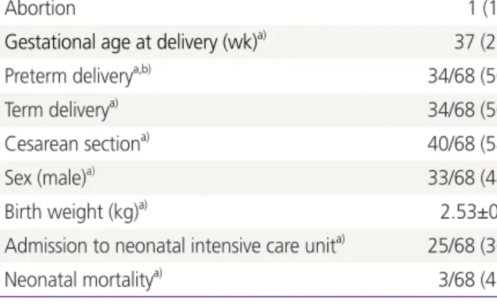
Table 3. Pregnancy outcome of patients who maintained the pregnancy (n=69)
Variable Value
Abortion 1 (1.4)
Gestational age at delivery (wk)a) 37 (25–40)
Preterm deliverya,b) 34/68 (50.0)
Term deliverya) 34/68 (50.0)
Cesarean sectiona) 40/68 (58.8)
Sex (male)a) 33/68 (48.5)
Birth weight (kg)a) 2.53±0.75
Admission to neonatal intensive care unita) 25/68 (36.8)
Neonatal mortalitya) 3/68 (4.4)
Data are expressed as number (%), median (range), or mean±standard deviation.
a)One case of abortion was excluded from the analysis; b)Both iatro- genic and spontaneous preterm delivery.
example, nausea and vomiting may be confused with morn- ing sickness and small breast masses may not be palpable due to breast engorgement during pregnancy. Also, headache and abdominal pain or discomfort are very common complaints during pregnancy. Therefore, it is difficult to differentiate these cancer symptoms from those of normal pregnancy, leading to delayed diagnosis. In our study, more than half of the GI cancers were diagnosed in the advanced stages and their prognosis was extremely poor. In contrast, all cervical and ovarian cancers were diagnosed in the early stages and their prognosis was good. This is thought to be due to the routine cervicovaginal cytology test and pelvic ultrasound included in the prenatal exams. Therefore, a physician should always be aware of the possibility of cancer in pregnant woman and initiate appropriate evaluations to diagnose the cancer in a timely manner.
Secondly, diagnostic tools available to detect cancer during pregnancy are limited and furthermore, both pregnant wom- en and physicians are, in many cases, reluctant to carry out such diagnostic tests. Ultrasound is considered safe for both mother and fetus [20], and it is the most commonly used test during pregnancy for diagnosing cancers such as breast and thyroid cancers. Magnetic resonance imaging is also safe for the fetus, but it is generally avoided during the first trimester [21]. X-ray exposure from a single diagnostic procedure, es- pecially exposure to less than 5 rad (50 mGy), does not result in any harm to the fetus [22,23]. However, high-dose ion- izing radiation tests, such as abdominal and pelvic computed tomography, should be avoided, especially during early preg- nancy [24]. Limited information is available regarding the fetal safety of nuclear medicine such as bone scans, thyroid scans and positron emission tomography scans. Upper GI endoscopy
Table 4. Comparisons of the stage and outcome among different cancer groupsBreast (n=20) GI
(n=17) Hematology
(n=13) Thyroid (n=11) CNS
(n=7) Cervix
(n=7) Ovary
(n=5) Lung
(n=3) Others
(n=4) P-value
Stagea) <0.001
I 4 (20.0) 3 (17.6) 2 (22.2) 10 (90.9) - 6 (85.7) 5 (100) 1 (33.3) 0 (0) -
II 9 (45.0) 3 (17.6) 3 (33.3) 1 (9.1) - 1 (14.3) 0 (0) 2 (66.7) 1 (25.0) -
III 5 (25.0) 3 (17.6) 2 (22.2) 0 (0) - 0 (0) 0 (0) 0 (0) 0 (0) -
IV 2 (10.0) 8 (47.1) 2 (22.2) 0 (0) - 0 (0) 0 (0) 0 (0) 3 (75.0) -
Cancer treatment strategy - 0.002
Termination of pregnancy
3 (15.0) 7 (41.2) 3 (23.1) 1 (9.1) 0 (0) 3 (42.9) 0 (0) 1 (33.3) 0 (0) - Treatment during
pregnancy 10 (50.0) 2 (11.8) 4 (30.8) 3 (27.3) 2 (28.6) 0 (0) 3 (60.0) 0 (0) 0 (0) -
Treatment after iatrogenic PTD
5 (25.0) 5 (29.4) 6 (46.2) 1 (9.1) 3 (42.9) 1 (14.3) 0 (0) 0 (0) 4 (100) - Delayed cancer
treatment until delivery
2 (10.0) 3 (17.6) 0 (0) 6 (54.5) 2 (28.6) 3 (42.9) 2 (40.0) 2 (66.8) 0 (0) -
PTDb,c,d) 6 (35.3) 6 (60.0) 9 (100) 1 (10.0) 5 (71.4) 1 (25.0) 1 (20.0) 1 (50) 4 (100) 0.001
Maternal outcomee) 0.005
CR and NED 13 (65.0) 8 (50.0) 8 (61.5) 10 (100) 0 (0) 6 (85.7) 4 (80.0) 1 (33.3) 2 (50.0) -
SD or PD 2 (10.0) 0 (0) 0 (0) 0 (0) 3 (50.0) 0 (0) 0 (0) 0 (0) 0 (0) -
Recurrence 0 (0) 0 (0) 1 (7.7) 0 (0) 0 (0) 0 (0) 0 (0) 0 (0) 0 (0) -
Death 5 (25.0) 8 (50.0) 4 (30.8) 0 (0) 3 (50.0) 1 (14.3) 1 (20.0) 2 (66.7) 2 (50.0) -
Data are expressed in number (%).
GI, gastrointestinal; CNS, central nervous system; PTD, preterm delivery; CR, complete response; NED, no evidence of disease; SD, stable dis- ease; PD, progressive disease.
a)Stage was unavailable for 11 cases (7 brain tumor cases and 4 leukemia cases); b)Only the patients who maintained the pregnancy were in- cluded in the analysis; c)Both iatrogenic and spontaneous preterm delivery; d)One case of abortion was excluded from the analysis; e)Three cases who were lost to follow up was excluded from the analysis.