Evaluation of bone healing in canine tibial defects filled with cortical autograft, commercial-DBM, calf fetal DBM, omentum and omentum-calf fetal DBM
7
0
0
전체 글
(2) 338 Amin Bigham-Sadegh et al.. xenogenic bovine fetal DBM, commercial DBM, omentum, omentum-DBM, cortical autograft and xenogenic cartilage powder on healing of an experimental bone defect in a dog model to determine the best material for bone healing. In the present study, cortical autograft and commercial DBM were used as positive standards to evaluate the effects of our home-made DBM and cartilage powder on bone healing procedures.. Materials and Methods Animals Seven male adult mongrel dogs that were 2 to 3 years old, 26.2 ± 2.5 kg and free of evident infectious or parasitic illnesses were used in this study. The experimental protocols were approved by the Animal Care and Experiment Committee of the University and were in accordance with the ethics standards of the Principles of Laboratory Animal Care. Preparation of calf fetal demineralized bone matrix DBM was prepared from the midshafts of the long bones of a 4-month-old Holstein calf fetus that were collected from a local slaughterhouse. The bones were collected aseptically, and the soft tissues were removed before o storage at −70 C. The bones were later cut into 1 cm pieces with a Stryker saw under saline (0.9% NaCl) o solution lavage, after which they were stored at −70 C until further use. The pieces were later thawed in 200-proof ethanol and air dried, after which they were milled (Universal Mill A-20; Tekmer, USA) and placed through a sieve to collect 2- to 4-mm pieces. Next, the pieces were decalcified in 0.6 mol/L HCL at 4ºC for 8 days under constant agitation. Demineralization was subsequently evaluated by radiography and calcium analysis [39]. Loss of density radiographically was used to subjectively evaluate demineralization. In addition, random samples of DBM were dried at 95ºC, weighed, and then ashed at o 600 C for 24 h. These samples were subsequently dissolved in 0.6 mol/L nitric acid and analyzed by atomic absorption spectrophotometry to determine the percent calcium per gram dry weight (% Ca: DW) [12,29]. Demineralization was considered adequate when samples were no longer visible radiographically and when the calcium content was less than 1% [38]. Following demineralization, all bone pieces were rinsed in sterile water and placed in phosphate buffer overnight. The bone pieces were then rinsed and the pH was adjusted to 7.3. Finally, the samples were placed in ethanol, the ethanol was allowed to evaporate overnight, and the pieces were o packaged aseptically and stored at 4 C. Preparation of bovine fetal cartilage powder Epiphyseal cartilage of the long bones of a 4-month-old. Holstein calf fetus were collected, washed three times in 95% ethanol for 15 min, rinsed in ether for 15 min, and finally air dried overnight. The cleaned and dried growth plate was then milled (Universal Mill A-20; Tekmer) to obtain 400∼700 μm granules, air dried and stored in o sterile plastic containers at 4 C until being used for implantation. The entire process was performed under sterile conditions (except for the milling) and a sample was cultured to demonstrate that the specimens contained no bacterial or fungal contamination.. Cortical autograft granules During tibial drilling to create defects, the protruding granules from beside the drill were collected for further use as autograft cortical granules. Omentum free graft For omental free graft preparation, the abdominal cavity was approached through a 3-cm ventral midline incision midway between the umbilicus and pelvic inlet, after which the free end of the greater omentum was located and exteriorized from the abdominal cavity. A 5 × 5-mm piece of the omentum was then isolated by two catgut ligatures and cut free from the remaining omentum. Surgical technique The dogs were sedated with acepromazine (0.05 mg/kg intrasubcuticularly) and anesthesia was induced with ketamine (10 mg/kg intravenously) and diazepam (0.25 mg/kg intravenously). The animals were then intubated and 1% to 2% halothane was used to maintain anesthesia with spontaneous breathing. During the procedure, saline (10 mL/kg per hour) was substituted through an intravenous catheter, and all operative procedures were conducted under general anesthesia. In all dogs, the left. Fig. 1. Seven bone defects were created for implantation of seven different biomaterials in tibial bone..
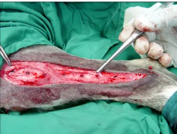
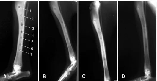
(3) Bone healing with various biomaterials in a canine model 339. hind limb from the stifle to the metatarsal region was prepared for aseptic surgery. The tibia was exposed via a medial approach and a circular bone defect of 4 mm in diameter was made (Fig. 1). Ostectomy was then performed with an electrical motor and seven 4-mm carbon burr under continuous irrigation with physiologic serum. Finally, the defects were filled with autograft, commercial DBM (Osteotech, USA), calf fetal DBM, omentum, omentum-calf fetal DBM and cartilage powder. The implanted site was changed between materials in each dog in a Latin square design.. Post operative evaluations Radiological evaluation: Lateral view radiographs were taken on the 1st day and then weeks 2, 4, 6 and 8 post injury using a step-wedge to calibrate the radiodensity. The radio-opacity of the implanted area was then scored using the range of 0 (minimally opaque) to 4 (most opaque) by an investigator blinded to treatment mode. Histopathological evaluation: Eight weeks after operation the dogs were euthanized for histopathological evaluation, which was carried out on all harvested specimens. Briefly, the left hind limb was harvested and dissected free of soft tissues. Sagital sections containing the defect were then cut with a slow speed saw, after which each slice was fixed in 10% neutral buffered formalin. The formalin-fixed bone samples were then decalcified in 15% buffered formic acid solution and processed for routine histological examination. Next, two 5 μm thick sections were cut from the centers of each specimen and stained with Hematoxylin and Eosin. Finally, the sections were blindly evaluated and scored by two pathologists according to Heiple’s scoring system [18] (Table 1).. Table 1. Lane and Sandhu histopathological scoring system* Union No evidence of union Fibrous union Osteochondral union Bone union Complete organization of shaft Cancellous bone No osseous cellular activity Early apposition of new bone Active apposition of new bone Reorganizing cancellous bone Complete reorganization of cancellous bone Cortical bone Non Early appearance Formation under way Mostly reorganized Completely formed Marrow None in resected area Beginning to appear Present in more than half of the defect Complete colonization by red marrow Mature fatty marrow Total points possible per category Distal union Cancellous bone Cortex Marrow Maximum score. 0 1 2 3 4 0 1 2 3 4 0 1 2 3 4 0 1 2 3 4 4 4 4 4 16. *Modified by Heiple et al. [18].. Fig. 2. Radiological evaluation on the 14th (A), 28th (B), 42nd (C) and 58th (D) postoperative days. 1: control, 2: autograft, 3: omentum, 4: omentum-calf fetal demineralized bone matrix (DBM), 5: commercial DBM, 6: calf fetal-DBM, 7: cartilage group..
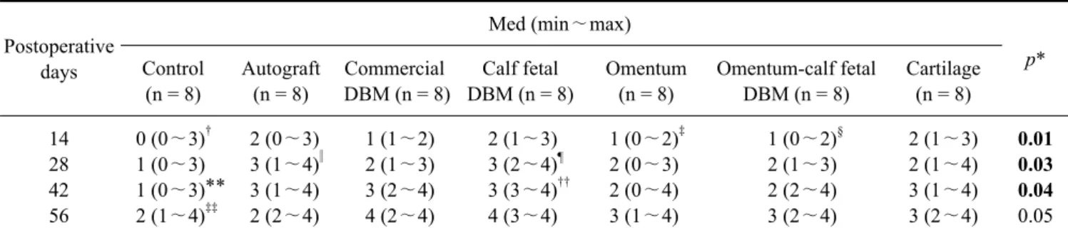
(4) 340 Amin Bigham-Sadegh et al.. Statistical analysis The radiological and histopathological data were compared by Kruskal-Wallis non- parametric ANOVA. When p values were found to be less than 0.05, pair wise group comparisons were performed by the Mann-Whitney U test (SPSS version 17 for windows; SPSS, USA).. Results There was no intraoperative and postoperative death during the study. None of the dogs sustained a fracture of the tibia.. Radiographic findings 14th postoperative day: On the 14th postoperative day, statistically significant differences (p < 0.05) were observed between the control group with autograft (p = 0.03), commercial DBM (p = 0.03), calf fetal DBM (p = 0.02) and cartilage (p = 0.01) groups, and the control group was significantly inferior to the other groups. Additionally,. the omentum group was significantly inferior to the autograft (p = 0.02), calf fetal DBM (p = 0.05) and cartilage (p = 0.03) groups. In addition, the omentum-calf fetal DBM was significantly inferior to the autograft (p = 0.02), calf fetal DBM (p = 0.03) and cartilage (p = 0.01) (Fig. 2, Table 2) groups. 28th postoperative day: Significant differences (p < 0.05) were observed on the 28th postoperative day, with the autograft group being significantly superior to the control (p = 0.03) and omentum (p = 0.05) groups. In addition, calf fetal DBM was significantly superior to the control group (p = 0.01) (Fig. 2, Table 2). 42nd postoperative day: On the 42nd postoperative day, statistically significant differences (p < 0.05) were observed, with the control group being significantly inferior to the autograft (p = 0.04), commercial DBM (p = 0.3), calf fetal DBM (p = 0.02), omentum (p = 0.05), omentum-calf fetal DBM (p = 0.05) and cartilage group (p = 0.04). At this stage, the calf fetal DBM group was significantly superior to the omentum (p = 0.01) and omentum-calf fetal DBM (p =. Table 2. Radiographical findings for bone healing at various post-operative intervals Postoperative days 14 28 42 56. Med (min∼max) Control (n = 8) 0 (0∼3)† 1 (0∼3) 1 (0∼3)** 2 (1∼4)‡‡. Autograft (n = 8) 2 (0∼3) 3 (1∼4)‖ 3 (1∼4) 2 (2∼4). Commercial Calf fetal DBM (n = 8) DBM (n = 8). Omentum (n = 8). 2 (1∼3) 3 (2∼4)¶ 3 (3∼4)†† 4 (3∼4). 1 (0∼2)‡ 2 (0∼3) 2 (0∼4) 3 (1∼4). 1 (1∼2) 2 (1∼3) 3 (2∼4) 4 (2∼4). Omentum-calf fetal DBM (n = 8) 1 (0∼2)§ 2 (1∼3) 2 (2∼4) 3 (2∼4). Cartilage (n = 8). p*. 2 (1∼3) 2 (1∼4) 3 (1∼4) 3 (2∼4). 0.01 0.03 0.04 0.05. Significant p values are presented in bold. *Kruskal-Wallis non-parametric ANOVA. †There were significant differences between the autograft (p = 0.03), commercial DBM (p = 0.03), calf fetal DBM (p = 0.02) and cartilage (p = 0.01) groups with the control group and the ‡ control group was significantly inferior to other groups. The lesion in the omentum implanted group was significantly inferior to those of the autograft (p = 0.02), calf fetal DBM (p = 0.05) and cartilage group (p = 0.03). §The omentum-calf fetal DBM implanted group was significantly ‖ inferior to those of the autograft (p = 0.02), calf fetal DBM (p = 0.03) and cartilage groups (p = 0.01). The autograft group was significantly superior to the control (p = 0.03) and omentum groups (p = 0.05). ¶The calf fetal DBM was significantly superior to the control group (p = 0.01). **The control group was significantly inferior to the autograft (p = 0.04), commercial DBM (p = 0.3), calf fetal DBM (p = 0.02), omentum †† (p = 0.05), omentum-calf fetal DBM (p = 0.05) and cartilage group (p = 0.04). The calf fetal DBM group was significantly superior to the ‡‡ omentum (p = 0.01) and omentum-calf fetal DBM (p = 0.03) groups. The control group was significantly inferior to the autograft (p = 0.04), commercial DBM (p = 0.3), calf fetal DBM (p = 0.02), and cartilage group (p = 0.03). DBM: demineralized bone matrix.. Table 3. Histopathological findings (sum of histopathological criteria) for bone healing at various groups Med (min∼max) Control (n = 8). Autograft (n = 8). Commercial Calf fetal Omentum Omentum-calf fetal Cartilage DBM (n = 8) DBM (n = 8) (n = 8) DBM (n = 8) (n = 8). Sum of histopathological 10 (8 ~ 12) 12 (12 ~ 13) 12 (11 ~ 13) 12 (11 ~ 13) 10 (8 ~ 12) criteria Significant p values are presented in bold. *Kruskal-Wallis non-parametric ANOVA.. 12 (10 ~ 13). p*. 11 (9∼12) 0.3.
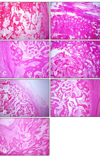
(5) Bone healing with various biomaterials in a canine model 341. 0.03) groups (Fig. 2, Table 2). 56th postoperative day: The control group was significantly inferior to the autograft (p = 0.04), commercial DBM (p = 0.3), calf fetal DBM (p = 0.02), and cartilage groups (p = 0.03) at the 56th postoperative day (Fig. 2, Table 2).. Histopathological findings There was no significant difference between the histopathological sections of the lesions of the animals of different groups in terms of bone healing criteria. None of the grafted materials elicited a significant inflammatory reaction. As shown in Fig. 3A-G, at 8 weeks post-surgery, histological examination demonstrated the presence of the regenerated bone with a typical structure of the trabecular bone in the defect site of all groups. Although the statistical analysis did not show any significant differences between. Fig. 3. Histopathological findings showed normal structure of trabecular bone in the defect area in all groups with various phases of remodeling. (A) Control. (B) Autograft. (C) Commercial-DBM. (D) Calf fetal-DBM. (E) Omentum. (F) Omentum-calf fetal DBM. (G) Cartilage group (H&E stain, ×10).. groups, the autograft, commercial-DBM and calf fetal DBM showed superior intense trabecular bone formation relative to the control, omentum and omentum-calf fetal DBM groups (Fig. 3, Table 3).. Discussion Autogenous bone still remains the “golden standard” of bone graft material in all facets of orthopedic surgery and is commonly used as a standard to which allografts and graft substitutes are compared [1,2,11,13,25,27]. In the present study, the autograft was found to be the best implant upon radiological evaluation. Additionally, histopathological evaluation showed that it had intensive proper thickened trabecular bone and did not elicit any inflammatory reaction. The bone inductive activity of the demineralized bone matrix (DBM) has been well established [33,34,37]. Additionally, the addition of the autologous bone marrow and/or autograft to DBM has been shown to provide an immediate source of osteogenic precursor cells at the implant site that is believed to provide an additional biochemical contribution to osteogenesis [6,34,37]. DBM also appears to support new bone formation through osteoconductive mechanisms [24]. The primary osteoinductive components of DBM are a series of low-molecular-weight glycoproteins that include bone morphogenetic proteins (BMPs). Decalcification of cortical bone exposes osteoinductive growth factors buried within the mineralized matrix, thereby enhancing the bone formation process [35]. Specifically, these proteins promote the chondroblastic differentiation of the mesenchymal cells, which is followed by new bone synthesis and endochondral osteogenesis [35,36]. In this study, the results of commercial-DBM and calf fetal DBM did not differ significantly from the autograft at 8 weeks post-injury. Overall, these results suggest that grafted xenogenic commercial-DBM and calf fetal DBM have osteoinductive activity, which may occur through the release of BMPs, similar to that of the autogenous cortical bone grafts. However, previous studies have shown that the cortical autograft has more osteoconductive properties and less osteoinductive activity than DBM material [4,21]. DBM also appears to support new bone formation through osteoconductive mechanisms [24]. No significant differences were found upon histopathological evaluation between the animals of different groups, and none of the graft material elicited a significant inflammatory reaction. It has been reported that the demineralization process destroys the antigenic materials in bone, making DBM less immunogenic than the mineralized allograft [17], and cortical autogenous bone graft does not induce an immunological reaction by the host [4], which was confirmed by the results of the present study..
(6) 342 Amin Bigham-Sadegh et al.. Omentum is considered a major source of supplying new vascularization into the implants [32]. Proper vascularization by omentum provides a valuable source of nutrients, oxygen, and angiogenic and growth factors, and creates a proper microenvironment for this graft implantation, further tissue maturation and bone formation [15,26]. A sufficient vascular flow effectively increases the oxygen concentration, and induces the production of osteoprogenitor cells from the perivascular mesenchymal cells [10,26]. During angiogenesis, the vascular endothelial growth factor (VEGF) increases capillary permeability, supplies hormones and growth factors [9] and maintains high levels of oxygen concentration, and all of these mechanisms [15] may have played a role in the ossification process of the grafted area of the animals investigated in the present study. Omentum and omentum-calf fetal DBM groups were found to be superior to the control group; however, the lesions of these two groups were inferior to those of the autograft, commercial-DBM and calf fetal DBM groups. These findings suggest that the osteoinductive and osteoconductive properties of the autograft, commercial-DBM and calf fetal DBM were more powerful than those of the omentum and omentum-calf fetal DBM. In the present study, it was expected that the omentum-calf fetal DBM would lead to better bone formation criterion, but its effects were not superior to those of the omentum group. Rather, the calf fetal DBM was covered with omentum, which obscured the proper action of the calf fetal DBM. It has been stated that the autogenous diced cartilage could act as a framework for deposition of bone in defects [16]. In the present study, the authors applied xenogenic processed cartilage as a graft material for bone healing and found that it led to satisfactory bone healing in comparison to the control, omentum and omentum-calf fetal DBM groups. These findings suggest that this technique has great potential for induction of bone formation in bone defects. Histopathological evaluation did not show any significant differences after 8 weeks in the present study even though statistical differences between groups were expected. There were likely earlier significant differences between the histological features of the lesions of different groups over the preceding postoperative intervals; however, histopathological evaluation was not conducted at earlier postoperative intervals because of limitations imposed by the ethics committee. Therefore, histopathological studies during the inflammatory, proliferative and remodeling phases of fracture healing as well as investigations of the type of the inflammatory cell constituents, osteoblasts proliferation and maturation, angiogenesis, collagen synthesis, presence or absence of cartilaginous materials, quantity and quality of mineralization and many other criteria are recommended. In the present study, the sites of material implantation. were changed in a chi square manner in the defected area, and each material was implanted in the same site at least once, but no differences related to the site of implantation were observed. Therefore, statistical analysis was not performed to identify differences related to the site. It is possible that the lack of significant differences in histopathological findings among materials may have been related to hole size (4 mm) in our study, and that large-sized holes (e.g., 10 mm in diameter) may show significant differences in histopathological findings. Based on the radiological findings of the present study, the autograft, commercial-DBM, calf fetal DBM and calf fetal cartilage demonstrated superior osteogenic potential in healing of the tibial bone defect in a dog model. The omentum and omentum-DBM groups were superior to the control group, but were inferior to the autograft, commercial-DBM, calf fetal DBM and calf fetal cartilage groups. The histological findings of the present study did not reveal any superior bone healing capability among groups at this stage.. Acknowledgments The authors would like to thank the authorities of the Veterinary School, Shahrekord University for their financial support and cooperation.. References 1. Alexander JW. Bone grafting. Vet Clin North Am Small Anim Pract 1987, 17, 811-819. 2. Alexander JW. Leonard’s Orthopedic Surgery of the Dog and Cat. 3rd ed. pp. 43-48, WB Sounders, Florida, 1985. 3. Arrington ED, Smith WJ, Chambers HG, Bucknell AL, Davino NA. Complications of iliac crest bone graft harvesting. Clin Orthop Relat Res 1996, 329, 300-309. 4. Bauer TW, Muschler GF. Bone graft materials: an overview of the basic science. Clin Orthop Relat Res 2000, 371, 10-27. 5. Bostrom MPG, Lane JM, Berberian WS, Missri AAE, Tomin E, Weiland A, Doty SB, Glaser D, Rosen VM. Immunolocalization and expression of bone morphogenetic proteins 2 and 4 in fracture healing. J Orthop Res 1995, 13, 357-367. 6. Burwell RG. The function of bone marrow in the incorporation of a bone graft. Clin Orthop Relat Res 1985, 200, 125-141. 7. Cook SD, Baffes GC, Wolfe MW, Sampath TK, Rueger DC. Recombinant human bone morphogenetic protein-7 induces healing in a canine long-bone segmental defect model. Clin Orthop Relat Res 1994, 301, 302-312. 8. Damien CJ, Parsons JR. Bone graft and bone graft substitutes: a review of current technology and applications. J Appl Biomater 1991, 2, 187-208. 9. Deckers MML, Karperien M, van der Bent C, Yamashita T, Papapoulos SE, Löwik CWGM. Expression of vascular endothelial growth factors and their receptors during osteoblast differentiation. Endocrinology 2000, 141,.
(7) Bone healing with various biomaterials in a canine model 343. 1667-1674. 10. Diaz-Flores L, Gutierrez R, Lopez-Alonso A, Gonzalez R, Varela H. Pericytes as a supplementary source of osteoblasts in periosteal osteogenesis. Clin Orthop 1992, 275, 280-286. 11. Fitch R, Kerwin S, Sinibaldi KR, Newman-Gage H. Bone autografts and allografts in dogs. Compend Contin Educ Pract Vet 1997, 19, 558-575. 12. Forell EB, Straw RC, Powers BE, Johnson J, Cooper MF, Withrow St J. Evaluation of the osteoinductive capacity of canine demineralized bone matrix in heterotopic muscle sites of athymic rats. Vet Comp Orthop Traumatol 1993, 1, 25-32. 13. Fox SM. Cancellous bone grafting in the dog: an overview. J Am Anim Hosp Assoc 1984, 20, 840-848. 14. Friedlaender GE. Bone grafts: the basic science rationale for clinical applications. J Bone Joint Surg Am 1987, 69, 786-790. 15. Glowacki J. Angiogenesis in fracture repair. Clin Orthop Relat Res 1998, 355 (Suppl), S82-89. 16. Gordon SD, Warren RF. Autogenous diced cartilage transplants to bone: an experimental study. Ann Surg 1947, 125, 237-240. 17. Guizzardi S, Di Silvestre M, Scandroglio R, Ruggeri A, Savini R. Implants of heterologous demineralized bone matrix for induction of posterior spinal fusion in rats. Spine (Phila Pa 1976) 1992, 17, 701-707. 18. Heiple KG, Goldberg VM, Powell AE, Bos GD, Zika JM. Biology of cancellous bone grafts. Orthop Clin North Am 1987, 18, 179-185. 19. Inoue K, Ohgushi H, Yoshikawa T, Okumura M, Sempuku T, Tamai S, Dohi Y. The effect of aging on bone formation in porous hydroxyapatite: biochemical and histological analysis. J Bone Miner Res 1997, 12, 989-994. 20. Jin DD. Bone matrix gelatin. Clinical application in 38 cases. Zhonghua Wai Ke Za Zhi 1991, 29, 312-314, 335. 21. Khan SN, Cammisa FPJ, Sandhu HS, Diwan AD, Girardi FP, Lane JM. The biology of bone grafting. J Am Acad Orthop Surg 2005, 13, 77-86. 22. Kirker-Head AC. Recombinant bone morphogenetic proteins: novel substances for enhancing bone healing. Vet Surg 1995, 24, 408-419. 23. Loredo GA, MacDonald MH, Benton HP. Regulation of glycosaminoglycan metabolism by bone morphogenetic protein-2 in equine cartilage explant cultures. Am J Vet Res 1995, 57, 554-559. 24. Martin GJ Jr, Boden SD, Titus L, Scarborough NL. New formulations of demineralized bone matrix as a more effective graft alternative in experimental posterolateral lumbar spine arthrodesis. Spine (Phila Pa 1976) 1999, 24, 637-645.. 25. McLaughlin RM, Roush JK. Autogenous cancellous and corticocancellous bone grafting. Vet Med 1998, 98, 10711074. 26. Moore MAS. Putting the neo into neoangiogenesis. J Clin Invest 2002, 109, 313-315. 27. Piermattei DL, Flo GL. Brinker, Piermattei, and Flo's Handbook of Small Animal Orthopedics and Fracture Repair. 3rd ed. pp. 147-153, WB Saunders, Philadelphia, 1997. 28. Reddi AH. Bone morphogenetic proteins, bone marrow stromal cells, and mesenchymal stem cells. Maureen Owen revisited. Clin Orthop Relat Res 1995, 313, 115-119. 29. Reddi AH, Huggins C. Biochemical sequences in the transformation of normal fibroblasts in adolescent rats. Proc Natl Acad Sci U S A 1972, 69, 1601-1605. 30. Riley EH, Lane JM, Urist MR, Lyons KM, Lieberman JR. Bone morphogenetic protein-2: biology and applications. Clin Orthop Relat Res 1996, 324, 39-46. 31. Tanaka T, Fujii K, Ohta M, Soshi S, Kitamura A, Murota K. Use of a guanidine extract of demineralized bone in the treatment of osteochondral defects of articular cartilage. J Orthop Res 1995, 13, 464-469. 32. Takada T, Kamei Y, Iwata T, Yokoi T, Torii S. Effect of omental lipid fraction on enhancement of skin flap survival. Ann Plast Surg 1998, 41, 70-74. 33. Tuli SM, Singh AD. The osteoinductive property of decalcified bone matrix: an experimental study. J Bone Joint Surg Br 1978, 60, 116-123. 34. Urist MR. Bone: formation by autoinduction. Science 1965, 150, 893-899. 35. Urist MR, Mikulski AJ, Lietz A. Solubilized and insolubilized bone morphogenetic protein. Proc Natl Acad Sci U S A 1979, 76, 1828-1832. 36. Urist MR, Sato K, Brownell AG, Malinin TI, Lietze A, Huo YK, Prolo DJ, Oklund S, Finerman GA, DeLange RJ. Human bone morphogenetic protein (hBMP). Proc Soc Exp Biol Med 1983, 173, 194-199. 37. Urist MR, Silverman BF, Büring K, Dubuc FL, Rosenberg JM. The bone induction principle. Clin Orthop Relat Res 1967, 53, 243-283. 38. Urist MR, Strates BS. Bone formation in implants of partially and wholly demineralized bone matrix: including observations on acetone-fixed intra and extracellular proteins. Clin Orthop Relat Res 1970, 71, 271-278. 39. Vail TB, Trotter GW, Powers BE. Equine demineralized bone matrix: relationship between particle size and osteoinduction. Vet Surg 1994, 23, 386-395. 40. Van Heest A, Swiontkowski M. Bone-graft substitutes. Lancet 1999, 353 (Suppl 1), S28-29..
(8)
수치
관련 문서
1 Division of Pulmonology & Allergy, Department of Internal Medicine, Departments of 2 Radiology, 3 Pathology, 4 Nuclear Medicine, Soonchunhyang University Bucheon
2 Departments of 1 Internal Medicine, 2 Pulmonary and Critical Care Medicine, 3 Radiology, Asan Medical Center, University of Ulsan College of Medicine, Seoul, 4
2 Department of Dermatology, Kangbuk Samsung Hospital, Sungkyunkwan University School of Medicine, Seoul, Korea. 3 Department of Orthopedic Surgery, Asan Medical Center,
Departments of Nephrology, 1 Pathology, 2 Plastic Surgery, Busan Paik Hospital, College of Medicine, Inje University, 3 Department of Internal Medicine, Bong Seng Hospital,
Departments of 1 Clinical Genetics, 2 Pediatrics, and 3 Pathology, Yonsei University College of Medicine, Seoul;. 4 Department of Pediatrics, Ajou University School of Medicine,
