© 2017 The Korean Ophthalmological Society
This is an Open Access article distributed under the terms of the Creative Commons Attribution Non-Commercial License (http://creativecommons.org/licenses /by-nc/3.0/) which permits unrestricted non-commercial use, distribution, and reproduction in any medium, provided the original work is properly cited.
Original Article
Analgesic Effect of Topical Sodium Diclofenac before Retinal Photocoagulation for Diabetic Retinopathy: A Randomized Double-
masked Placebo-controlled Intraindividual Crossover Clinical Trial
Alireza Ramezani1,2,3, Morteza Entezari2,3, Mohammad Mehdi Shahbazi2, Yosef Semnani4, Homayoun Nikkhah2,3, Mehdi Yaseri2,5
1Ophthalmic Epidemiology Research Center, Shahid Beheshti University of Medical Sciences, Tehran, Iran
2Ophthalmic Research Center, Shahid Beheshti University of Medical Sciences, Tehran, Iran
3Imam Hossein Medical Center, School of Medicine, Shahid Beheshti University of Medical Sciences, Tehran, Iran
4Department of Psychiatry, Imam Hossein Medical Center, Shahid Beheshti University of Medical Sciences, Tehran, Iran
5Department of Biostatistics and Epidemiology, Tehran University of Medical Sciences, Tehran, Iran
Purpose: To evaluate the analgesic effect of topical sodium diclofenac 0.1% before retinal laser photocoagula- tion for diabetic retinopathy.
Methods: Diabetic patients who were candidates for peripheral laser photocoagulation were included in a ran- domized, placebo-controlled, intraindividual, two-period, and crossover clinical trial. At the first session and based on randomization, one eye received topical sodium diclofenac 0.1% and the other eye received an ar- tificial tear drop (as placebo) three times before laser treatment. At the second session, eyes were given the alternate drug. Patients scored their pain using visual analogue scale (max, 10 cm) at both sessions. Patients and the surgeon were blinded to the drops given. Difference of pain level was the main outcome measure.
Results: A total of 200 eyes of 100 patients were enrolled. Both treatments were matched regarding the applied laser. Pain sensation based on visual analogue scale was 5.6 ± 3.0 in the treated group and 5.5 ± 3.0 in the control group. The calculated treatment effect was 0.15 (95% confidence interval, –0.27 to 0.58; p = 0.486).
The estimated period effect was 0.24 (p = 0.530) and the carryover effect was not significant (p = 0.283).
Conclusions: Pretreatment with topical sodium diclofenac 0.1% does not have any analgesic effect during pe- ripheral retinal laser photocoagulation in diabetic patients.
Key Words: Analgesic effect, Diabetic retinopathy, Laser photocoagulation, Topical sodium diclofenac
The annual incidence of new cases of proliferative dia- betic retinopathy (PDR) is 2.7% to 4% for patients with
type 1 diabetes and 0.6% to 3.2% for patients with type 2 diabetes [1]. Untreated high risk characteristic PDR results in a 33% risk of severe vision loss at 3 years [2]. This risk, however, is reduced by 50% by adequate panretinal laser photocoagulation (PRP) [2]. The procedure can be painful, leading to undertreatment in a large number of pain intol- erant patients [3].
Non-steroidal anti-inflammatory drugs (NSAIDs) are a
Received: June 4, 2016 Accepted: August 22, 2016
Corresponding Author: Alireza Ramezani, MD. Ophthalmic Research Center, Shahid Beheshti University of Medical Sciences, No. 23, Boostan 9 St., Pasdaran Ave., Tehran 16666, Iran. Tel: 98-21-22584733, Fax: 98-21- 22562138, E-mail: [email protected]
group of compounds used systemically or locally as anal- gesic, antipyretic, and anti-inflammatory agents. They have been used after cataract [4,5] and photorefractive sur- gery [6-8] as well as in patients with corneal abrasions [9].
Topical NSAID diclofenac was found to have greater anal- gesic action than topical betamethasone 0.1% in patients undergoing scleral buckling and vitrectomy [10]. There- fore, it might be preferred to use topical NSAIDs before performing PRP in order to diminish pain sensation during the procedure. This hypothesis has been investigated in a number of PRP or macular grid laser therapy cases and seems to be effective [11].
In this trial, we evaluated the analgesic effect of topical diclofenac sodium as a pretreatment drug for diabetic pa- tients undergoing PRP. Pain sensation, the main outcome measure of this study, is susceptible to subjectivity bias, and we therefore designed this placebo-controlled trial as an intraindividual, two-period, and crossover study.
Materials and Methods
This study was performed as a randomized, dou- ble-blinded, placebo-controlled, intraindividual, two-peri- od, and crossover clinical trial. After fully explaining the study protocol and its probable safety and efficacy, written informed consent was obtained from all patients. This clinical trial was approved by the review board/ethics committee of the Ophthalmic Research Center of Shahid Beheshti University of Medical Sciences.
All patients with severe non-PDR and early or high-risk characteristic PDR who were considered as candidates for bilateral PRP by a retina specialist were included in this trial. Patients with a history of intraocular surgery, periph- eral retinal laser therapy, or any eye disease that may inter- fere with the study protocol in either eye were excluded.
Vitreous hemorrhage or other media opacity precluding proper laser therapy in either eye was also used as exclu- sion criteria. A data sheet was completed for all included patients.
Enrolled patients were scheduled to receive three or four sessions of PRP. However, the study protocol was per- formed only for the first two sessions of treatment. For each person, randomization was performed before the first session. The right eye of each person was randomly as- signed to one group based on a randomized permuted
block with randomly selected block lengths of 4 and 6. The left eye was assigned to the other group. At the first session and based on randomization, one eye received topical sodi- um diclofenac ophthalmic solution 0.1% and the other eye topical artificial tear drop (as placebo) three times, 5 min- utes apart, at least 20 minutes before laser treatment. At the second session, the eyes were given the alternate drug with the same schedule. An argon laser was used with a wavelength of 532 nm, spot size of 200 μm, exposure time of 0.2 ms, and energy level of 100 to 500 mW.
In this study, pain level was scored using a visual ana- logue scale (VAS). It consisted of a 10 cm scale arranged from no pain to the worst pain imaginable. Markings of the subjects on the scale are translated to a number from 0 to 10. Before applying the laser, patients were instructed on how VAS works. Patients unable to cooperate with the VAS test were excluded.
Laser therapy was performed based on the Early Treat- ment Diabetic Retinopathy Study protocol, and started with the right eyes. Surgeons were encouraged to use near- ly the same power and amount of laser for both eyes at each stage and to apply the laser on the same quadrants of both eyes, avoiding the area of long ciliary nerves. At the end of laser treatment in each session, patients scored the pain in each eye.
Patients, the nurse who instilled the drops, and the sur- geons who performed the laser operation and asked about the pain were all blinded to the drops given. The difference of pain level was compared between the two drugs as the main outcome measure.
Statistical analysis
To describe data, we used the mean ± standard devia- tion, median (range), and frequency (%). To evaluate dif- ferences between the groups in each session, we used Mc- Nemar and Wilcoxon signed-rank tests. We evaluated the carryover, period (session), and treatment effect by means of a generalized linear mixed model. All statistical analy- ses were performed using SAS ver. 9.2 (SAS Institute Inc., Cary, NC, USA).
Results
A total of 200 eyes of 100 diabetic patients enrolled in
and completed the study. The mean age was 54 ± 10 years (range, 26 to 75) and 63% were female. The laser proce- dure data for each group are presented in Table 1. Groups were comparable regarding the mean number of laser ap- plications, mean energy level of laser, and frequency of the treated quadrants. The mean time interval between instil- lation of drops and laser procedure was 119 ± 55 minutes, with 114 ± 55 minutes for the first session and 124 ± 55 minutes for the second session.
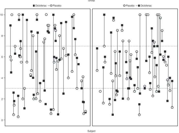
Pain sensation according to VAS at the first session was 5.9 ± 3.1 in the diclofenac group and 5.5 ± 3.0 in the place- bo group (Table 1). The corresponding values for the sec- ond session were 5.4 ± 2.8 and 5.5 ± 2.9. Fig. 1 demon- strates change in pain scores in each subject in every session. Collecting both sessions together, pain sensation was 5.6 ± 3.0 and 5.5 ± 3.0 based on VAS in the treated and control groups, respectively. In total, the calculated treat- ment effect was 0.15 (95% confidence interval [CI], –0.27 to 0.58; p = 0.486) and therefore, there was no significant difference between the treatments. Carryover effect was statistically insignificant (p = 0.283) and the estimated pe- riod effect was 0.24 (95% CI, –0.50 to 0.98; p = 0.530).
Discussion
This trial demonstrated that pretreatment with topical sodium diclofenac ophthalmic solution 0.1% did not dimin- ish pain during PRP in diabetic patients. Pain experienced during PRP is very variable but appears to be more com- mon with retreatment and with treatment applied anterior to the equator, especially in the 3, 6, 9, and 12 o’clock posi- tions (corresponding to the location of the long ciliary nerves) [3]. As suggested by various authors, there are sev- eral ways of mitigating this pain. Some patients respond to pretreatment oral analgesia. Others may even require local anesthetic blockade or general anesthesia in order to toler- ate treatment. However, these interventions carry the risk of side effects and require additional monitoring of the pa- tient [3]. Inhaled Entonox was also evaluated as an analge- sic during PRP in a randomized, crossover, and dou- ble-blinded pilot study. Mean pain scores, based on VAS, from Entonox treatment compared to air inhalation (as a placebo) were 2.94 ± 2.73 vs. 3.73 ± 3.20, respectively (p <
0.03). The investigators concluded that Entonox can be used as a safe and effective analgesic agent during PRP Table 1. Laser characteristics and pain level of the two groups in each session separately and in both sessions together First sessionSecond sessionBoth sessions
Diclofenac (n = 100) Placebo (n = 100)
p-value*
Diclofenac (n = 100) Placebo (n = 100)
p-value
Diclofenac (n = 200) Placebo (n = 200)
p-value† No. of applications
361 ± 66 (200–510) 372 ± 58 (204–528)
0.098
377 ± 89 (200–720) 374 ± 80 (200–600)
0.792
368 ± 77 (3–720) 373 ± 68 (200–600)
0.331 Energy level (mW)
372 ± 164 (100–900) 372 ± 153 (100–900)
0.673
376 ± 168 (140–900) 364 ± 162 (102–800)
0.648
374 ± 165 (14–900) 368 ± 157 (100–900)
0.485 Time interval (min)‡
115 ± 56 (20–310) 113 ± 54 (15–240)
0.571
124 ± 55 (40–270) 123 ± 56 (40–260)
0.842
120 ± 56 (20–310) 118 ± 55 (15–260)
0.241 Treated quadrants (%)0.6490.3060.428 ST161813141516 SN252426302527 IT 6 751472625 IN535010 93432 Pain score
5.9 ± 3.1 (0.3–1
1.5)
5.5 ± 3.0 (0.3–1
1.5)0.215
5.4 ± 2.8 (0.5–1
1.8)
5.5 ± 2.9 (0.3–1
1.6)0.918
5.6 ± 3.0 (0.3–1
1.8)
5.5 ± 3.0 (0.3–1 1.6)0.486 Values are presented as mean ± standard deviation (range). ST = supratemporal; SN = supranasal; IT = infratemporal; IN = infranasal. * Based on McNemar and Wilcoxon signed-rank test;† Based on generalized linear mixed model; ‡ Times between drop administration and laser applications.
treatment [3]. The feasibility of manual acupuncture for reducing pain during PRP treatment was also investigated in a prospective, comparative nonrandomized study and was found to be helpful [12]. In a recent published random- ized controlled trial, single spot short duration time (20 ms) was compared with conventional laser therapy as an- other option for diabetic retinopathy treatment. The inves- tigators found that short pulse laser was significantly less painful but just as effective as conventional laser during 6 months of follow-up [13].
The analgesic effect of NSAIDs is believed to apply through inhibition of the arachidonic acid cascade. This cascade divides into the cyclo-oxygenase and the lipo-oxy- genase pathways. The main products of the cyclo-oxygen- ase pathway are prostaglandins that take part in maintain- ing and amplifying the cellular and humoral phases of the inflammatory response. During an inflammatory response, several mediators are released that stimulate pain produc-
ing nerve fibers [11].
Sodium diclofenac 0.1% belongs to the phenylacetic acid chemical class and appears to have a dual effect on both cyclo-oxygenase and lipo-oxygenase pathways [14]. It is believed that it acts on the posterior segment of the eye ei- ther by diffusion into the vitreous from the aqueous or by
“desensitization” of the entire distribution of the trigemi- nal nerve fibers around the globe [11].
The analgesic effect of topical sodium diclofenac 0.1%
during PRP and macular laser photocoagulation was eval- uated in a prospective, double-blinded, crossover, random- ized, and clinical study [11]. In this report, the study popu- lation included 87 patients, 45 with PDR treated with PRP (group A) and 42 with non-PDR and clinically significant macular edema (group B) who received grid treatment of the posterior pole. Treatment of the posterior pole was as- sociated with no, mild, or negligible pain and this was at- tributed to the lower power levels used for grid photocoag-
10
8
6
4
2
0
Group
Subject
█ Diclofenac → O Placebo O Placebo → █ Diclofenac
Fig. 1. Drop lines showing the changes in pain scores in each subject for the two treatment groups in every session, separately.
ulation. However, they found a statistically significant effect (p = 0.01 by paired t-test) in group A by using topi- cal diclofenac. It should be noted that even in this group, nine patients out of 45 had more pain when using di- clofenac than using sodium chloride and seven patients had similar levels of pain using both treatments. Neverthe- less, the mean reported level of pain was 44.2% when so- dium diclofenac 0.1% drops were used and 53.1% when so- dium chloride 0.9% drops were used. Although the difference between the groups was statistically significant, the reported level of pain sensation did not differ signifi- cantly from the clinical point of view. The mean pain score in our study was similar to the sodium chloride group of this study. It was 5.4 in the treated group and 5.3 in the control group, and we found no significant difference be- tween the groups. The difference between two studies might be due to the different times of drop installations before PRP and the various characteristics of laser applica- tion.
In another randomized, double-masked, and place- bo-controlled clinical trial, the authors compared oral di- clofenac, topical diclofenac, and placebo in pain reduction during PRP and concluded that a single dose of oral di- clofenac was an effective pretreatment analgesic agent for reducing pain experienced during PRP for PDR. They found a significantly lower pain level in patients receiving pretreatment of oral diclofenac compared to the controls.
However, the difference between topical diclofenac and placebo was not significant in univariate analysis [15]. Al- though this latter result was comparable to our result, mul- tivariate regression analysis for age, gender, and total laser energy in their study demonstrated a significantly lower pain level for topical diclofenac versus placebo [15].
Despite the above reported beneficial effect of the topi- cal sodium diclofenac 0.1% during PRP, there is another study with a conclusion similar to our results. In this trial, the investigators evaluated the effect of topical ketorolac 0.5%, another topical NASAID, for ocular pain relief during PRP [16]. It was a prospective, randomized, dou- ble-blinded, and controlled study with 60 eyes of 30 con- secutive PDR patients. One hour before laser treatment, ketorolac tromethamine 0.5% was instilled to one eye and artificial tear drop to the fellow eye. Directly after treat- ment, patients were asked for the severity of pain in both eyes using VAS. Mean pain level for placebo-instilled eyes was 4.8 and 4.4 for ketorolac-instilled eyes. There was no
significant difference for pain levels between the groups (p
= 0.29). The conclusion of this study was parallel to ours, but instead using a different type of topical NSAID.
In the present trial, the mean time interval between in- stillation of the drops and laser treatment was about 115 minutes. Although this waiting time would not be conve- nient for daily practice, it was an appropriate interval in or- der to achieve near maximum analgesic effect of topical so- dium diclofenac 0.1%. The highest average concentration of this drug to be found in the aqueous humor was 82 ng/mL at 2.4 hours after instillation [17]. This time interval was 68.3 minutes in the study using diclofenac [11] and 1 hour in the trial using ketorolac 0.5% [16].
Our study was sufficiently powered with 100 cases, 200 eyes, and 400 interventions. Performing a randomized double-masked placebo-controlled intraindividual cross- over clinical trial enabled us to overcome many biases and limitations as the subjective-dependent main outcome of this study, which was pain sensation. Nonetheless, we used the VAS test, which has been found to be correlative and reproducible [18]. It is an acceptable and widely used meth- od for measuring pain sensation. Additionally, surgeons were encouraged to try to apply the laser on the same quadrants of both eyes in each session since the level of pain sensation may vary based on different quadrants re- ceiving the laser.
In summary, we report that using topical diclofenac so- dium as an analgesic before PRP in patients with diabetic retinopathy was not helpful in pain reduction during the procedure. As previously reported, the drug is supposed to act on the posterior segment of the eye either by diffusion into the vitreous from the aqueous or by “desensitization”
of the entire trigeminal nerve fibers around the globe [11].
However, we did not observe any effect using this type of intervention in our study. We believe that the concentra- tion of the drug around the corresponding nerves with the aforementioned method of administration would not be enough to diminish pain level during a relatively painful procedure like PRP. Further studies evaluating other mo- dalities and routes of administration are warranted.
Conflict of Interest
No potential conflict of interest relevant to this article was reported.
References
1. Klein R, Klein BE, Moss SE, Cruickshanks KJ. The Wis- consin Epidemiologic Study of diabetic retinopathy. XIV:
ten-year incidence and progression of diabetic retinopathy.
Arch Ophthalmol 1994;112:1217-28.
2. Photocoagulation treatment of proliferative diabetic reti- nopathy: clinical application of Diabetic Retinopathy Study (DRS) findings, DRS report number 8. The Diabetic Reti- nopathy Study Research Group. Ophthalmology 1981;88:583- 600.
3. Cook HL, Newsom RS, Mensah E, et al. Entonox as an an- algesic agent during panretinal photocoagulation. Br J Ophthalmol 2002;86:1107-8.
4. Fry LL. Efficacy of diclofenac sodium solution in reducing discomfort after cataract surgery. J Cataract Refract Surg 1995;21:187-90.
5. Herbort CP, Jauch A, Othenin-Girard P, et al. Diclofenac drops to treat inflammation after cataract surgery. Acta Ophthalmol Scand 2000;78:421-4.
6. Epstein RL, Laurence EP. Effect of topical diclofenac solu- tion on discomfort after radial keratotomy. J Cataract Re- fract Surg 1994;20:378-80.
7. Sher NA, Golben MR, Bond W, et al. Topical bromfenac 0.09% vs. ketorolac 0.4% for the control of pain, photopho- bia, and discomfort following PRK. J Refract Surg 2009;25:214-20.
8. Durrie DS, Kennard MG, Boghossian AJ. Effects of non- steroidal ophthalmic drops on epithelial healing and pain in patients undergoing bilateral photorefractive keratectomy (PRK). Adv Ther 2007;24:1278-85.
9. Salz JJ, Reader AL 3rd, Schwartz LJ, Van Le K. Treatment of corneal abrasions with soft contact lenses and topical di- clofenac. J Refract Corneal Surg 1994;10:640-6.
10. Lesnoni G, Coppe AM, Manni G, et al. Analgesic effect of topical diclofenac versus betamethasone after posterior segment surgery. Retina 1995;15:34-6.
11. Weinberger D, Ron Y, Lichter H, et al. Analgesic effect of topical sodium diclofenac 0.1% drops during retinal laser photocoagulation. Br J Ophthalmol 2000;84:135-7.
12. Chiu HH, Wu PC. Manual acupuncture for relieving pain associated with panretinal photocoagulation. J Altern Complement Med 2011;17:915-21.
13. Mirshahi A, Lashay A, Roozbahani M, et al. Pain score of patients undergoing single spot, short pulse laser versus conventional laser for diabetic retinopathy. Graefes Arch Clin Exp Ophthalmol 2013;251:1103-7.
14. Ku EC, Lee W, Kothari HV, Scholer DW. Effect of di- clofenac sodium on the arachidonic acid cascade. Am J Med 1986;80:18-23.
15. Zakrzewski PA, O’Donnell HL, Lam WC. Oral versus top- ical diclofenac for pain prevention during panretinal photo- coagulation. Ophthalmology 2009;116:1168-74.
16. Esgin H, Samut HS. Topical ketorolac 0.5% for ocular pain relief during scatter laser photocoagulation with 532 nm green laser. J Ocul Pharmacol Ther 2006;22:460-4.
17. Ellis PP, Pfoff DS, Bloedow DC, Riegel M. Intraocular di- clofenac and flurbiprofen concentrations in human aqueous humor following topical application. J Ocul Pharmacol 1994;10:677-82.
18. Scott J, Huskisson EC. Graphic representation of pain.
Pain 1976;2:175-84.