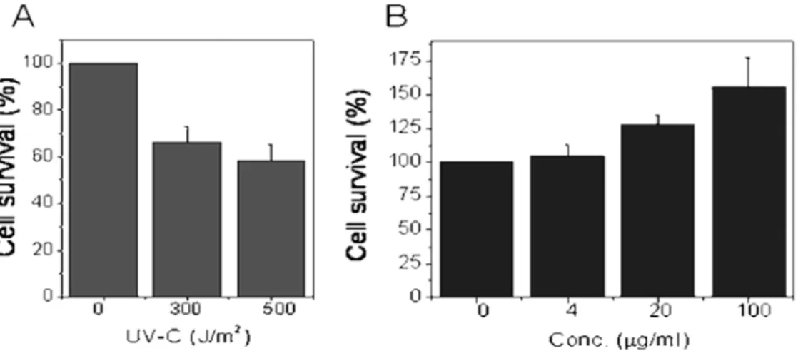
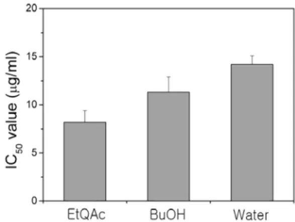
Betula Platyphylla var. Japonica Extract Prevent Ultraviolet C Light-induced Cell Damage in Chinese Hamster Fibroblast (V79-4) Cells
5
0
0
전체 글
(2)
(3)
(4)
(5)
수치
관련 문서
