© Copyright
Keimyung University School of Medicine 2015 Received: April 27, 2015
Accepted: May 19, 2015
Corresponding Author: Sung Ken Yu, M.D., Division of Pulmonary and Critical Care Medicine, Department of Internal Medicine,
Daegu Fatima Hospital,
99 Ayang-ro, Dong-gu, Daegu 701-724, Korea Tel: +82-53-940-7411
E-mail: [email protected]
• The authors report no conflict of interest in this work.
A 77-year-old male patient was hospitalized due to dyspnea and cough. At chest auscultation, Rhonchi was heard from both lung fields. The chest computed tomography (CT) observed nodular lesions within mid-trachea. Bronchoscope observed salient mass from the membranous portion in the mid-trachea, and after taking a biopsy, it was diagnosed as hamartoma. Tracheal hamartoma is a rare benign tumor of lung. Similar way to the endoscopic mucosal resection (EMR), we did endoscopic resection of tracheal hamartoma. We report a case of tracheal hamartoma treated with Endoscopic mucosal resection via flexible bronchoscopy.
Key Words :
Bronchoscopy, Endoscopic mucosal resection, Tracheal hamartomaIntroduction
Pulmonary hamartomas are benign lung tumors, with an incidence between 0.25% and 0.32% according to different necreopsy studies [1,2]. From a previous paper reviwing a total of 215 cases of hamartoma reported in the literature, the endobronchial location was found in only 1.4% of cases [3]. In contrast, other studies found an incidence of endobronchial location in 10 and 20% of all pulmonary hamartoma [4,5]. Pulmonary hamartoma is most common among benign tumors in the lung. It is observed in the lung parenchyme for the most cases, but it is rarely observed in trachea.
Several techniques are available for the bronchoscopic treatment of obstructing tissue in the tracheobronchial tree, including electrosurgical snaring, laser therapy, cryosurgery, airway stents, Division of Pulmonary and Critical Care Medicine, Department of Internal Medicine,
Daegu Fatima Hospital, Daegu, Korea
Ji Na Kim, M.D., Bi Na Jeong, M.D., Sung Ken Yu, M.D., Yeon Jae Kim, M.D., Byung Ki Lee, M.D.
A Case of Tracheal Hamartoma Treated with Endoscopic Mucosal
Resection
brachytherapy, and balloon dilation [6,7].
Endoscopic mucosal resection (EMR) is widely used for gastrointestinal tumors, but rarely for endobronchial lesions.
We report a case of tracheal hamartoma treated with endoscopic mucosal resection (EMR) in a 77-y e a r-o l d m a l e p r e s e n t i n g a s a n a c u t e exacerbation of chronic airway disease.
Case Report
A 77-year-old male patient was hospitalized due to dyspnea. The patient had complained about shortness of breath at work and coughs since 1 month ago and got worse 1 day before his hospitalization. He had a history of hypertension and cerebral infarction. At chest auscultation, Rhonchi was heard from both lung fields. The symptoms improved with conservative treatment, but he continued to complain about being uncomfortable in upper chest when breathing, and the chest computed tomography (CT) observed nodular lesions within mid-trachea (Fig. 1).
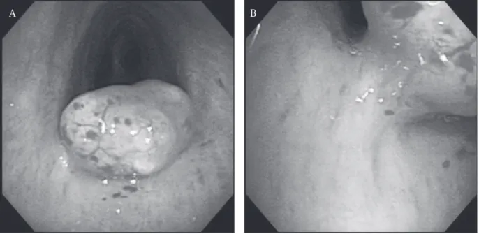
Bronchoscope observed salient mass from the membranous portion in the mid-trachea (Fig. 2), and after taking a biopsy, it was diagnosed as hamartoma. Tumor resection is considered to be available and the hamartoma was removed by endoscopic mucosal resection (EMR) via flexible bronchoscopy.
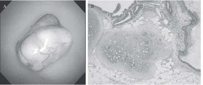
Similar way to the endoscopic mucosal resection (EMR), we did endoscopic resection of tracheal hamartoma. First, submucosal injection was done to separate mucosal and submucosal lesions from the muscularis propria. Once the lesion has been lifted away from the muscularis propria, it is resected by electrosurgical snaring and removed by forcep (Fig. 3). The size of removed mass was 0.8X1X1.3 cm. This mass consisted of
cartilage and adipose tissue (Fig. 4).
After the procedure, there were no complications and his respiratory symptoms improved and he could be discharged from the hospital. Repeat chest CT scan done after three weeks showed normal airway (Fig. 5).
Discussion
Pulmonary hamartoma is observed in the lung parenchyme for the most cases , but it is rarely observed in trachea. Clinically most parenchymal hamartomas are asymptomatic. But endobronchial hamartomas present usually with symptoms of airway obstruction with wheezing and stridor.
Many cases have been misdiagnosed as asthma until the lesions were identified at bronchoscopy or at Chest CT scan [3,8-9]. So if localized wheezing or stridor are long-standing and uncontrolled symptoms of chronic pulmonary disease, Chest CT scan or bronchoscopy is necessary.
In this case, the patient was hospitalized with acute exacerbation of chronic pulmonary disease.
In spite of conservative treatment, his symptoms Fig. 1. Chest CT scan showed a mid-tracheal polypoid mass (arrow).
persisted, so we did Chest CT scan. At the Chest CT scan, Nodular lesion was observed in mid- trachea. This nodular lesion was dignosed as a hamartoma by bronchoscopy and biopsy.
Histologically the mesenchymal components of the endobronchial hamartomas are highly varied.
There is predominance of adipose tissue over other
mesenchymal components. They have also been found to contain cartilage, myxomatous connective tissue, smooth muscle and epithelial structures [10].
In this case, mass was consisted of cartilage and adipose tissue.
The traditional surgical treatment (thoracotomy and bronchotomy) is currently indicated only in cases where the Fig. 2. Flexible bronchoscopy showed a tracheal mass with smooth surface attached on the mucosa of left lateral wall.
A B
Fig. 3. An endoscopic mucosal resection (EMR) was done. (A) Epinephrine/normal saline solution (1:10000) fluid (2 cc) was injected into the stalk. (B) Resection with snare was done. (C) After EMR, there was no bleeding on pale mucosa at resection site.
A B C
endobronchial hamartoma cannot be approached through endoscopy, or when lung resection is indicated due to irreverible parenchymal damage from long standing airway obstruction [11,12]. Recently, bronchoscopic intervention has been shown to be a safe and effective tool for the treatment of a benign tracheobronchial tumor [13]. Many diffent interventional bronchoscopic techniques has been used, such as resection by forceps, Argon plasma coagulation, Nd-YAG laser, cryosurgery or
electrosurgical snaring [6,7]. Electrocautery seems more cost-effective than the Nd-YAG laser for palliative bronchoscopic inter-vention to debulk intraluminal tumor in patients with NSCLC.
Electrocautery equipment is less expensive, the application technique is simple, and it is more easily accessible for emergency use in most hospitals [14]. In this case, the tracheal hamartoma was removed by fibroptic broncho-scopy electrosurgial snaring.
There are differences between bronchoscopic and gastrointestinal endoscopic tumor removal by snare. Endoscopic mucosal resection (EMR) and endoscopic submucosal dissection (ESD) were developed for minimally invasive, organ-sparing endoscopic removal of benign and early malignant lesions in the gastrointestinal tract. Endoscopic mucosal resection (EMR) is typically used for removal of lesions smaller than 2 cm or piecemeal removal of larger lesions. On gastrointestinal endoscopy, it is possible that the tumor base can be separated by injection of a solution into the submucosal space unrer the lesions, so complete Fig. 4. (A) Gross finding shows a yellowish white colored hard tissue measuring 1.3×1×0.8 cm. (B) Microscopic finding shows a normal epithelial lining with submucosal pathology composed of cartilage and adipose tissue (H&E stain).
A B
Fig. 5. After bronchoscopic resection, chest CT scan was noramal in trachea.
removal of a tumor including the base can be accomplished [15]. The tracheal hamartoma having a narrow based stalk that can be removed using a electrosurgical snaring. But we removed the tracheal hamartoma in the same way as the endoscopic mucosal resection (EMR) for complete removal of tumor including the base.
Given that tracheal hamartoma in an elderly patient was removed completely with endoscopic m u c o s a l r e s e c t i o n (E M R) u s i n g f l e x i b l e bronchoscope without no complications like bleeding and that general anesthesia is not necessary and the procedure time and recovery period after the procedure is so fast, this case is considered to be better attempted for endobron- chial benign tumors, and further researches and attempts on intervention using flexible broncho- scope for the treatment of intra-airway benign tumors are necessary.
References
1. Mcdonald JR, Hrrington SW, Clagett OT. Hamartoma (often called chondroma) of the lung. J Thorac Surg 1945;14:128-43.
2. Murray J, Kielkowski D, Leiman G. The prevalence and age distribution of peripheral pulmonary hamartomas in adult males: an autopsy-based study. S Afr Med J 1991;79:247-9.
3. Gjevre JA, Myers JL, Prakash UB. Pulmonary hamartomas. Mayo Clin Proc 1996;71:14-20.
4. Sibala JL. Endobronchial hamartomas. Chest 1972;62:631-4.
5. Tomashefski JF Jr. Benign endobronchial mesenchymal tumors: their relationship to parenchymal pulmonary hamartomas. Am J Surg Pathol 1982;6:531-40.
6. Bolliger CT, Mathur PN, Beamis JF, Becker HD,
Cavaliere S, Colt H, et al. ERS/ATS statement on interventional pulmonology. European Respiratory Society/American Thoracic Society. Eur Respir J 2002;19:356-73.
7. Ernst A, Silvestri GA, Johnstone D, American College of Chest Physicians. Interventional pulmonary procedures:
Guidelines from the American College of Chest Physicians. Chest 2003;123:1693-717.
8. Ortiz-Saracho J, Picher J, Garcia-Rull S, Reboiras SD, Perez I. Endobronchial hamartoma resected by rigid bronchoscope. Eur J Cardiothorac Surg 1993;7:445-6.
9. Sahin AA, Aydiner A, Kalyoncu F, Tokgozoglu L, Baris YI. Endobronchial hamartoma removed by rigid bronchoscope. Eur Respir J 1989;2:479-80.
10. van den Bosch JM, Wagenaar SS, Corrin B, Elbers JR, Knaepen PJ, Westermann CJ. Mesenchymoma of the lung (so called hamartoma): a review of 154 parenchymal and endobronchial cases. Thorax 1987;42:790-3.
11. Cheu HW, Grishkin BA, Linville WK. Endobronchial hamartoma treated by bronchoscopic excision. South Med J 1993;86:1164-5.
12. Na W, Shinn SH, Paik SS. Dumbbell shaped exophytic and endobronchial lipomatous hamartoma. Thorac Cardiovasc Surg 2009;57;122-4.
13. Kim SA, Um SW, Song JU, Jeon K, Koh WJ, Suh GY, et al. Bronchoscopic features and bronchoscopic intervention for endobronchial hamartoma. Respirology 2010;15:150-4.
14. Boxem T, Muller M, Venmans B, Postmus P, Sutedja T.
Nd-YAG laser vs bronchoscopic electrocautery for palliation of symptomatic airway obstruction: a cost- effectiveness study. Chest 1999;116:1108-12.
15. Asge Technology Committee, Kantsevoy SV, Adler DG, Conway JD, Diehl DL, Farraye FA, et al.
Endoscopic mucosal resection and endoscopic submucosal dissection. Gastrointest Endosc 2008;68:11-8.