Review https://doi.org/10.14478/ace.2017.1003
방사성 폐기물의 생물정화를 위한
극한세균 데이노코쿠스 라디오두란스의 연구적 고찰
정선욱⋅최용준†
서울시립대학교 환경공학부
(2017년 1월 10일 접수, 2017년 1월 18일 심사, 2017년 1월 31일 채택)
Research Perspective of an Extremophilic Bacterium,
Deinococcus radiodurans on Bioremediation of Radioactive Wastes
Sun-Wook Jeong and Yong Jun Choi†
School of Environmental Engineering, University of Seoul, 163, Seoulsiripdae-ro, Dongdaemun-gu, Seoul 02504, Republic of Korea (Received January 10, 2017; Revised January 18, 2017; Accepted January 31, 2017)
초 록
방사성 폐기물에 대한 우려가 증대됨에 따라 생물정화 기술에 대한 관심이 고조되고 있다. 병원과 원자력발전 등에서 발생되는 많은 양의 방사성 폐기물이 환경에 직접 노출됨에 따라, 이를 정화하기 위한 다양한 물리화학적 기술이 보 고되고 있다. 하지만, 이러한 방법은 고비용 및 고위험성 과정이 수반되기 때문에 미생물을 이용한 친환경적 생물정화 기술이 요구되고 있다. 최근, 고방사선 노출 등과 같은 극한환경에서 서식할 수 있는 방사선저항성 미생물에 대한 연구가 많이 보고되고 있으며, 이를 이용한 방사성 폐기물 정화에 대한 연구적 관심이 높아지고 있다. 데이노코쿠스 라디오두란스는 대표적인 방사선저항성 미생물로써 높은 방사선에 저항성을 갖는 특성으로 인해 방사성 폐기물 등의 유해물질 정화에 이용될 수 있다. 본 총설에서는 데이노코쿠스 라디오두란스의 방사선내성과 관련한 일반적 기작에 대해 소개하고, 방사성 폐기물의 생물정화 활용 가능성에 대해 논의한다.
Abstract
Increasing concerns on radioactive wastes have drawn much attention on the development of remediation technologies.
Massive amounts of radioactive wastes generated from hospital and nuclear power plants were exposed to our environment.
Although physicochemical removal methods were developed, an eco-friendly remediation method has not yet been demonstrated. Recently, an extremophilic bacterium has received much attention due to their extraordinary characteristics.
Among them, Deinococcus radiodurans (D. radiodurans) strain was regarded as the best host organism for the removal of radioactive heavy metals and radionuclides, because of their superb characteristics like tolerance against the high level of radioactivity. In this article, we briefly introduced the extraordinary nature of D. radiodurans and also discussed the potential use of D. radiodurans strain for the removal of radioactive wastes.
Keywords: Deinococcus radiodurans, radioactive waste, bioremediation, antioxidation, DNA repair
1. Introduction
1)
Over the past few years, radiation and radioactive materials are re- garded as an indispensable source to human beings on daily life such as medical application, academic research, and energy generation. The X-ray cinefluography and radiation therapy are commonly applied to patients to diagnose medical treatment condition. And also, huge amount of electricity is generated by nuclear power plant by using ra-
† Corresponding Author: University of Seoul,
School of Environmental Engineering, 163, Seoulsiripdae-ro, Dongdaemun-gu, Seoul 02504, Republic of Korea
Tel: +82-2-6490-2873 e-mail: [email protected]
pISSN: 1225-0112 eISSN: 2288-4505 @ 2017 The Korean Society of Industrial and Engineering Chemistry. All rights reserved.
dioactive materials such as uranium as an energy source. In spite of those of advantages, concerns on the radioactive substance and waste has been gradually increased due to the threat of radioactivity to public and environment as appears by serious accidental events. The huge amount of radioactive materials is leaked to environment after the cata- strophic nuclear accidents such as the Chernobyl disaster and the Fukushima Daiichi nuclear disaster[1,2]. Not all radioactivity comes from disastrous accidents. The tremendous amount of radioactive waste which is believed to trigger development of cancer and chronic disease also has occurred from biomedical and industrial applications.
However, despite the risk of radioactivity, a definite answer for re- mediation method of radioactive polluted environment has not yet been addressed.
Radionuclides Organism Mechanism Reference
Uranium
Deinococcus radiodurans
Bioprecipitation [73]
Bioaccumulation [74]
Bioprecipitation [59]
Desulfovibrio desulfuricans G20 Bioreduction [75]
Desulfovibrio desulfuricans Bioreduction [76]
Sphingomonas sp. BSAR-1 Bioprecipitation [77]
Thermoanaerobacter sp. TOR-39 Bioreduction [78]
Thermoterrabacterium ferrireducens Bioreduction [79]
Citrobacter sp. Biomineralisation [80]
Cobalt
Rhodopseudomonas palustris CGA0009
Bioreduction [9]
Novosphingobium aromaticivorans F-199
Deinococcus radiodurans [62]
Neptunium(V) Shewanella putrefaciens
Bioreduction [82]
Citrobacter sp.
Technetium
Desulfovibrio desulfuricans Bioreduction [83]
Thiobacillus spp Bioreduction [84]
Anaeromyxobacter dehalogenans 2CP-C Bioreduction [85]
Plutonium
Geobacter metallireducens GS15
Bioreduction Shewanella oneidensis [86]
Bacillus mycoides Serratia marcescens [87]
Americium Serratia sp Biomineralisation [88]
Iodine Desulfovibrio desulfuricans Bioreduction [89]
Strontium
Pseudomonas fluorescens
biomineralisation
[90]
Halomonas sp. [91]
Sporosarcina pasteurii [92]
Table 1. The Microbial Sources for Bioremediation of Radioactive Substances
There are several techniques to remove toxic materials from environ- ment such as chemical precipitation, oxidation or reduction, and elec- trochemical treatments[3]. However, most of these techniques entails expensive physicochemical process as well as secondary contamination.
To overcome these shortcomings, there has been attracting increasing interest in bio-based treatment, called “Bioremediation”[4]. Aside from the other physicochemical processes, bioremediation is also commonly used in clean-up contaminated water and soil as it has several advan- tages over physicochemical processes in terms of less side effect, cost-effectiveness and complete degradation of contaminants through natural processes such as biosorption, bioaccumulation, bio- transformation, and biomineralization, which can be exploited for either ex situ or in situ (Table 1)[5,6]. For examples, Desulfovibrio de- sulfuricans can couple the oxidation of a range of electron donors to reduction of Tc(VII) so that it was precipitated as an insoluble oxide at cell peripheries[7]. The Pseudomonas aeruginosa and Pseudomonas putida strains can remove Cd2+, Zn2+, Cu2+, Fe2+, Co2+, and Ni2+ from solution by incorporation of inorganic phosphate as a precipitant[8].
Genetically engineered Escherichia coli strain expressing Ni/Co trans- porter (NiCoT) genes from Rhodopseudomonas palustris and
Novosphingobium aromaticivorans was able to remove trace cobalt from aqueous solutions[9]. However, such radiosensitive organisms cannot be effectively used for remediation of radioactive heavy metals and radionuclide. From this point of view, the extremophile, micro- organisms possess extraordinary characteristics originated from various genetic diversity evolved over a long period time, has been receiving much attentions as best candidate organism for bioremediation of toxic contaminants[10,11].
Among them, Deinococcus is well-known microorganism for highly resistance to environmental hazards[12]. Deinococcus radiodurans has been extensively studied to elucidate its extraordinary capacity to sur- vive against various environmental stresses such as high level of gam- ma-irradiation, UV irradiation, and oxidative stresses[13-16]. This ex- treme resistance is mainly originated from a powerful DNA repair and efficient antioxidation mechanism[16]. Thus, the superior properties of D. radiodurans make it an ideal candidate for bioremediation of radio- active heavy metals and radionuclides. In this article, we reviewed re- markable characteristics of D. radiodurans and demonstrated the poten- tial use of D. radiodurans for remediation of radioactive polluted envi- ronments throughout the previous observations.
Heavy metals Organism Mechanism Reference
Mercury Deinococcus geothermalis Bioreduction [11]
Fe(III) Geobacter sulfurreducens Bioreduction [81]
Cadmium Pseudomonas veronii Biosorption [93]
Lead Bacillus sp. L14 Biosorption [94]
Copper Kocuria flava Bioprecipitation [95]
Zinc Bacillus cereus, Pseudomonas veronii Biosorption [93,96]
Arsenic Sporosarcina ginsengisoli Biomineralisation [97]
Chromium Bacillus cereus Bioreduction [98]
Table 2. The Microbial Sources for Bioremediation of Non-radioactive Substances
2. Common Features of D. radiodurans
In this section, we introduce the unique properties of an ex- tremophilic bacterium Deinococcus radiodurans to survive under the acute exposure of irradiation[17]. D. radiodurans is first discovered in gamma irradiated canned meat. This bacterium initially named Micrococcus radiodurans because of morphological similarity to mem- bers of the genus Micrococcus. After that, this bacterium was re- classified to Deinococcus-Thermus phylum and renamed as Deinococcus radiodurans[15,18]. Since its discovery, Deinococcus spp.
was also found in diverse environments, such as air sample[19], acti- vated sludge[20], soil[21,22], water[23], hot spring[24], Antarctica[25], and surface on paper machine[26] etc. Up to now, 47 species of Deinococcus spp. were reported and the genetic information and ge- nome sequence of some strains such as D. radiodurans[27,28], D. geo- thermalis[29], D. deserti[30], D. marcopensis[31], D. gobiensis[32], D.
proteolyticus[33], D. wulumuqiensis[34], D. xibeiensis[35], D. phoeni- cis[36], and D. grandis[37] had completely been revealed in public. It is aerobic, non-motile, non-spore forming, and red pigmented tetrad forming bacterium and well known for its supreme resistance to ioniz- ing radiation.
2.1. DNA damage responses in D. radiodurans
The acute DNA damage is usually occurred by hydroxyl radical (⋅OH) and the quintessential reactive oxygen species (ROSs) from ionizing radiation. As the DNA damage appears to be lethal to living organ- isms, cells possess various mechanisms to repair damaged DNA to sur- vive[38]. The extreme radiation resistance of D. radiodurans is mainly originated from a powerful DNA repair system that accomplishes pre- cise and an efficient reassembly of shattered chromosomal DNA by ionizing radiation through excision, mismatch, and recombination proc- ess[16,39]. Although DNA repair system of D. radiodurans relatively simple compare to those of reported other radiosensitive bacteria such as E. coli and Shewanella oneidensis, they have remarkable DNA re- pair capacity[40]. Among the various DNA repair mechanisms in D.
radiodurans, double strand break (DSB) repair is mainly accomplished by extended synthesis-dependent strand annealing (ESDSA) and some DNA repair proteins such as RecFOR, UvrD, RecJ, RecA, PolI, and PolIII are involved in ESDSA DNA repair mechanism[16,41]. In addi-
tion, a large number of radiation induced DNA repair related proteins such as DdrBCDO and PprA are identified in Deinococcaceae[42].
PprI, novel regulatory protein, plays an important role in DNA dam- age response as well as various cellular metabolisms such as stress re- sponse, transcription, translation, cell cycle control, and signal transduction. Under the exposure to radiation, the expression of recA and pprA gene were positively controlled by PprI to enhance DNA re- pair capacity[43-45]. In addition, DdrO is an another essential protein able to bind specific consensus nucleotide sequence called Radiation/
Desiccation Response Motif (RDRM) of genes[46]. Thus, the ex- pression of genes containing RDRM sequence genes involved in DNA repair mechanism is tightly repressed by DdrO under the normal con- dition[46]. Recently, Devigne et al. and Ludanyi et al. suggested that PprI-DdrO pathway has critical roles in repairing mechanism for dam- aged DNA in D. radiodurans and D. deserti[47,48]. Moreover, it has been reported that DrRRA[49], RadS/RadR[50], and several small non-coding RNAs (sRNAs)[51] found in D. radiodurans may play an important role for withstanding high dose of radiation.
2.2. Antioxidation mechanism in D. radiodurans
Various reactive oxygen species (ROSs) including hydroxyl radical, superoxide anion radical, and hydrogen peroxide are generated by the ionizing radiation from radioactive wastes[10]. During the treatment of radioactive waste, the organism was suggested to be exposed to the ROSs which trigger DNA breaks and protein oxidation resulting in le- thal damage to the cell. Therefore, the organism used for bio- remediation of radioactive polluted environment should have cellular mechanisms to protect and repair cells from cellular damages induced by ROSs[38].
There are mainly two types of scavenge mechanisms, enzymatic and non-enzymatic mechanisms in D. radiodurans. First, enzymatic ROS scavenging is mediated by three catalases, four SODs, two peroxidases, and two DNA protection (Dps) proteins during starvation[16].
Superoxide dismutase (SOD) alternatively removes or adds electrons from superoxide to make the molecules encounter subsequently stable species such as O2 and hydrogen peroxide (H2O2). Dps proteins protect DNA from oxidative stress by chelating Fe2+ and reducing H2O2[52].
Second, divalent manganese complexes and carotenoids are also re- sponsible for non-enzymatic scavenging mechanisms. Previous studies reported that high ratio of Mn2+/Fe2+ was observed in radio-tolerant mi-
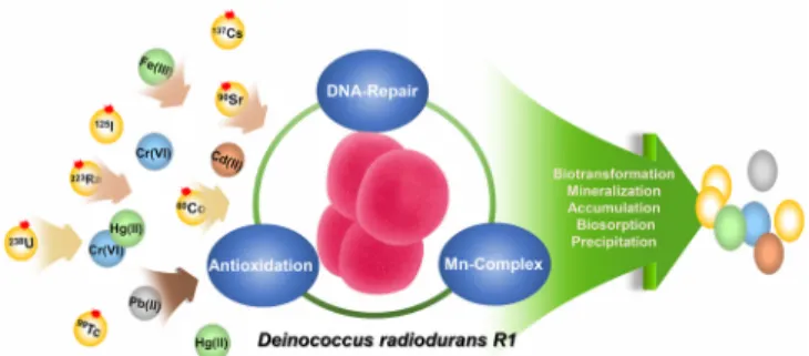
Figure 1. The scheme of bioremediation process for the removal of radioactive wastes using D. radiodurans R1 strain. The various radioactive wastes represented in the colored small circles are managed by D. radiodurans R1 strain through bioremediation process.
croorganisms exposed to high dose of ionizing radiation[53,54]. It is interesting to note that intracellular level of Mn2+ is higher than that of radiosensitive bacteria such as E. coli, Enterococcus faecium, P. pu- tida, and S. oneidensis, whereas relatively lower in ferrous ion[54].
The complex of free amino acids or peptides with Mn2+ and orthophos- phate (or bicarbonate) catalytically decompose H2O2 and scavenge su- peroxide radicals[55]. It has been reported that D. radiodurans was in- herently able to produce large amount of new carotenoid-like pigment, named as deinoxanthin, acts as a hydroxyl radical scavenger[56]. The deletion study of crtB and crtI which are involved in deinoxanthin bio- synthetic pathway revealed that deinoxanthin plays important roles in scavenging ROSs induced by ionizing radiation, UV, and H2O2[57]. In vitro test of scavenging activity of deinoxanthin have also shown that it showed decomposing activity of H2O2 as well as singlet oxygen, and the scavenging activity is much higher than those of carotenes (lycopene and β-carotene) and xanthophylls (zeaxanthin and lutein)[58].
3. Potential Application of D. radiodurans on Bioremediation of Radioactive Polluted Environment
Due to the unique DNA repair system and strong anti-oxidation mechanism described above, D. radiodurans can survive against acute exposure to more than 10-20 kGy which is lethal to living organisms without any genotypic and phenotypic changes[16]. Therefore, D. ra- diodurans has been attempted to clean-up radioactive contaminated toxic compounds. Genetically engineered D. radiodurans harboring phoN from Salmonella entarica and phoK from Sphigomonas sp. which encodes the nonspecific acid phosphatase and ribose-phosphate py- rophosphokinase, respectively, was able to efficiently precipitate ura- nium from diluted nuclear waste[59]. The genetically engineered D. ra- diodurans expressing the merA gene encoding mercuric ion reductase from E. coli strain BL308 was able to efficiently reduce toxic Hg2+, which is main component of radioactive waste sites, to monoatomic volatile Hg(0)[60]. There was also previous report on recombinant D.
radiodurans expressing toluene dioxygenase (tod) from P. putida capa- ble of degrading indole, toluene, chlorobenzene, and 3, 4-di- chloro-1-butene under high level of chronic ionizing radiation (60 Gy/h)[61]. The recombinant D. radiodurans overexpressed with the
nxiA and nvoA genes from Rhodopseudomonas palustris CGA009 and Novosphingobium aromaticivorans F-199, respectively, showed effi- cient removal efficacy of 60Co (> 60%) from spent decontamination solution[62].
Recently, the new possibility of nanotechnology on environmental remediation for increasing number of toxic compounds has been addressed. Nanoparticles have been used to cover the treatment of in- dustrial wastewater contaminated by toxic metals, radionuclides, organ- ic, and inorganic solutes[63,64]. Recently, Choi et al reported that ra- dioactive iodine (125I) was efficiently absorbed by chemically synthe- sized gold nanoparticles (AuNPs) embedded desalting column under the various aqueous media[65]. There are also several studies on the microbial synthesis of various nanoparticles[66-72]. Microbial synthesis of gold and silver nanoparticles using D. radiodurans has been re- ported and considered it as an antimicrobial reagent[71,72]. Although there had been a few reports on microbial synthesis of nanoparticles, direct use of biogenic nanoparticles on bioremediation has not yet been reported. Therefore, development of genetically engineered D. radio- durans strain capable of synthesizing nanoparticles and its application on removal of radioactive wastes is worth investigating for the ad- vances on bioremediation technology.
4. Conclusion
During the last few decades, concerns on the treatment of radio- active waste has been gradually increased as the radioactive materials are significant part of our life and the management of radioactive waste is critical for sustainability of public and environment health.
Recent advances in biotechnology, especially focus on extremophilic bacterium, allow us to develop bioremediation processes which has ad- vantages over the currently used physicochemical processes in terms of 1) cost-effectiveness, 2) natural process without less side effects, 3) sufficiently applicable to inaccessible contaminated areas. Therefore, D.
radiodurans has potential to be used as promising host microorganism for removal of radioactive waste (Figure 1). Furthermore, advances in bioremediation will be achieved by engineering of microorganisms to have novel abilities worth investigating for industrial applications.
Acknowledgement
This work has been performed with the financial support in Seoul Green Environment Center (SGEC), KOREA.
The authors declare no financial and commercial conflict of interest.
References
1. N. Yoshida and J. Kanda, Tracking the Fukushima radionuclides, Science, 336, 1115-1116 (2012).
2. K. O. Buesseler, S. R. Jayne, N. S. Fisher, I. I. Rypina, H.
Baumann, Z. Baumann, C. F. Breier, E. M. Douglass, J. George, A. M. Macdonald, H. Miyamoto, J. Nishikawa, S. M. Pike, and S.
Yoshida, Fukushima-derived radionuclides in the ocean and biota
off Japan, Proc. Natl. Acad. Sci., U. S. A., 109, 5984-5988 (2012).
3. F. F. Evans, S. Rosado, G. V. Sebastian, R. Casella, PLOA Machado, C. Holmstrom, S. Kjelleberg, J. D. Van Elsas, and L.
Seldin, Impact of oil contamination and biostimulation on the di- versity of indigenous bacterial communities in soil microcosms, FEMS Microbiol. Ecol., 49, 295-305 (2004).
4. S. K. Brar, M. Verma, R. Y. Surampalli, K. Misra, R. D. Tyagi, N. Meunier, and J. F. Blais, Bioremediation of hazardous wastes:
A review, Pract. Period. Hazard. Toxic Radioact. Waste Manag., 10, 59-72 (2006).
5. D. Prakash, P. Gabani, A. K. Chandel, Z. Ronen, and O. V. Singh, Bioremediation: a genuine technology to remediate radionuclides from the environment, Microb. Biotechnol., 6, 349-360 (2013).
6. G. M. Gadd, Bioremedial potential of microbial mechanisms of metal mobilization and immobilization, Curr. Opin. Biotechnol., 11, 271-279 (2000).
7. J. R. Lloyd, J. Ridley, T. Khizniak, N. N. Lyalikova, and L. E.
Macaskie, Reduction of technetium by Desulfovibrio desulfuricans:
biocatalyst characterization and use in a flow-through bioreactor, Appl. Environ. Microbiol., 65, 2691-2696 (1999).
8. R. AP. Thomas, A. J. Beswick, G. Basnakova, R. Moller, and L.
E. Macaskie, Growth of naturally occurring microbial isolates in metal-citrate medium and bioremediation of metal-citrate wastes, J.
Chem. Technol. Biotechnol., 75, 187-195 (2000).
9. G. Raghu, V. Balaji, G. Venkateswaran, A. Rodrigue, and P. M.
Mohan, Bioremediation of trace cobalt from simulated spent de- contamination solutions of nuclear power reactors using E. coli ex- pressing NiCoT genes, Appl. Microbiol. Biotechnol., 81, 571-578 (2008).
10. M. Daly, Engineering radiation-resistant bacteria for environmental biotechnology, Curr. Opin. Biotechnol., 11, 280-285 (2000).
11. H. Brim, A. Venkateswaran, H. M. Kostandarithes, J. K.
Fredrickson, and M. J. Daly, Engineering Deinococcus geo- thermalis for bioremediation of high-temperature radioactive waste environments, Appl. Environ. Microbiol., 69, 4575-4582 (2003).
12. E. Gerber, R. Bernard, S. Castang, N. Chabot, F. Coze, A.
Dreux-Zigha, E. Hauser, P. Hivin, P. Joseph, C. Lazarelli, G.
Letellier, J. Olive, and J.-P. Leonetti, Deinococcus as new chassis for industrial biotechnology: biology, physiology and tools, J.
Appl. Microbiol., 119, 1-10 (2015).
13. D. M. Sweet and B. E. Moseley, The resistance of Micrococcus radiodurans to killing and mutation by agents which damage DNA, Mutat. Res., 34, 175-186 (1976).
14. J. R. Battista, Against all odds: the survival strategies of Deinococcus radiodurans, Annu. Rev. Microbiol., 51, 203-224 (1997).
15. M. M. Cox and J. R. Battista, Deinococcus radiodurans-the con- summate survivor, Nat. Rev. Microbiol., 3, 882-892 (2005).
16. D. Slade and M. Radman, Oxidative stress resistance in Deinococcus radiodurans, Microbiol. Mol. Biol. Rev., 75, 133-191 (2011).
17. C. C. Lange, L. P. Wackett, K. W. Minton, and M. J. Daly, Engineering a recombinant Deinococcus radiodurans for organo- pollutant degradation in radioactive mixed waste environments, Nat. Biotechnol., 16, 929-933 (1998).
18. B. W. Brooks and R. G. E. Murray, Nomenclature for Micrococcus radiodurans and other radiation resistant cocci:
Deinococcaceae fam. nov. and Deinococcus gen. nov., including
five species, Int. J. Syst. Bacteriol., 31, 353-360 (1981).
19. S. H. Yoo, H. Y. Weon, S. J. Kim, Y. S. Kim, B. Y. Kim, and S. W. Kwon, Deinococcus aerolatus sp. nov. and Deinococcus aerophilus sp. nov., isolated from air samples, Int. J. Syst. Evol.
Microbiol., 60, 1191-1195 (2010).
20. W. T. Im, H. M. Jung, L. N. Ten, M. K. Kim, N. Bora M.
Goodfellow, S. Y. Lim, J. W. Jung, and S. T. Lee, Deinococcus aquaticus sp. nov., isolated from fresh water, and Deinococcus caeni sp. nov., isolated from activated sludge, Int. J. Syst. Evol.
Microbiol., 58, 2348-2353 (2008).
21. A. D. Groot, V. Chapon, P. Servant, R. Christen, M. F. Saux, S.
Sommer, and T. Heulin, Deinococcus deserti sp. nov., a gam- ma-radiation-tolerant bacterium isolated from the Sahara Desert, Int. J. Syst. Evol. Microbiol., 55, 2441-2446 (2005).
22. F. A. Rainey, K. Ray, M. Ferreira, B. Z. Gatz, M. F. Nobre, D.
Bagaley, B. A. Rash, M. J. Park, A. M. Earl, N. C. Shank, A. M.
Small, M. C. Henk, J. R. Battista, P. Kämpfer, and M. S. da Costa, Extensive diversity of ionizing-radiation-resistant bacteria recovered from Sonoran Desert soil and description of nine new species of the genus Deinococcus obtained from a single soil sam- ple, Appl. Environ. Microbiol., 71, 5225-5235 (2005).
23. K. Suresh, G. S. Reddy, S. Sengupta, and S. Shivaji, Deinococcus indicus sp. nov., an arsenic resistant bacterium from aquifer in West Bengal, India, Int. J. Syst. Evol. Microbiol., 54, 457-461 (2004).
24. A. C. Ferreira, M. F. Nobre, F. A. Rainey, M. T. Silva, R. Wait, J. Burghardt, A. P. Chung, and M. S. da Costa, Deinococcus geo- thermalis sp. nov. and Deinococcus murrayi sp. nov., two ex- tremely radiation-resistant and slightly thermophilic species from hot springs, Int. J. Syst. Bacteriol., 47, 939-947 (1997).
25. P. Hirsch, C. A. Gallikowski, J. Siebert, K. Peissl, R.
Kroppenstedt, P. Schumann, E. Stackebrandt, and R. Anderson, Deinococcus frigens sp. nov., Deinococcus saxicola sp. nov., and Deinococcus marmoris sp. nov., low temperature and draught-tol- erating, UV resistant bacteria from continental Antarctica, Syst.
Appl. Microbiol., 27, 636-645 (2004).
26. M. Kolari, U. Schmidt, E. Kuismanen, and M. S.
Salkinoja-Salonen, Firm but slippery attachment of Deinococcus geothermalis, J. Bacteriol., 184, 2473-2480 (2002).
27. O. White, J. A. Eisen, J. F. Heidelberg, E. K. Hickey, J. D.
Peterson, R. J. Dodson, D. H. Haft, M. L. Gwinn et al., Genome sequence of the radioresistant bacterium Deinococcus radiodurans R1, Science, 286, 1571-1577 (1999).
28. X. Hua and Y. Hua, Improved complete genome sequence of the extremely radioresistant bacterium Deinococcus radiodurans R1 obtained using PacBio single-molecule sequencing, Genome Announc., 4, e00886-16 (2016).
29. K. S. Makarova, M. V. Omelchenko, E. K. Gaidamakova, V. Y.
Matrosova, A. Vasilenko, M. Zhai, A. Lapidus, A. Copeland, E.
Kim, M. Land, K. Mavromatis, S. Pitluck, P. M. Richardson, and M. J. Daly, Deinococcus geothermalis: The pool of extreme radia- tion resistance genes shrinks, PLoS One, 2, e955 (2007).
30. A. de Groot, R. Dulermo, P. Ortet, L. Blanchard, P. Guérin, B.
Fernandez, B. Vacherie, and C. Dossat, E. Jolivet, P. Siguire, M.
Chandler, M. Barakat, A. Dedieu, and J. Armengaud, Alliance of proteomics and genomics to unravel the specificities of Sahara bacterium Deinococcus deserti, PLoS Genet., 5, e1000434 (2009).
31. R. Pukall, A. Zeytun, S. Lucas, A. Lapidus, N. Hammon, S.
Deshpande, M. Nolan, J. F. Cheng, S. Pitluck, K. Liolios, L.
Pagani, N. Mikhailova, N. Ivanova, K. Mavromatis, A. Pati, R.
Tapia, C. Han, L. Goodwin, A. Chen, K. Palaniappan, M. Land, L. Hauser, Y. J. Chang, C. D. Jeffries, E. M. Brambilla, M.
Rohde, M. Goker, J. C. Detter, T. Woyke, J. Bristow, J. A. Eisen, V. Markowitz, P. Hugenholtz, N. C. Kyrpides, and H. P. Klenk, Complete genome sequence of Deinococcus maricopensis type strain (LB-34T), Stand. Genomic Sci., 4, 163-172 (2011).
32. M. Yuan, M. Chen, W. Zhang, W. Lu, J. Wang, M. Yang, P.
Zhao, R. Tang, X. Li, Y. Hao, Z. Zhou, Y. Zhan, H. Yu, and M.
Lin, Genome sequence and transcriptome analysis of the radio- resistant bacterium Deinococcus gobiensis: Insights into the ex- treme environmental adaptations, PLoS One, 7, e34458 (2012).
33. A. Copeland, A. Zeytun, M. Yassawong, M. Nolan, S. Lucas, N.
Hammon, S. Deshpande, J. F. Cheng, C. Han, R. Tapia, L. A.
Goodwin, S. Pitluck, K. Mavromatis, K. Liolios, I. Pagani, N.
Ivanova, A. Pati, A. Chen, K. Palaniappan, M. Land, L. Hauser, C. D. Jeffries, E. M. Brambilla, M. Rohde, J. Sikorski, R. Pukall, M. Goker, J. C. Detter, T. Woyke, J. Bristow, J. A. Eisen, V.
Markowitz, N. C. Kyrpides, H. P. Klenk, and A. Lapidus, Complete genome sequence of the orange-red pigmented, radio- resistant Deinococcus proteolyticus type strain (MRPT), Stand.
Genomic Sci., 6, 240-250 (2012).
34. X. Xu, L. Jiang, Z. Zhang, Y. Shi, and H. Huang, Genome se- quence of a gamma- and UV-ray-resistant Strain, Deinococcus wu- lumuqiensis R12, Genome Announc., 1, e00206-13 (2013).
35. Y. Hu, X. Xu, P. Song, L. Jiang, Z. Zhang, and H. Huang, Draft genome sequence of Deinococcus xibeiensis R13, a new car- otenoid-producing strain, Genome Announc., 1, e00987-13 (2013).
36. V. G. Stepanov, P. Vaishampayan, K. Venkateswaran, and G. E.
Fox, Draft genome sequence of Deinococcus phoenicis, a novel strain isolated during the phoenix lander spacecraft assembly, Genome Announc., 2, e00301-14 (2014).
37. K. Satoh, T. Onodera, K. Omoso, K. T. Yano, T. Katayama, Y.
Oono, and I. Narumi, Draft genome sequence of the radioresistant bacterium Deinococcus grandis, isolated from freshwater fish in Japan, Genome Announc., 4, e01631-15 (2016).
38. J. A. Imlay, Cellular defenses against superoxide and hydrogen peroxide, Annu. Rev. Biochem., 77, 755-776 (2008).
39. M. M. Cox, J. L. Keck, and J. R. Battista, Rising from the Ashes:
DNA Repair in Deinococcus radiodurans, PLoS Genet., 6, e1000815 (2010).
40. D. Ghosal, M. V. Omelchenko, E. K. Gaidamakova, V. Y.
Matrosova, A. Vasilenko, A. Venkateswaran, M. Zhai, H. M.
Kostandarithes, H. Brim, K. S. Makarova, L. P. Wackett, J. K.
Fredrickson, and M. J. Daly, How radiation kills cells: survival of Deinococcus radiodurans and Shewanella oneidensis under oxida- tive stress, FEMS Microbiol. Rev., 29, 361-375 (2005).
41. K. Zahradka, D. Slade, A. Bailone, S. Sommer, D. Averbeck, M.
Petranovic, A. B. Lindner, and M. Radman, Reassembly of shat- tered chromosomes in Deinococcus radiodurans, Nature, 443, 569-573 (2006).
42. E. Griffiths and R. S. Gupta, Identification of signature proteins that are distinctive of the Deinococcus-Thermus phylum, Int.
Microbiol., 10, 201-208 (2007).
43. A. M. Earl, M. M. Mohundro, I. S. Mian, and J. R. Battista, The IrrE protein of Deinococcus radiodurans R1 is a novel regulator of recA expression, J. Bacteriol., 184, 6216-6224 (2002).
44. Y. Hua, I. Narumi, G. Gao, B. Tian, K. Satoh, S. Kitayama, and B. Shen, PprI: a general switch responsible for extreme radio- resistance of Deinococcus radiodurans, Biochem. Biophys. Res.
Commun., 306, 354-360 (2003).
45. H. Lu, G. Gao, G. Xu, L. Fan, L. Yin, B. Shen, and Y. Hua, Deinococcus radiodurans PprI switches on DNA damage-response and cellular survival networks after radiation damage, Mol. Cell.
Proteom., 8, 481-494 (2009).
46. Y. Wang, Q. Xu, H. Lu, L. Lin, L. Wang, H. Xu, X. Cui, H.
Zhang, T. Li, and Y. Hua, Protease activity of PprI facilitates DNA damage response: Mn(2+)-dependence and substrate se- quence-specificity of the proteolytic reaction, PLoS One, 10, e0122071 (2015).
47. M. Ludanyi, L. Blanchard, R. Dulermo, G. Brandelet, L. Bellanger, D. Pignol, D. Lemaire, and A. de Groot, Radiation response in Deinococcus deserti: IrrE is a metalloprotease that cleaves repress- or protein DdrO, Mol. Microbiol., 94, 434-449 (2014).
48. A. Devigne, S. Ithurbide, T. C. Bouthier, F. Passot, M. Mathieu, S. Sommer, and P. Servant, DdrO is an essential protein that regu- lates the radiation desiccation response and the apoptotic-like cell death in the radioresistant Deinococcus radiodurans bacterium, Mol. Microbiol., 96, 1069-1084 (2015).
49. L. Wang, X. Guangzhi, H. Chen,Y. Zhao, N. Xu, B. Tian, and Y.
Hua, DrRRA: a novel response regulator essential for the extreme radioresistance of Deinococcus radiodurans, Mol. Microbiol., 67, 1211-1222 (2008).
50. S. S. Desai, Y. S. Rajpurohit, H. S. Misra, and D. N. Deobagkar, Characterization of the role of the RadS/RadR two-component sys- tem in the radiation resistance of Deinococcus radiodurans, Microbiology, 157, 2974-2982 (2011).
51. C. H. Tsai, R. Liao, B. Chou, and L. M. Contreras, Transcriptional analysis of Deinococcus radiodurans reveals novel small RNAs that are differentially expressed under ionizing radiation, Appl.
Environ. Microbiol., 81, 1754-1764 (2015).
52. A. Martinez and R. Kolter, Protection of DNA during oxidative stress by the nonspecific DNA-binding protein Dps, J. Bacteriol., 179, 5188-5194 (1997).
53. J. K. Fredrickson, S. M. Li, E. K. Gaidamakova, V. Y. Matrosova, M. Zhai, H. M. Sulloway, J. C. Scholten, M. G. Brown, D. L.
Balkwill, and M. J. Daly, Protein oxidation: key to bacterial desic- cation resistance?, ISME J., 2, 393-403 (2008).
54. M. J. Daly, E. K. Gaidamakova, V. Y. Matrosova, A. Vasilenko, M. Zhai, A. Venkateswaran, M. Hess, M. V. Omelchenko, H. M.
Kostandarithes, K. S. Makarova, L. P. Wackett, J. K. Fredrickson, and D. Ghosal, Accumulation of Mn(II) in Deinococcus radio- durans facilitates gamma-radiation resistance, Science, 306, 1025-1028 (2004).
55. M. J. Daly, E. K. Gaidamakova, V. Y. Matrosova, J. G. Kiang, R.
Fukumoto, D. Y. Lee, N. B. Wehr, G. A. Viteri, B. S. Berlett, and R. L. Levine, Small-molecule antioxidant proteome-shields in Deinococcus radiodurans, PLoS One, 5, e12570 (2010).
56. L. Lemee, E. Peuchant, M. Clerc, M. Brunner, and H. Pfander, Deinoxanthin: a new carotenoid isolated from Deinococcus radio- durans, Tetrahedron, 53, 919-926 (1997).
57. L. Zhang, Q. Yang, X. Luo, C. Fang, Q. Zhang, and Y. Tang, Knockout of crtB or crtI gene blocks the carotenoid biosynthetic pathway in Deinococcus radiodurans R1 and influences its resist- ance to oxidative DNA-damaging agents due to change of free rad-
icals scavenging ability, Arch. Microbiol., 188, 411-419 (2007).
58. B. Tian, Z. Xu, Z. Sun, J. Lin, and Y. Hua, Evaluation of the anti- oxidant effects of carotenoids from Deinococcus radiodurans through targeted mutagenesis, chemiluminescence, and DNA dam- age analyses, Biochim. Biophys. Acta, 1770, 902-911 (2007).
59. D. Appukuttan, A. S. Rao, and S. K. Apte, Engineering of Deinococcus radiodurans R1 for bioprecipitation of uranium from dilute nuclear waste, Appl. Environ. Microbiol., 72, 7873-7878 (2006).
60. H. Brim, S. C. McFarlan, J. K. Fredrickson, K. W. Minton, M.
Zhai, L. P. Wackett, and M. J. Daly, Engineering Deinococcus ra- diodurans for metal remediation in radioactive mixed waste envi- ronments, Nat. Biotechnol., 18, 85-90 (2000).
61. C. C. Lange, L. P. Wackett, K. W. Minton, and M. J. Daly, Engineering a recombinant Deinococcus radiodurans for organo- pollutant degradation in radioactive mixed waste environments, Nat. Biotechnol., 16, 929-933 (1998).
62. G. Raghu, S. S Singh, S. K. Lunavat, M. M. Pamarthi, A.
Rodrigue, B. Vadivelu, P. B. Phanithi, V. Gopala, and S. K. Apte, Engineered Deinococcus radiodurans R1 with NiCoT genes for bi- oremoval of trace cobalt from spent decontamination solutions of nuclear power reactors, Appl. Microbiol. Biotechnol., 99, 9203-9213 (2015).
63. L. Newsome, K. Morris, and J. R. Lloyd, The biogeochemistry and bioremediation of uranium and other priority radionuclides, Chem.
Geol., 363, 164-184 (2014).
64. L. Xiangqian, X. Huizhong, Z. S. Chen, and G. Chen, Biosynthesis of Nanoparticles by Microorganisms and Their Applications, J.
Nanomater, 2011, 1-16 (2011).
65. M. H. Choi, H. E. Shim, S. J. Yun, S. H. Park, D. S. Choi, B.
S. Jang, Y. J. Choi, and J. J. Jeon, Gold-nanoparticle-immobilized Desalting columns for highly efficient and specific removal of ra- dioactive iodine in aqueous media, ACS Appl. Mater. Interfaces, 8, 29227-29231 (2016).
66. L. Du, H. Jiang, X. Liu, and E. Wang, Biosynthesis of gold nano- particles assisted by Escherichia coli DH5α and its application on direct electrochemistry of hemoglobin. Electrochem. Commun., 9, 1165-1170 (2007).
67. M. I. Husseiny, M. A. El-Aziz, Y. Badr, and M. A. Mahmoud, Biosynthesis of gold nanoparticles using Pseudomonas aeruginosa, Spectrochim. Acta A, 67, 1003-1006 (2007).
68. S. Bose, M. F. Hochella, Y. A. Gorby, D. W. Kennedy, D. E.
McCready, A. S. Madden, and B. H. Lower, Bioreduction of hem- atite nanoparticles by the dissimilatory iron reducing bacterium Shewanella oneidensis MR-1, Geochim. Cosmochim. Acta, 73, 962-976 (2009).
69. M. M. G. Babu and P. Gunasekaran, Production and structural characterization of crystalline silver nanoparticles from Bacillus cereus isolate, Colloids Surf. B, 74, 191-195 (2009).
70. S. He, Z. Guo, Y. Zhang, S. Zhang, J. Wang, and N. Gu, Biosynthesis of gold nanoparticles using the bacteria Rhodopseudomonas capsulate, Mater. Lett., 61, 3984-3987 (2007).
71. R. R. Kulkarni, N. S. Shaiwale, D. N. Deobagkar, and D. D.
Deobagkar, Synthesis and extracellular accumulation of silver nanoparticles by employing radiation-resistant Deinococcus radio- durans, their characterization, and determination of bioactivity, Int.
J. Nanomed., 10, 963-974 (2015).
72. J. Li, Q. Li, X. Ma, B. Tian, T. Li, J. Yu, S. Dai, Y. Weng, and
Y. Hua, Biosynthesis of gold nanoparticles by the extreme bacte- rium Deinococcus radiodurans and an evaluation of their anti- bacterial properties, Int. J. Nanomed., 11, 5931-5944 (2016).
73. C. S. Misra, D. Appukuttan, V. S. Kantamreddi, A. S. Rao, and S. K. Apte, Recombinant D. radiodurans cells for bioremediation of heavy metals from acidic/neutral aqueous wastes, Bioeng. Bugs, 3, 44-48 (2012).
74. J. K. Fredrickson, H. M. Kostandarithes, S. W. Li, A. E. Plymale, and M. J. Daly, Reduction of Fe(III), Cr(VI), U(VI), and Tc(VII) by Deinococcus radiodurans R1, Appl. Environ. Microbiol., 66, 2006-2011 (2000).
75. R. B. Payne, D. M. Gentry, B. J. Rapp-Giles, L. Casalot, and J.
D. Wall, Uranium reduction by Desulfovibrio desulfuricans strain G20 and a cytochrome c3 mutant, Appl. Environ. Microbiol., 68, 3129-3132 (2002)
76. D. R. Lovley and E. J. Phillips, Reduction of uraniumby Desulfovibrio desulfuricans, Appl. Environ. Microbiol., 58, 850-856 (1992).
77. K. S. Nilgiriwala, A. Alahari, A. S. Rao, and S. K. Apte, Cloning and overexpression of alkaline phosphatase PhoK from Sphingomonas sp. strain BSAR-1 for bioprecipitation of uranium from alkaline solutions, Appl. Environ. Microbiol., 74, 5516-5523 (2008).
78. A. S. Madden, A. I. Swindle, M. J. Beazley, J. W. Moon, B.
Ravel, and T. J. Phelps, Longterm solid-phase fate of co-pre- cipitated U(VI)-Fe(III) following biological iron reduction by Thermoanaerobacter, Am. Mineral., 97, 1641-1652 (2012).
79. T. V. Khijniak, A. I. Slobodkin, V. Coker, J. C. Renshaw, F. R.
Livens, E. A. Bonch-Osmolovskaya, N. K. Birkeland, N. N.
Medvedeva-Lyalikova, and J. R. Lloyd, Reduction of uranium(VI) phosphate during growth of the thermophilic bacterium Thermoterrabacterium ferrireducens, Appl. Environ. Microbiol., 71, 6423-6426 (2005).
80. L. E. Macaskie, R. M. Empson, A. K. Cheetham, C. P. Grey, and A. J. Skarnulis, Uranium bioaccumulation by a Citrobacter sp. as a result of enzymically mediated growth of polycrystalline HUO2PO4, Science, 257, 782-784 (1992).
81. J. R. Lloyd, C. Leang, C., A. L. Hodges Myerson, M. V. Coppi, S. Cuifo, B. Methe, S. J. Sandler, and D. R. Lovely, Biochemical and genetic characterization of PpcA, a periplasmic c-type cyto- chrome in Geobacter sulfurreducens, Biochem. J., 369, 153-161 (2003).
82. J. R. Lloyd, P. Yong, and L. E. Macaskie, Biological reduction and removal of Np(V) by two microorganisms, Environ. Sci.
Technol., 34, 1297-1301 (2000).
83. J. R. Lloyd, J. Ridley, T. Khizniak, N. N. Lyalikova, and L. E.
Macaskie, Reduction of technetium by Desulfovibrio desulfuricans:
biocatalyst characterization and use in a flowthrough bioreactor, Appl. Environ. Microbiol., 65, 2691-2696 (1999).
84. N. N. Lyalikova and T. V. Khizhnyak, Reduction of heptavalent technetium by acidophilic bacteria of the genus Thiobacillus, Microbiology, 65, 468-473 (1996).
85. M. J. Marshall, A. C. Dohnalkova, D. W. Kennedy, A. E. Plymale, S. H. Thomas, F. E. Loffler, R. A. Sanford, J. M. Zachara, J. K.
Fredrickson, and A. S. Beliaev, Electron donordependent radio- nuclide reduction and nanoparticle formation by Anaeromyxobacter dehalogenans strain 2CP-C, Environ. Microbiol., 11, 534-543 (2009).
86. H. Boukhalfa, G. A. Icopini, S. D. Reilly, and M. P. Neu, Plutonium (IV) reduction by the metal-reducing bacteria Geobacter metallireducens GS15 and Shewanella oneidensis MR1, Appl.
Environ. Microbiol., 73, 5897-5903 (2007).
87. B. Luksiene, R. Druteikiene, D. Peciulyte, D. Baltrunas, V.
Remeikis, and A. Paskevicius, Effect of microorganisms on the plutonium oxidation states, Appl. Radiat. Isot., 70, 442-449 (2012).
88. L. E. Macaskie, B. C. Jeong, and M. R. Tolley, Enzymically accel- erated biomineralization of heavy metals: Application to the re- moval of americium and plutonium from aqueous flows, FEMS Microbiol. Rev., 14, 351-367 (1994).
89. T. Councell, E. Landa, and D. Lovley, Microbial reduction of io- date, Water Air Soil Pollut., 100, 99-106 (1997).
90. S. Anderson and V. D. Appanna, Microbial formation of crystal- line strontium carbonate, FEMS Microbiol. Lett., 116, 43-48 (1994).
91. V. Achal, X. Pan, and D. Zhang, Bioremediation of strontium (Sr) contaminated aquifer quartz sand based on carbonate precipitation induced by Sr resistant Halomonas sp, Chemosphere, 89, 764-768 (2012).
92. F. G. Ferris, C. M. Fratton, J. P. Gerits, S. Schultze‐Lam, and B. S. Lollar, Microbial precipitation of a strontium calcite phase at a groundwater discharge zone near Rock Creek, British
Columbia, Canada, Geomicrobiol. J., 13, 57-67 (1995).
93. D. L. Vullo, H. M. Ceretti, E. A. Hughes, S. Ramyrez, and A.
Zalts, Cadmium, zinc and copper biosorption mediated by Pseudomonas veronii 2E, Bioresour. Technol., 99, 5574-5581 (2008).
94. H. Guo, S. Luo, L. Chen, X. Xiao, Q. Xi, W. Wei, G. Zeng, C.
Liu, Y. Wan, J. Chen, and Y. He, Bioremediation of heavy metals by growing hyperaccumulator endophytic bacterium Bacillus sp.
L14, Bioresour. Technol., 101, 8599-8605 (2010).
95. V. Achal, X. Pan, and D. Zhang, Remediation of copper-con- taminated soil by Kocuria flava CR1, based on microbially in- duced calcite precipitation, Ecol. Eng., 37, 1601-1605 (2011).
96. K. Hrynkiewicz, G. Dabrowska, C. Baum, K. Niedojadlo, and P.
Leinweber, Interactive and single effects of ectomycorrhiza for- mation and Bacillus cereus on metallothionein MT1 expression and phytoextraction of Cd and Zn by Willows, Water Air Soil Pollut., 223, 957-968 (2012).
97. V. Achal, X. Pan, Q. Fu, and D. Zhang, Biomineralization based remediation of As (III) contaminated soil by Sporosarcina ginsengisoli. J. Hazard. Mater., 201-202, 178-184 (2012).
98. P. Kanmani, J. Aravind, and D. Preston, Remediation of chromium contaminants using bacteria, Int. J. Environ. Sci. Technol., 9, 183-193 (2012).