- 419 -
Bactericidal effect of 461 nm blue light emitting diode on pathogenic bacteria
Jung Sun Do1,2, Woo Suk Bang1,2*
1
LED-IT Fusion Technology Research Center, Yeungnam University, Gyeongsan 712-749, Korea
2Department of Food and Nutrition, Yeungnam University, Gyeongsan 712-749, Korea
461nm 청색 LED를 이용한 식중독세균의 살균효과
도정선1,2․방우석1,2*
1영남대학교 LED-IT융합산업화연구센터, 2영남대학교 식품영양학과
Abstract
The objective of this study was to characterize the bactericidal effect of 461nm visible-light LED on three common foodborne bacteria: Escherichia coli O157:H7, Staphylococcus aureus and Vibrio parahaemolyticus. Tests were conducted against pathogen strains that were treated with 461nm LED for 10 h at 15°C. The E. coli (ATCC 43894, ATCC 8739 and ATCC 35150) and the S. aureus (ATCC 27664, ATCC 19095 and ATCC 43300) had average reductions of 2.5, 6.6, 1.5, 2.5 and 2.0 log CFU/mL, respectively, after they were exposed for 10 h to 461nm LED light (p<0.05). In contrast, V. parahaemolyticus (ATCC 43969) had 6 log CFU/mL reductions after it was exposed for 4 h to 461nm LED light. The results showed that both the Gram-positive and Gram-negative bacteria were inactivated with 461nm LED light exposure. Also, the Gram-negative bacteria were more sensitive to the LED treatment than the Gram-positive bacteria. These results show the potential use of 461nm LED as a food preservation and application technology.
Key words:461 nm LED, blue light, microorganism, photosensitization
서 론
1)
식품은 생물학적 오염이 부패와 변질의 주요 원인이다.
우리나라 식중독 발생 통계에 따르면 Norovirus를 제외한 Escherichia coli, Staphylococcus aureus, Vibrio parahaemolyticus, Salmpnella sp., Clostridium perfringens등의 병원성 미생물 이 세균성 식중독의 주요원인으로 57%를 차지하며 바이러 스성 식중독이 27.9% 이외 불검출(불명), 자연독, 화학물질 에 의해 2011년 271건과 환자수 7,218명으로 세균성 식중독 은 해마다 증가하고 있는 추세이다(1).
세균에 의한 식품의 오염을 방지하기 위하여 초기에 생 육을 억제하고 식품저장기간 연장을 위해 물리적 방법인 가열, 냉동, 방사선을 이용하거나 화학적 합성품인 보존제, 방부제, 훈증제 등이 이용되고 있다. 그러나 이러한 방법들
*Corresponding author. E-mail:[email protected] Phone:82-53-810-2877, Fax:82-53-810-4768
은 영양손실, 품질저하, 내성화, 환경오염, 독성 및 제어기 술에 따른 문제점을 지닌다(2,3). 따라서 식품고유의 영양 성분을 유지하며 오염과 변형이 없는 고품질의 식품 생산을 위하여 다양한 비가열가공기술(non-thermal process)의 연 구가 활발히 이루어지고 있다(4). 최근 이러한 기술의 일환 으로 LED 기술 분야가 주목받고 있다. LED(light emitting diode)는 화합물 반도체 기술로써 스스로 빛을 발생하는 다이오드이다. 1879년에 발명된 백열전구를 시작으로 형광 램프, HID 램프에 이은 제 4의 광원으로 기존광원에 비해 고효율, 친환경, 반영구적 수명 등의 장점을 지니고 있다.
LED는 반도체 칩의 화합물 구성원소 배합에 의해 여러가지 발광색상과 파장을 나타낼 수 있으며 청색은 400∼500 nm 의 파장을 형성 할 수 있다(5,6).
현재 100∼400 nm 사이의 파장규모를 가진 UV-LED (Ultra Violet-LED)는 물과 공기의 살균, 폐수처리, 탈취, 피부병 치료 및 의료용으로 생활 보건 분야에서 이용되고 있다(7). 특히 UV-C영역은 살균선이라 불리며 220∼280
nm의 파장을 가진다. 직접 조사 시 미생물의 DNA, RNA에 광반응에 따른 손상을 일으켜 불화성화를 통한 직접적인 살균효과를 타나낸다. 특히 253.7 nm 의 경우 미생물의 DNA에 가장 치명적인 효과를 주는 파장이라고 알려져 있 다(8). 그러나 자외선(UV)은 미생물 살균과 phototherapy에 서 특정 파장의 효과는 뛰어나지만 여러 가지 파장의 선택 과 조사시간에 의해 눈의 망막과 각막, 피부세포의 DNA손 상 및 면역억제 등을 유발한다(9-11). 이러한 단점을 보완하 여 가시광선 파장과 광감작제를 이용한 PDI(photodynamic inactivation) 및 PDT(photodynamic therapy)가 의학적 용도 로 이용되고 있다. 또한 채소, 과실 등의 생육 및 저장기간 중 변화, 품질향상에 이용되고 있으며 최근 405 nm 청색 LED를 이용하여 세균 자체에 효과적인 살균효과가 입증되 고 있다(12,13). 이는 PDI, PDT의 원리와 같은 photosensitization 에 기초 하며 기존의 UV와는 다른 매커니즘을 가지며, 산소 의 존재하에서 무독성의 광활성화합물과 가시광선에 의해 세포의 선택적 사멸을 유도하게 된다. 이는 바이러스, yeasts, parasites 등에 효과적이며 균종에 따른 독립적인 저항패턴을 가지므로 활용이 유용하다(14).
그람양성균(Gram-positive bacteria)과 그람음성균(Gram- negative bacteria)의 물리적인 구조차이에 의해서 Photosensitization의 민감성은 다르게 나타나며 내인성 porphyrin 생산의 차이에 의해 광감작제로써의 효과가 다르 게 나타난다(15,16). 현재 LED시장의 90∼95%를 차지하고 있는 V-LED(Visible-LED)의 경우 400∼700 nm 사이의 파 장 규모를 가지며 각종 실내외조명, 휴대폰, 노트북, LCD BLU, 자동차 등에 이용되고 있다(7). 최근 의료, 농업, 환경, 해양 등 응용범위가 넓어져 응용제품의 연구와 개발이 확대 되고 있다(17-20). 그러나 현재 405 nm를 제외한 다양한 파장 및 균종에 대한 효과 연구가 미비한 실정이다. 따라서 본 연구는 청색영역의 461 nm LED를 이용하여 저온환경에 서E. coliO157:H7,S. aureus그리고V. parahaemolyticus의 살균효과를 알아보고자 한다.
재료 및 방법
Microorganisms
본 연구에 사용된E. coli O157:H7(ATCC 8739, ATCC 35150, ATCC 43894), S. aureus(ATCC 19095, ATCC 27664, ATCC 43300)는 실험 전 영하 70℃에 보관된 균을 사용하였 다. 멸균된 tryptic soy broth(TSB, Difco Laboratories, Detroit, MI, USA)에 접종하여 37℃의 배양기에서 20∼24시간 배양 하였으며 적어도 3번 이상 계대배양을 거친 정지기 균을 실험에 사용하였다. Tryptic soy agar(TSA, Merck Co., Draft, Germany)로 주입평판배양을 하여 24∼48시간 동안 배양기 에서 배양한 후, 25∼250 사이의 집락을 계수하여 109
CFU/mL로 생장하는 것을 확인하였다.V. parahaemolyticus (ATCC 43969)는 실험 전 실온에서 mineral oil(Sigma Chemical Co., St. Louis, MO, USA)이 첨가된 2%
NaCl(Sigma) 사면배지에 보관된 균을 사용하였다. 2%
NaCl을 첨가한 멸균된 tryptic soy broth(TSB, Difco)에 접종 하여 37℃의 배양기에서 20∼24시간 배양하였으며 적어도 3번 이상 계대배양을 거친 정지기 균을 실험에 사용하였다.
2% NaCl이 첨가된 tryptic soy agar(TSA, Difco)로 주입평판 배양을 하여 24∼48시간 동안 배양기에서 배양한 후, 그 집락을 계수하여 109 CFU/mL로 생장하는 것을 확인 하였다.
461nm LED light source
실험에 사용된 461 nm blue light LED는 X.H.Y OPTOELECTRONICS(GX-HP10W-B, China)에서 생산된 제품이며 InGaN의 화합물 재료로 제작되었다. 파장은 최대 461 nm(±3)이며 열 발생에 따른 cooling fan과 heat sink가 부착되었다. 실험장비는 10.4×10.4×7 cm3크기의 ABS수지 (acrylonitrile butadiene styrene copolymer) 박스를 제작한 후 박스의 상부에 LED를 부착하여 제작하였다. 461 nm LED는 9개의 칩이 직렬 및 병렬구조로 연결되어 있는 하나 의 패키지 형태로 구성되었다. 전류 전원은 AC adapter(0∼2 A, 0∼12 V)를 이용하여 공급하였으며 전압은 10 V(±2)에 서 1 A(±0.05)로 사용되었다.
Exposure experiments
모든 실험은 15℃로 맞춰진 저온 배양기(Sanyo Electric Co. Ltd., Tokyo, Japan)에서 실험을 실시하였다. 배지와 LED간의 조사거리는 3.3 cm로 상부 중앙에 위치되어 샘플 의 중앙으로 빛이 잘 전달되도록 하였고 배지의 깊이는 12 mm이며 평균온도는 15±1℃를 유지하였다. 방사조도 (irradiance)는 22.1 mW/cm2로 S130C 400∼1100 nm 센서가 부착된 Thorlabs Laser Power and Energy Meter Console PM100D을 이용하여 측정되었다. 같은 방사조도 조건에서 각각의 노출된(dose) 샘플은 다음 식을 이용하여 계산되었다.
E = pt
E는 Dose(energy density, 에너지 밀도)로 J/cm2으로 나타 내며, p는 irradiance(방사조도)로 W/cm2, t는 시간(second) 으로 정의된다.
Plating and enumeration
461 nm LED 조사에 따른 살균효과를 평가하기 위해서 각각의 균은 109CFU/mL의 정지기 상태에서 TSB에 106 CFU/mL 수준으로 희석한 후 60×15 mm petridish에 10 mL 분주하여 15℃에서 10시간 동안 조사한 후 0시간부터 1시
간 마다 0.1 mL씩 sampling 하였다. 각각의 sampling 시점에 서 조사되지 않은 0시간의 대조구와 실험군의 sample들은 TSA로 pour plate 한 후 37℃의 배양기에서 24∼48시간 배양 하여 생성된 집락의 수를 계산하였다.
Statistical analysis
각 실험은 3회 반복 수행되었으며 통계분석 결과는 SAS program, Version 9.2(SAS Institute Inc., Cary, NC, USA)을 이용하여 회귀분석으로 D값(일정한 온도에서 90%의 미생 물을 사멸하는데 걸리는 시간)을 계산하였다. 각 처리군 간의 유의적인 차이는 SPSS program, Version 19(SPSS Inc., Chicago, IL, USA)을 이용하여 Duncan's test로 검증하였고, 5% 수준(p<0.05)에서 이루어졌다.
결과 및 고찰
Inactivation effects of treatment of 461 nm blue light LED 본 연구는 461 nm blue LED를 이용하여 10시간 동안 bacterial cultures에 조사한 결과를 Fig. 1∼3에 나타냈다.
그람음성균인E. coli의 경우 세 균주 모두 3시간 되는 시점 에서 급격하게 감소하기 시작하였으며 ATCC 43894는 10 시간 동안 조사 되어진 후 약 2.5 log CFU/mL의 감소를 보였으며 ATCC 8739, 35150은 9시간 이후 사멸되었다.
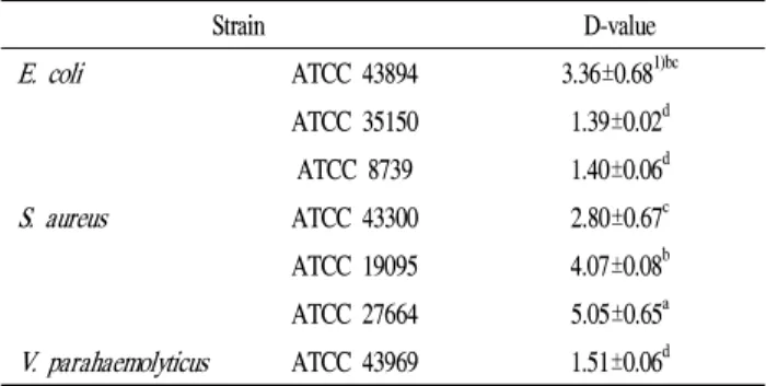
그람양성균인S. aureus의 경우E. coli균종과 마찬가지로 3시간 되는 시점에서 감소가 시작되며 10시간 조사 되어진 후 ATCC 27664, 19095, 43300은 각각 약 1.5, 2, 2.5 log CFU/mL의 감소를 나타냈다. 그람음성균의V. parahaemolyticus 또한 2.5시간 되는 지점에서 급격한 감소가 시작되어 조사 5시간 만에 사멸되었다. 세 균주 각각의 균종에 대한 D값은 Table 1과 같다. S. aureusATCC 27664는 5.05시간의 가장 높은 값을 나타내어 살균효과가 가장 낮음을 보였으며E.
Fig. 1. Inactivation effects of Escherichia coli O157:H7.
(○: ATCC 43894, ●: ATCC 8739, ▼: ATCC 35150) after 10 h irradiation using 461 nm blue light LED.
Fig. 2. Inactivation effects of Staphylococcus aureus.
(○: ATCC 19095, ●: ATCC 43300(MRSA), ▼: ATCC 27664) after 10 h irradiation using 461 nm blue light LED.
Fig. 3. Inactivation effects of Vibrio parahaemolyticus (ATCC 43969) after 5 hr irradiation using 461 nm blue light LED.
coliATCC 35150은 1.39시간의 가장 낮은 값을 나타내어 살균효과가 가장 뛰어났으며 두 균종간에 유의적인 차이가 나타났다(p<0.05).E. coliATCC 8739,V. parahaemolyticus ATCC 43969 또한 각각 1.40, 1.51시간으로 높은 살균효과 를 나타냈다. 본 실험에서 461 nm LED에서 10시간 동안 조사된E. coli와V. parahaemolyticus의 경우S. aureus에 비해 높은 사멸률을 나타내어 각각의 균주에 따라 살균효과 에 차이가 있으며 이는 그람양성균과 그람음성균의 차이에 따른 photosensitization 현상에 의한 결과로 판단된다(14).
Photosensitization은 무독성의 광활성 화합물과 가시광선이 산소의 존재 하에서 세포의 선택적 사멸을 일어나게 하는 것으로 기존의 UV 살균 기작과는 차이가 있는 것으로 알려 져 있다(21). Kim(22)은 425 nm blue LED에서 E. coli와 S. aureus를 12시간 조사시킨 결과 30%, 10%의 감소를 보고 하였다. 그람음성균인 E. coil의 살균효과가 높게 나타난 연구결과는 461 nm blue LED를 이용한 본 실험결과와 유사 한 경향을 보였다. 반면에 Murdoch 등(23)은 그람음성균인 Salmonella, Shigella, E. coli균종과 그람양성균인Listeria
균종을 405 nm blue LED에서 평균 71 mW/cm2로 조사한 결과Listeria균종은 조사 30분 시점(180 J/cm2)에서 사멸되 었으며 나머지 그람음성균은 45분(270 J/cm2) 시점에서 대 부분 사멸되어 그람양성균의 살균효과가 높다고 보고하였 다. 405 nm blue LED를 10 mW/cm2로 조사하여 그람양성균 과 음성균을 비교한 결과(24), 그람양성균에서Enterococcus 균종을 제외한S. aureus, Streptococcus, Clostridium, MRSA (Methicillin resistantStaphylococcus aureus) 균종이 약 70분 조사된 시점에서 사멸되었다. 그람음성균인Acinetobacter 의 경우 180분 시점에서 101CFU/mL 수준으로 감소하였고 Pseudomonas 균종은 240분에 101 CFU/mL, E. coli와 Proteus, Klebsiella 균종은 각각 300분 시점에 102CFU/mL, 101CFU/mL 수준으로 감소하여Enterococcus균종 외 그람 양성균이 그람음성균에 비해 우수한 살균효과를 나타냈다.
이는 461 nm blue LED를 사용한 본 실험결과와는 상반된 결과로써 405 nm blue light의 경우 일반적으로 그람음성균 에 비해 그람양성균의 살균효과가 우수한 것으로 나타났 다. 같은 blue light 영역이지만 wavelength의 차이에 의해 상반된 살균효과가 나타난 결과로 판단된다. 이 외 그람양 성균과 그람음성균의 3차원 물리적 구조 및 photosensitization 에 영향을 미치는 인자로써 박테리아 내에서 생산되어 내인 성 광감작제로 이용되는 porphyrin의 유형, 생산량, 파장에 따른 흡수영역, 일중항 산소 발생 및 의존 차이가 있으며 LED 광원의 세기 및 외부환경의 작용 등이 영향을 미친 것으로 사료된다(25,26).
Table 1. D-values of the three Microorganism after exposure to 461 nm blue light LED
Strain D-value
E. coli ATCC 43894 3.36±0.681)bc ATCC 35150 1.39±0.02d
ATCC 8739 1.40±0.06d S. aureus ATCC 43300 2.80±0.67c ATCC 19095 4.07±0.08b ATCC 27664 5.05±0.65a V. parahaemolyticus ATCC 43969 1.51±0.06d
1)Mean±SD of triplicated experiments.
2)a-dSignificance among different strains against the same wavelength (p<0.05).
Nitzan 등(27)은 407∼420 nm사이의 blue light metal halide lamp를 이용하여 4시간 동안 조사 후 그람양성균과 그람음성균의 porphyrin 유형과 생산량을 살펴본 결과 Staphylococcus, Bacillus의 그람양성균은E. coli, Acinetobacter, Aeromonas의 그람음성균에 비해 uroporphyrin, coproporphyrin 의 유형이 가장 높았으나 porphyrin유도체의 경우 그람음성 균이 높은 경향을 나타내었으며 생산량은 porphyrin 유형 과 유사하게 나타났다. 이는 균종에 따른 porphyrin의 존재
가 빛의 흡수에 따른 불활성화에 영향을 미치는 것으로 사료된다. 따라서 461 nm blue LED가 그람음성균에서 우수 한 살균효과를 나타낸 본 실험의 결과와 차이가 있으며 이는 조사광원의 종류 및 세기, 외부환경에 따른 porphyrin 유형과 생산량에 차이가 있을 것으로 판단된다.
이상의 연구결과를 종합하여 볼 때 본 실험에 이용된 461 nm blue LED는 15℃의 저온환경에서 그람음성균에서 뛰어난 살균효과를 나타내어 그람음성균을 효과적으로 제 어할 수 있다고 판단되었다. 하지만 연구결과 사용된 균종 외에 다양한 균종에 대한 살균효과 연구 및 살균기작에 의한 porphyrin 생산과 존재량에 대한 추가적인 연구가 필 요한 것으로 사료된다.
요 약
이 연구는 3가지 유형의 식중독균인 E. coli O157:H7, S. aureus그리고V. parahaemolyticus의in vitro를 통해 461 nm LED의 살균효과를 입증하였다. Bacterial cultures는 약 15℃에서 10시간동안 LED에 노출되었다. 3종의 E. coli, ATCC 8739, ATCC 43894, ATCC 35150 과 3 종의S. aureus, ATCC 43300, ATCC 19095, ATCC 27664의 bacteria cultures 는 각각 461 nm LED에서 10시간 조사된 후 평균적으로 6, 2.5, 6, 2.5, 2, 1.5 log CFU/mL 감소하였다(p<0.05). 반면에 V. parahaemolyticus, ATCC 43969는 461 nm에서 4시간 노 출된 후 6 log CFU/mL 감소하였다. 이 결과는 그람양성균과 그람음성균 모두 불활성화 되었으며 461 nm LED 노출에 의해 그람양성균보다 그람음성균이 더 민감함을 보여준다.
그러므로 이 결과는 LED의 사용이 식품보존과 응용기술로 써 잠재력이 있음을 나타낸다.
감사의 글
본 논문은 산업통상자원부 산업기술연구기반조성사업 (산업융합기반구축사업, 10033630)의 지원에 의해 수행되 었으며 이에 감사드립니다.
References
1. Korea Food and Drug Administration (2012) Available from: http://www.mfds.go.kr/e-stat/index.do Accessed on 2012
2. Kim YS, Shin DH (2003) Researches on the volatile antimicrobial compounds from edible plants and their food application. Korean J Food Sci Technol, 35, 159-165
3. Kim YS, Park IS, Ha SD (2009) Application sanitizer for the control of microorganisms in Food. Food Sci Indus, 42, 26-35
4. Shin JK, Kim BR, Kim AJ (2010) Nonthermal food processing technology using electric Power. Food Sci Indus, 43, 21-34
5. Um GY (2008) display engineering. gijeon, Seoul, Korea p 320-368
6. Nam SY, Park MS, Gang JG (2010) The LED technology and application of green energy. SangHakdang, Seoul, Korea p 11-114
7. BIR research group (2010) Eco-friendly, high-efficient LED technology development trends and market outlook.
BIR Inc, Seoul, Korea p 27-188
8. United States Environmental Protection Agency (2003) Ultraviolet disinfection guidance manual. NSCEP, EPA 815-D-03-007
9. Young AR (2006) Acute effects of UVR on human eyes and skin. Prog Biophys Mol Biol, 92, 80-85 10. Halliday GM, Norval M, Byrne SN, Huang XX, Wolf
P (2008) The effects of sunlight on the skin. Drug discovery today: Disease Mechanisms, 5, 201-209 11. Ikehata H, Ono T (2011) The mechanisms of UV
mutagenesis. J Radiat Res, 52, 115-125
12. Maisch T (2007) Anti-microbial photodynamic therapy:
useful in the future? Lasers Med Sci, 22, 83-91 13. Endarko E, Maclean M, Timoshkin IV, Macgregor SJ,
Anderson JG (2012) High-Intensity 405 nm Light Inactivation of Listeria monocytogenes. J photochem photobiol, 88, 1280-1286
14. Luksiene Z (2005) New approach to inactivation of harmful and pathogenic microorganisms by Photosensitization.
Food Technol Biotechnol, 43, 411-418
15. Luksiene Z, Zukauskas A (2009) Prospects of photosensitization in control of pathogenic and harmful micro-organisms. J Appl Microbiol, 107, 1415-1424 16. Buchovec I, Paskeviciute E, Luksiene Z (2010)
Photosensitization-based inactivation of food pathogen Listeria monocytogenes in vitro and on the surface of packaging material. J Photochem Photobiol B, 99, 9-14 17. Barolet D (2008) Light-emitting diodes (LEDs) in
dermatology. Semin Cutan Med Surg, 27, 227-238 18. Baek KH, Jang MH, Kwack YB, Lee SW, Yun HK (2010)
Regulation of acid contents in kiwifruit irradiated by various wavelength of light emitting diode during postharvest storage. Clean Tech, 16, 88-94
19. Oh MS, Lee HS (2010) Development of phototactic test apparatus equipped with light source for monitoring Pests.
J Appl Biol Chem, 53, 248-252
20. Oh SJ, Park DS, Yang HS, Yoon YH, Honjo T (2007) Bioremediation on the benrhic layer in polluted inner bay by promotion of microphytobenthos growth using Light Emitting Diode (LED). J Kor Soc MEE, 10, 93-101 21. Durantini EN (2006) Photodynamic inactivation of
bacteria. Curr Bioactive Compounds, 2, 127-142 22. Kim SW (2011) In vitro bactericidal effect of red, green
and blue (RGB) light emitting diode (LED) irradiation.
MS thesis Chonnam National University Gwangju, Korea 23. Murdoch LE, Maclean M, Endarko E, MacGregor SJ, Anderson JG (2012) Bactericidal effects of 405 nm light exposure demonstrated by inactivation of Escherichia, Salmonella, Shigella, Listeria, and Mycobacterium species in liquid suspensions and on exposed surfaces.
The Scientific World Journal, 137805, 1-8
24. Maclean M, MacGregor SJ, Andersom JG, and Woolsey G (2009) Inactivation of bacterial pathogens following exposure to light from a 405-nanometer light-emitting diode array. Appl Environ Microbiol, 75, 1932-1937 25. Maclean M, Macgregor SJ, Anderson JG, Woolsey
(2008) The role of oxygen in the visible-light inactivation of Staphylococcus aureus. J Photochem Photobiol, 92, 180-184
26. Fotinos N, Convert M, Piffaretti JC, Gurny R, Lange N (2008) Effects on gram-negative and gram-positive bacteria mediated by 5-aminolevulinic Acid and 5-aminolevulinic acid derivatives. Antimicrob Agents Chemother, 52, 1366-1373
27. Nitzan Y, Salmon DM, Shporen E, Malik Z (2004) ALA induced photodynamic effects on Gram positive and negative bacteria. Photochem Photobiol Sci, 3, 430-435 (접수 2013년 5월 2일 수정 2013년 5월 20일 채택 2013년 6월 5일)