• 검색 결과가 없습니다.

Effect of Skin Fat Lipid Barrier Formation on Hataedock with Coptis Japonica & Glycyrrhiza Uralensis

N/A
N/A
Protected

Academic year: 2021

Share "Effect of Skin Fat Lipid Barrier Formation on Hataedock with Coptis Japonica & Glycyrrhiza Uralensis"

Copied!
10
0
0

로드 중.... (전체 텍스트 보기)

전체 글

(1)

황련감초 하태독법의 피부 지방장벽형성 증진효과

안상현1․김기봉2,3

1세명대학교 한의과대학 해부학교실,

2부산대학교 한의학전문대학원 한방소아과교실, 3부산대학교한방병원 한방소아과

Received: July 12, 2017 ∙ Revised: August 8, 2017 ∙ Accepted: August 10, 2017 Corresponding Author: Kibong Kim

Department of Korean Pediatrics, Pusan National University Korean Medicine Hospital, Geumo-ro 20, Mulgeum-eup, Yangsan-si, Gyeongsangnam-do, 50612, Republic of Korea

Tel: +82-55-360-5952 / Fax: +82-55-360-5952 E-mail: [email protected]

ⓒ The Association of Pediatrics of Korean Medicine. All rights reserved. This is an open-access article distributed under the tenus of the Creative Commons Attribution Non-Commercial License (http://creativecommons.org/licenses/by-nc/3.0/), which permits unrestricted non-commercial use, distribution, and reproduction in any medium, provided the original work is properly cited.

Abstract

Effect of Skin Fat Lipid Barrier Formation on Hataedock with Coptis Japonica & Glycyrrhiza Uralensis

Ahn Sang Hyun

1

․Kim Ki Bong

2,3

1

Department of Anatomy, College of Korean Medicine, Semyung University

2

School of Korean Medicine, Pusan National University

3

Department of Korean Pediatrics, Korean Medicine Hospital, Pusan National University

Objectives

This study is conducted to evaluate skin fat barrier formation of Hataedock using the Coptis japonica & Glycyrrhiza uralensis extract.

Methods

The 3-week-old NC/Nga mice were divided into 3 groups: control group (Ctrl), Hataedock-treated group that uses the Coptis japonica & Glycyrrhiza uralensis (CGT) extract, and Hataedock-treated group that uses Bifidobacterium (BBT). After 2 weeks, changes in immunohistochemicals, and skin-lipid-barrier regulators were observed for the effects of Hataedock.

Results

In CGT group, loricrin-positive reaction has been increased by 231%, along with involucrin-positive reaction by 90%, filaggrin-positive reaction by 143%, and ASM-positive reaction by 341% in the stratum corneum.

Conclusions

Hataedock, using the extract of Coptis japonica & Glycyrrhiza uralensis, increased the expression of proteins promoting keratinocyte differentiation. This leads into conclusion that Hataedock may increase the keratinocyte formation and function which promotes skin barrier formation.

Key words: Hataedock, Filaggrin, Loricrin, Involucrin, Skin

ISSN 1226-8038(Print), 2287-9463(Online), https://doi.org/10.7778/jpkm.2017.31.3.014

(2)

Ⅰ. Introduction

표피 (epidermis)는 체내의 수분을 지키고 항원이나 감염원 등의 외부 침입인자에 대한 일차적인 장벽기능 및 면역반응을 나타내는 피부장벽이다

1,2)

. 표피의 장벽 기능은 각질세포 (corneocyte), 각질세포외막 (cornified envelope), 각질세포간 지질막 (lamellar membrane lipid, intercorneocyte lipid), 각질교소체 (corneodesmosome)로 구성된 각질층 구조체를 통해 일어난다

3)

. 각질세포외 막 중 각질세포 단백질외막 (cornified protein envelope) 은 involucrin, loricrin, trichohyalin, small proline-rich proteins 등의 단백질 교차 결합으로 형성되어 물리적 장벽의 역할을 담당하게 되고

4,5)

, 각질세포 지질외막 (cornified lipid envelope)은 각질세포간 지질의 다중 층 상 구조 형성을 유도하는 scaffold 역할을 하며 완전한 피부장벽구조를 형성하도록 한다

6)

. 또한 각질세포외막 과 keratin 중간 미세섬유를 연결하는 단백질 중 하나인 filaggrin은 keratin 사이의 접착제 역할을 하여 keratin 섬유들을 견고하게 붙도록 할 뿐 아니라 involucrin, lor- icrin 등의 단백질 결합에도 기여한다. 이를 통해 각질 세포의 형태도 점점 편평해져 피부장벽의 강력한 물리 적인 지지력을 높여 준다

7)

.

피부장벽 기능 손상은 염증성 피부질환 유발과 상 관관계를 가진다. 최근에는 아토피피부염에서 Th2 분 화관련 cytokine인 IL-4, IL-13, IL-31 등이 피부장벽의 주요 구성 단백인 involucrin, loricrin, filaggrin의 발현 감소

8)

, 아토피피부염과 filaggrin 유전자 변이의 관련성

9-12)

, filaggrin의 발현 증가로 피부장벽 개선을 통한 아 토피피부염 완화 효과

13)

등의 보고가 있다. 이는 Th2 skewed condition 억제와 항염증 중심의 아토피피부염 치료에서 피부장벽 개선 중심의 예방적 치료로의 전환 을 의미한다.

하태독 (下胎毒)은 영유아에서 발생하는 다양한 질 환들의 원인을 태독 (胎毒)으로 인식하고 출생 직후 시 행된 한의학 고유의 치료법으로, 동의보감 (東醫寶鑑) 에서는 아이가 태어나면 부드러운 비단을 한약재를 달 인 약물에 담갔다가 입 속의 더러운 것을 닦아주며 소 량 먹이는 방법이라고 설명하고 있다

14)

. 이중 황련과 감초를 사용한 하태독법이 있다. 황련 (黃連)은 청열조 습 (淸熱燥濕), 사화해독 (瀉火解毒), 청심제번 (淸心除 煩), 청열명목 (淸熱明目)하는 효능이 있으며

15)

, 주요 활성성분인 berberine은 isoquinoline alkaloid계열로 항

16-18)

, 항바이러스

19)

, 항균

20)

작용 등의 약리 효능이

있다. 감초 (甘草)는 감평무독 (甘平無毒)하여 청열해 독 (淸熱解毒), 윤폐지해 (潤肺止咳), 보비익기 (補脾益 氣)하는 효능이 있으며, 간염이나 호흡기 질환, 소화기 질환, 종양, 면역질환 등에 사용되었다

21,22)

. 감초의 성분 인 glycyrrhizin, liquiritin, liquiritigenin, isoliquiritigenin 은 항균효과가 있다

23,24)

.

이전 연구에서 황련감초 하태독법 시행 후 아토피 피부염 유발 조절을 확인할 수 있었다

25)

. 하태독법이 실시된 NC/Nga 생쥐에서는 IL-4 생성 증가에 따른 과 도한 Th2 분화가 일어나지 않았다. 또한 연쇄적으로 일어나는 iNOS와 COX-2을 비롯한 염증성 cytokine에 의한 조직 손상과 비만세포 활성으로 유도되는 edema 와 itching도 적었다. 이 중 피부각질층의 유지와 이에 따른 protein kinase C (PKC) 활성이 적었다는 결과는 주목할 만한 내용이었다

25)

. 이것은 황련감초 하태독법 이 피부의 지방장벽 생성을 유도하여 아토피피부염 유 발을 조절한 것으로 생각된다. 그러나 하태독법 실시 후 아토피피부염이 유발되기까지 그 사이 피부장벽이 어떤 변화를 하는지에 관한 관련연구는 전무하여 확인 할 수 없었다.

프로바이오틱스 (probiotics)는 인체에 이로움을 주 는 살아있는 미생물로서, 장내에서 증식하여 과민성 대장 증후군, 염증성 장질환 등의 질병 개선에 효과가 있다

26)

.

이에 황련감초 하태독법 실시에 따른 피부의 지방 장벽 생성 유도 효과를 검증하기 위해 하태독법 실시 이후 일어나는 피부각질층내 involucrin, loricrin, filag- grin, acid sphingomyelinase (ASM)의 면역조직학 변화 를 프로바이오틱스 투여와 비교하여 관찰하였다. 그 결과 피부 지방장벽 형성 증진에 대한 유의한 결과를 얻었기에 보고하는 바이다.

Ⅱ. Method and Materials

1. 황련감초 추출물의 제조

황련 (Coptis japonica Makino) 100 g과 감초 (Glycyrrhiza

uralensis Fischer) 100 g을 증류수 1000 ㎖에 넣고 3시간

동안 전탕한 후 여과하였다. 그 여액을 rotary evaporator

를 이용하여 50 ㎖으로 감압 농축한 후 동결 건조하여

추출물을 31 g (수득률 15.5%) 획득했다.

(3)

2. 하태독법 실시

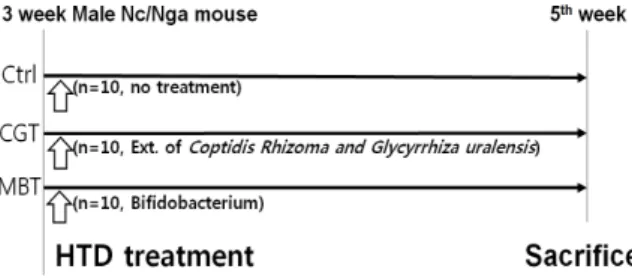
실험동물은 중앙실험동물 (Seoul, Korea)에서 분양 받 은 태령 3주된 NC/Nga 수컷 생쥐 (13~15 g)를 사용하였 다. 5주령군 (Ctrl), 황련감초 하태독법 시행군 (CGT), 프 로바이오틱스 처리군 (BBT)으로 나누었으며, 각 군에 10마리씩 배정하였다. 황련감초 하태독법 시행군은 황 련감초 추출물 10 ㎎/㎏을 경구 투여하는 하태독법을 실시하였다. 프로바이오틱스 처리군에는 Bifidobacterium (BB12) 20 ㎎/㎏을 경구 투여하였다. 본 연구과정은 부산 대학교 IACUC 승인을 받아 시행되었으며 (IACUC num- ber: PNU-2015-0924), 실험실 동물의 관리와 사용에 대해 서는 NIH 가이드라인에 따라 시행되었다.

Fig. 1. Protocol of Hataedock treatment

In the CGT group, 5-week-old mice were given the extract of Coptis japonica Makino and Glycyrrhiza uralensis Fischer. In the BBT group, 5-week-old mice were given a Bifidobacterium. On 5th week, the mice were deeply anesthetized and killed. Abbreviation. HTD: Hataedock.

3. 조직절편제작

하태독법 실시 2주 후 sodium pentobarbital 용액으 로 마취하여 처치하였다. 얻어진 등쪽 피부를 10%

NBF에 실온에서 24시간 동안 고정한 후 통상적인 방 법으로 paraffin에 포매하고 5 ㎛ 두께로 연속절편을 만 들었다.

4. 조직화학

피부각질층 내 지방장벽의 분포를 관찰하기 위해 Wright-green method를 실시하였다. Wright stain을 통 해 핵 염색 후 Methyl green solution에 10분간 노출시켜 각질층주변을 염색하였다.

5. 면역조직화학

피부각질층 내 지방장벽 구성성분의 변화를 관찰하 기 위해 면역조직화학을 실시하였다. 피부절편을 pro- teinase K (20 ㎍/㎖)에 5분 동안 proteolysis 과정을 거친

후 blocking serum인 10% normal mouse serum에서 2시 간 동안 반응시켰다. 그리고 1차 항체인 rabbit an- ti-Loricrin (1:50, Santa Cruz Biotec, USA), rabbit an- ti-Involucrin (1:50, Santa Cruz Biotec, USA), rabbit an- ti-Filaggrin (1:100, Santa Cruz Biotec, USA), rabbit an- ti-Acid sphingomyelinase (ASM; 1:50, Santa Cruz Biotec) 에 4 ℃ humidified chamber에서 72시간 동안 반응시켰 다. 그런 다음 2차 항체인 biotinylated mouse anti-rabbit IgG (1:100, Santa Cruz Biotec, USA)에 실온에서 24시 간 link한 후, avidin biotin complex kit (Vector Lab, USA)에 1시간 동안 실온에서 반응시켰다. 0.05%

3,3'-diaminobenzidine과 0.01% HCl이 포함된 0.05M tris-HCl 완충용액 (pH 7.4)에서 발색시킨 후, hematox- ylin으로 대조 염색하였다.

6. 영상분석

조직화학과 면역조직화학 결과는 image Pro Plus (Media cybernetics, USA)를 이용한 영상분석을 통해 수 치화 (means ± standard error)했다. 각 군의 표본에서 임 의로 선정된 피부절편을 x400 배율에서 촬영한 다음 positive pixels / 10,000,000 pixels로 영상분석 하였다.

7. 통계처리

영상분석 결과의 통계는 SPSS software (SPSS 23, SPSS Inc., USA)로 이루어졌으며, one-way ANOVA 시 행을 통해 유의성 (P<0.05)을 검증하고 Duncan’s multi- ple range test로 사후 검증하였다.

Ⅲ. Results

1. 지방장벽 형성

CGT군의 각질층 내에서 관찰된 Wright-green 양성 반응은 Ctrl군에 비해 193% 증가된 199,570 ± 2,489 / 10,000,000 pixel로 나타났다. BBT군는 CGT군보다 37% 적은 126,705 ± 2,721 / 10,000,000 pixel로 관찰되 었다 (Fig. 2).

2. 각질세포 단백질외막 형성

CGT군의 각질층 내에서 관찰된 involucrin 양성반응

은 Ctrl군에 비해 90% 증가된 138,197 ± 2,088 /

10,000,000 pixel로 나타났다. BBT군는 CGT군보다

(4)

21% 적은 109,140 ± 2,254 / 10,000,000 pixel로 관찰되 었다 (Fig. 3).

CGT군의 각질층 내에서 관찰된 loricrin 양성반응은 Ctrl

군에 비해 231% 증가된 154,850 ± 2,350 / 10,000,000 pixel 로 나타났다. BBT군는 CGT군보다 28% 적은 112,203 ± 3,181 / 10,000,000 pixel로 관찰되었다 (Fig. 3).

Fig. 2. Activation of lipid barrier's generation by extract of the extract of Coptis japonica Makino and Glycyrrhiza uralensis Fischer (CGE) and Bifidobacterium

The Wright-green positive reaction increased in SC (arrow) of the CGT & BBT (Wright-green method; Bar size, 50 ㎛). Data of Wright-green image analysis was also shown same result (p<0.05). Abbreviations. SC, stratum corneum; Ctrl, no treated 5th week ages; CGT, CGE of Hatedock treated 5th week ages; BBT, Bifidobacterium of Hatedock treated 5th week ages; *, p<0.05, compared with the Ctrl; #, p<0.05, compared with the BBT.

Fig. 3. Activation of involucrin & loricrin production by CGE and Bifidobacterium

The loricrin positive reaction increased in SC (arrow) of the CGT & BBT group compared with the Ctrl group (involucrin & loricrin immunohistochemistry;

Bar size, 50 ㎛). Data of involucrin & loricrin image analysis was also shown same result (p<0.05). Abbreviations same as Fig. 2.

(5)

3. 각질세포사이 matrix 단백질 생성

CGT군의 각질세포사이 matrix 단백질인 filaggrin 양성반응은 Ctrl군에 비해 143% 증가된 116,617 ± 3,060 / 10,000,000 pixel로 나타났다. BBT군는 CGT군 보다 29% 적은 83,244 ± 1,739 / 10,000,000 pixel로 관찰되었다 (Fig. 4).

4. Ceramide 형성

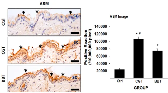
Ceramide 전환효소인 ASM 양성반응은 CGT군 에서 Ctrl군에 비해 341% 증가된 105,649 ± 2,145 / 10,000,000 pixel로 나타났다. BBT군는 CGT군보다 30% 적은 74,140 ± 1,823 / 10,000,000 pixel로 관찰되 었다 (Fig. 5).

Fig. 4. Activation of filaggrin production by CGE and Bifidobacterium

The filaggrin positive reaction increased in SC (arrow) of the CGT & BBT group compared with the Ctrl group (filaggrin immunohistochemistry;

Bar size, 50 ㎛). Data of filaggrin image analysis was also shown same result (p<0.05). Abbreviations same as Fig. 2.

Fig. 5. Activation of acid sphingomyelinase (ASM) by CGE and Bifidobacterium

The ASM positive reaction increased in SC (arrow) of the CGT & BBT group compared with the Ctrl group (ASM immunohistochemistry; Bar size, 50 ㎛). Data of ASM image analysis was also shown same result (p<0.05). Abbreviations same as Fig. 2.

(6)

Ⅳ. Discussion

피부의 가장 중요한 기능은 수분유지 및 병원균 및 유해물질의 침입을 방어하는 피부장벽으로서의 역할 이다

27)

. 최근 피부질환과 피부장벽기능의 관련성에 대 한 연구들이 보고되었으며

28,29)

, 피부장벽 및 수분유지 기능강화를 위한 피부질환 치료제 개발연구가 지속적 으로 진행되고 있다

30)

.

피부장벽기능은 주로 표피층이 담당한다. 표피의 기 저층에 분포된 각질형성세포는 분열 후 상층으로 이동 하며 분화되고, 이러한 각질세포의 분화에 의해 조절 받는 유전자의 발현에 따라 변화가 일어나게 된다

31)

. 발현을 촉진하는 유전자로는 transglutaminase 1 및 3, involucrin, loricrin, cornifin, filaggrin, small proline-rich protein (SPR) 등이 있다

32)

.

피부 각질층에 존재하는 ceramide, cholesterol, free fatty acid를 포함하는 각질세포간 지질은 일반적으로 피부장벽 및 수분유지에 중요한 것으로 알려져 있다

33)

.

ceramide는 각질세포의 외부를 둘러싸고 있는 대표 적인 수분 보존물질로서

34)

, 아토피피부염 환자의 피부 에서 발현이 감소되어 있다

35)

. ceramide가 감소하는 원 인으로는 sphingomyelin deacylase와 glucosyl ceramide deacylase의 증가에 따른 ceramide의 비정상적인 대사 증가, glucosylceramidase 및 sphingomyelinase에 의한 ce- ramide의 합성이상, ceramidase에 의한 ceramide의 과도 한 분해 등이 있다

36)

.

free fatty acid은 표피의 pH를 4.5-5.5의 약산성으로 유지하며 피부장벽의 항상성 유지에 기여하는 물질로 서

37)

, phospholipids가 표피의 과립층과 각질층의 경계 선상에 분비된 secretory phospholipase A2 (sPLA2)에 의 해 대사되어 생성된다

38)

. 산성 pH에서는 피부 손상 후 정상적인 피부장벽의 회복과정을 보이지만 pH가 증가 하면 회복이 지연될 뿐 아니라 serine protease의 활성 증가 및 각질교소체 (corneodesmosome)의 분해 촉진을 유도하여 각질층의 견고함을 감소시킨다. 또한 β -glucocerebrosidase와 ASM (acidic sphingomyelinase)의 기능 감소를 유발하여 각질세포의 지질막 형성에 중요 한 역할을 하는 ceramide의 생성 또한 감소시킨다

39)

. 또한 각질세포는 피부 분화과정의 최종 산물로 지 속적으로 탈락되고 새로운 분화과정을 거친 각질세포 로 대치되는데, 각질세포의 탈락에 관여하는 단백질 분해효소의 경우에도 각질층의 pH에 의해 조절된다

40,41)

. 피부표면의 pH가 증가하면 serine protease의 활성 이 증가하게 되고, 이는 비정상적으로 각질교소체의 구성 성분을 분해하여 각질층의 응집력 이상을 초래함 으로써 각질세포의 탈락을 촉진시켜 피부장벽 기능의 이상을 초래하게 된다

42-44)

. 아토피피부염에서 비병변 부위보다 병변 부위 피부의 pH가 더 높게 측정되며, 이로 인해 정상적인 피부장벽 기능을 수행하지 못하게 된다

45)

.

cholesterol은 노화 피부에서 나타나는 합성 장애가 가장 두드러지는 특징이며, 이로 인한 피부장벽 기능 저하와 연관성이 보고된 바 있다

46)

.

본 연구에서는 하태독법의 아토피피부염 유발 조절 효과, 각질층 손상, 표피세포의 과형성 감소, 피부 상피 장벽의 유지효과 증진 등 기존 연구를 기반으로 황련 감초 하태독법의 피부장벽형성 증진효과에 미치는 영 향을 확인하고자 하였다.

표피 각질세포의 분화 촉진인자 중 하나인 in- volucrin은 α-helix 구조를 갖는 가용성 단백질로 각질 세포막 (cornified cell envelope, CE)을 구성하는 전구단 백질 중 처음으로 기술된 단백질이다. 완성된 각질세 포막의 가장 바깥쪽에 밀집해 있고, 표피의 분화 표지 자로 사용되며, 여러 전구단백질이 cross-linking되는 과정에서 scaffold 역할을 수행한다

47)

.

loricrin은 과립층에서 발현되며 각질세포막을 구성 하는 주요 단백질로서, 총단백 무게의 70-85%를 차지 한다. loricrin-loricrin, loricrin-SPRs 간의 cross-linking이 일어나 각질세포막을 강화하는 역할을 한다

47)

.

involucrin과 loricrin은 표피 분화와 피부장벽형성의 중요한 단백질로 알려져 있으며

31)

, 아토피피부염 환자 의 경우 과발현된 Th2 cytokines에 의해 유발된 IL-4, IL-13이 STAT-6 신호경로를 활성화하여 involucrin, loricrin을 down-regulation시킴으로써 발현이 현저하게 감소함을 확인하였다

48)

.

filaggrin은 keratin을 응집시키는 matrix 단백질로 작 용하며, 여러 분해효소에 의해 자유 아미노산으로 분 해되어 물과 결합함으로써 천연보습인자로 작용할 뿐 아니라 각질층의 pH 조절 및 UV 필터기능을 갖는다

49)

. filaggrin은 아토피피부염의 대표적 유전인자이다. fi- laggrin 변이는 아토피피부염에서 피부장벽 손상을 유 발하고, 손상된 피부장벽을 통해 IgE 감작이 용이해져 궁극적으로 피부 염증 반응을 유발시킨다

50)

.

ASM (acid sphingomyelinase)은 sphingomyelin을 ce-

ramide로 전환하는 분해효소로서, 피부장벽의 발달에

(7)

주요한 역할을 한다

51)

. 아토피피부염에서 ASM의 활성 이 감소되면 ceramide, involucrin, loricrin, filaggrin 등 이 감소되어 피부장벽 회복의 장애 원인이 된다

52)

.

전통적으로 아토피피부염의 기전은 Type 2 immune response가 관여하는 염증이라는 견해가 우세하였으나, 표피의 투과성과 항균장벽의 기능 손상에 의한 피부장 벽 이상으로 기인한 결과라는 outside-inside 개념으로 바뀌고 있다

53)

. 더 나아가 면역학적 기전에 의해 발생 한 피부의 염증 반응이 또다시 피부장벽의 손상을 유 발한다는 outside-inside-outside 가설이 제시되었고, 아 토피피부염의 피부장벽 손상은 Type 2 cytokine medi- ated 염증 반응을 유발할 뿐 아니라 유발된 Type 2 cyto- kine mediated 염증 반응은 다시 피부장벽을 손상시킨 다는 연구

54)

를 통해 피부장벽 이상과 면역학적 이상에 대한 이해를 새롭게 하고 있다. 실제로 Th2 cytokine은 직접적인 피부장벽 손상 후 회복률을 저하시키고, 분 화된 각질세포의 주요 성분인 filaggrin, loricrin, in- volucrin의 발현을 억제하며, 표피 지질인 ceramide의 합성 및 선천 면역을 담당하는 항균 펩타이드의 발현 을 억제하는 것으로 알려져 있다

48,55,56)

.

본 연구는 이러한 피부장벽의 변화를 폐주피모 (肺 主皮毛) 이론과 폐-대장의 한의학적 개념과 연관이 있 을 것으로 생각되어 프로바이오틱스 처리군을 대조군 으로 하여 연구를 진행하였다. 실제 프로바이오틱스 (probiotics)는 인체에 이로움을 주는 살아있는 미생물 로서, 장내에서 증식하여 과민성 대장 증후군, 염증성 장질환 등의 질병 개선에 효과가 있다

26)

고 보고되고 있다.

본 연구에서는 황련감초 하태독법의 피부장벽형성 에 대한 효과를 확인하기 위해 각질층 내 involucrin, loricrin, filaggrin, ASM의 양성반응을 프로바이오틱스 와 비교하여 확인하였다. 황련감초 하태독법 시행 후 관찰한 각질층 내 양성반응에서는 involucrin, loricrin, filaggrin, ASM 모두에서 통계적으로 유의하게 증가하 였음을 확인하였다. 이러한 결과는 황련감초 추출물이 각질형성세포의 분화를 촉진시켜 피부장벽을 개선 및 강화시킬 수 있음을 의미한다.

이상의 결과로 황련감초 하태독법은 각질형성세포 분화시 촉진되는 단백질의 발현을 증가시켜 각질세포 막의 형성과 기능유지를 통해 피부장벽형성을 증진시 키는 것으로 생각된다. 아울러 향후 후속연구를 통해 하태독법의 각질형성세포 분화와 관련된 기전 및 유효 성의 검증이 필요할 것으로 생각된다.

Ⅴ. Conclusion

황련감초 하태독법의 피부장벽형성에 대한 증진효 과를 확인하기 위한 본 연구는 태령 3주의 약물 처리된 NC/Nga 생쥐의 피부 조직의 각질세포 형성 조절인자 의 변화 및 피부 조직 형성 정도를 면역조직학적 변화 를 통해 관찰한 결과 다음과 같은 결과를 얻었다.

1. 각질층 내 involucrin 양성반응은 황련감초 하태 독법 시행군에서 유의하게 증가하였다.

2. 각질층 내 loricrin 양성반응은 황련감초 하태독법 시행군에서 유의하게 증가하였다.

3. 각질층 내 filaggrin 양성반응은 황련감초 하태독 법 시행군에서 유의하게 증가하였다.

4. 각질층 내 ASM 양성반응은 황련감초 하태독법 시행군에서 유의하게 증가하였다.

Ⅵ. Acknowledgement

이 연구는 2016년도 정부 (교육부)의 재원으로 한국 연구재단의 지원을 받아 수행된 기초연구사업임 (No.

NRF-2016R1D1A1B03930474).

References

1. Kim YJ, Ahn JY, Seo SJ, Hong CK. The effect of retinoic acid on expression of human beta defensin-2 and LL-37 in keratinocyte. J Korean Invest Dermatol.

2007;14(1):29-35.

2. Kwon MS, Choi TB, Kim GY. The effect on the skin barrier function of ceramide. Korean J Aesthet Soc.

2005;3(1):131-7.

3. Yoon YM, Yoon JY, Lim KM, Hahn HJ, Kim YR,

Ahn KJ, An SK. Effects of the complex containing

Centella asiatica - and folic acid-fermented extracts, acetyl

glutamine, and nicotinic acid adenine dinucleotide phos-

phate on the inhibition of senescence and melanogenesis,

promotion of collagen expression, cellular regeneration,

and keratinocyte differentiation, and anti-inflammation.

(8)

Korean J Aesthet Soc. 2013;11(4):675-84.

4. Steinert PM, Marekov LN. The proteins elafin, filaggrin, keratin intermediate filaments, loricrin, and small pro- line-rich proteins 1 and 2 are isodipeptide cross-linked components of the human epidermal cornified cell envelope. J Biol Chem. 1995;270(30):17702-11.

5. Steven AC, Steinert PM. Protein composition of cornified cell envelopes of epidermal keratinocytes. J Cell Sci.

1994;107(2):693-700.

6. Elias PM, Menon GK. Structural and lipid biochemical correlates of the epidermal permeability barrier. Adv Lipid Res. 1991;24:1-26.

7. Steinert PM, Cantieri JS, Teller DC, Lonsdale-Eccles JD, Dale BA. Characterization of a class of cationic proteins that specifically interact with intermediate filaments. Proc Natl Acad Sci USA. 1981;78(7):4097-101.

8. Cornelissen C, Marquardt Y, Czaja K, Wenzel J, Frank J, Luscher-Firzlaff J, Lüscher B, Baron J. IL-31 regulates differentiation and filaggrin expression in human organo- typic skin models. J Allergy Clin Immunol. 2012;

129(2):426-33.

9. Irvine AD, McLean WH. Breaking the (un)sound barrier:

filaggrin is a major gene for atopic dermatitis. J Invest Dermatol. 2006;126(6):1200-2.

10. Palmer CN, Irvine AD, Terron-Kwiatkowski A, Zhao Y, Liao H, Lee SP, Goudie DR, Sandilands A, Campbell LE, Smith FJ, O'Regan GM, Watson RM, Cecil JE, Bale SJ, Compton JG, DiGiovanna JJ, Fleckman P, Lewis-Jones S, Arseculeratne G, Sergeant A, Munro CS, Houate BE, McElreavey K, Halkjaer LB, Bisgaard H, Mukhopadhyay S, McLean WH. Common loss-of-func- tion variants of the epidermal barrier protein filaggrin are a major predisposing factor for atopic dermatitis.

Nat Genet. 2006;38(4):441-6.

11. O'Regan GM, Irvine AD. The role of filaggrin in the atopic diathesis. Clin Exp Allergy. 2010;40(7):965-72.

12. Barnes KC. An update on the genetics of atopic dermati- tis: scratching the surface in 2009. J Allergy Clin Immunol. 2010;125(1):16-29.

13. Kim BB, Kim JR, Kim JH, Kim YA, Park JS, Yeom MH, Lee HJ, Lee KW, Kang NJ. 7,3’,4’-Trihydroxyisoflavone ameliorates the development of Dermatophagoides far- inae-induced atopic dermatitis in NC/Nga mice. Evid

Based Complement Alternat Med. 2013;2013:1-10.

14. Kang MY, Jang GT, Kim JH. A study on fetal toxicosis removal therapy. J Pediatr Korean Med. 2003;17(1):

9-51.

15. Tang J, Feng YB, Tsao S, Wang N, Curtain R, Wang YW. Berberine and Coptidis rhizoma as novel antineo- plastic agents: a review of traditional use and biomedical investigations. J Ethnopharmacol. 2009;126(1):5-17.

16. Kaboli PJ, Rahmat A, Ismail P, Ling KH. Targets and mechanisms of berberine, a natural drug with poten- tial to treat cancer with special focus on breast cancer.

Eur J Pharmacol. 2014;740:584-95.

17. Ortiz LM, Lombardi P, Tillhon M, Scovassi AI. Berberine, an epiphany against cancer. Molecules. 2014;19(8):

12349-67.

18. Yu M, Tong X, Qi B, Qu H, Dong S, Yu B. Berberine enhances chemosensitivity to irinotecan in colon cancer via inhibition of NF-κB. Mol Med Rep. 2014;9(1):

249-54.

19. Chin LW, Cheng YW, Lin SS, Lai YY, Lin LY, Chou MY, Chou MC, Yang CC. Anti-herpes simplex virus effects of berberine from Coptidis rhizoma, a major compo- nent of a Chinese herbal medicine, Ching-Wei-San. Arch Virol. 2010;155(12):1933-41.

20. Yu YE, Park EY, Jung DH, Byun SH, Kim SC, Park SM. Antibacterial activity of oriental medicinal herb extracts against skin pathogens. J Life Sci. 2010;20(7):

1143-50.

21. Miao KL, Zhang JZ, Dong Y, Xi YF. Research progress on the chemical compounds and pharmacology of Sophora flavescens. Nat Prod Res Dev. 2001;13(2):69-73.

22. Kusano G, Shibano M, Watanabe H. Pharmaceutical botanical studies on some Glycyrrhiza species. Yakugaku Zasshi. 2003;123(8):619-31.

23. Kim HJ, Bae JY, Jang HN, Park SN. Comparative study on antimicrobial activity of Glycyrrhiza uralensis and Glycyrrhiza glabra extracts in various country of origin as natural antiseptics. J Microbiol Biotechnol. 2013;

41(3):358-66.

24. Kim HJ, Jang HN, Bae JY, Ha JH, Park SN.

Antimicrobial activity, quantification and bactericidal

activities of licorice active ingredients. J Microbiol

Biotechnol. 2014;42(2):386-92.

(9)

25. Cha HY, Ahn SH, Jeong AR, Cheon JH, Park SY, Choi JY, Kim KB. Anti-inflammatory effects of Hataedock extracted from Coptidis rhizoma and Glycyrrhiza uralensis on atopic dermatitis-like skin lesions of NC/Nga mouse. J Int Korean Med. 2015;36(4):486-97.

26. Nobaek S, Johansson ML, Molin G, Ahrne S, Jeppsson B. Alteration of intestinal microflora is associated with reduction in abdominal bloating and pain in patients with irritable bowel syndrome. Am J Gastroenterol.

2000;95(5):1231-8.

27. Forslind B. A domain mosaic model of the skin barrier.

Acta Derm Venereol. 1994;74(1):1-6.

28. Mohsenzadeh M. Evaluation of antibacterial activity of selected Iranian essential oils against Staphylococcus aureus and Escherichia coli in nutrient broth medium. Pak J Biol Sci. 2007;10(20):3693-7.

29. Rasul A, Akhtar N, Khan BA, Mahmood T, Uz Zaman S, Khan HM. Formulation development of a cream con- taining fennel extract: in vivo evaluation for anti-aging effects. Pharm. 2012;67(1):54-8.

30. Yu HY, Yang IJ, Lincha VR, Park IS, Lee DU, Shin HM. The effects of the fruits of Foeniculum vulgare on skin barrier function and hyaluronic acid production in HaCaT keratinocytes. J Life Sci. 2015;25(8):880-8.

31. Steinert PM, Marekov LN. The proteins elafin, filaggrin, keratin intermediate filaments, loricrin, and small pro- line-rich proteins 1 and 2 are isodipeptide cross-linked components of the human epidermal cornified cell envelope. J Biol Chem. 1995;270(30):17702-11.

32. Kwon YB, Choi DK, Sohn KC, Jeon EK, Nam MS, Lee JH, Kim CD. Effects of colostrum on keratinocyte differentiation and wound healing. Korean J Invest Dermatol. 2007;14(2):45-50.

33. Imokawa G, Abe A, Jin K, Higaki Y, Kawashima M, Hidano A. Decreased level of ceramides in stratum cor- neum of atopic dermatitis: an etiologic factor in atopic dry skin?. J Invest Dermatol. 1991;96(4):523-6.

34. Choi MJ, Maibach HI. Role of ceramides in barrier function of healthy and diseased skin. Am J Clin Dermatol. 2005;6(4):215-23.

35. Hara J, Higuchi K, Okamoto R, Kawashima M, Imokawa G. High-expression of sphingomyelin deacylase is an important determinant of ceramide deficiency leading

to barrier disruption in atopic dermatitis. J Invest Dermatol. 2000;115(3):406-13.

36. Murata Y, Ogata J, Higaki Y, Kawashima M, Yada Y, Higuchi K, Tsuchiya T, Kawaminami S, Imokawa G. Abnormal expression of sphingomyelin acylase in atopic dermatitis: an etiologic factor for ceramide defi- ciency?. J Invest Dermatol. 1996;106(6):1242-9.

37. Fluhr JW, Kao J, Jain M, Ahn SK, Feingold KR, Elias PM. Generation of free fatty acids from phospholipids regulates stratum corneum acidification and integrity.

J Invest Dermatol. 2001;117(1):44-51.

38. Song JH, Ahn SH, Cheon JH, Park SY, Kim HH, Kim KB. Effects of Hataedock with Douchi on 2,4-dini- trofluorobenzene-induced atopic dermatitis-like skin le- sion in NC/Nga mice. J Physiol Pathol Korean Med.

2016;30(2):109-15.

39. Hachem JP, Crumrine D, Fluhr J, Brown BE, Feingold KR, Elias PM. pH directly regulates epidermal perme- ability barrier homeostasis and stratum corneum in- tegrity/cohesion. J Invest Dermatol. 2003;121(2):345-53.

40. Proksch E, Brandner JM, Jensen JM. The skin: an indis- pensable barrier. Exp Dermatol. 2008;17(12):1063-72.

41. Baroni A, Buommino E, De Gregorio V, Ruocco E, Ruocco V, Wolf R. Structure and function of the epi- dermis related to barrier properties. Clin Dermatol.

2012;30(3):257-362.

42. Hachem JP, Man MQ, Crumrine D, Uchida Y, Brown BE, Rogiers V, Roseeuw D, Feingold KR, Elias PM.

Sustained serine proteases activity by prolonged increase in pH leads to degradation of lipid processing enzymes and profound alterations of barrier function and stratum corneum integrity. J Invest Dermatol. 2005;125(3):510-20.

43. Hachem JP, Crumrine D, Fluhr J, Brown BE, Feingold KR, Elias PM. pH directly regulates epidermal perme- ability barrier homeostasis and stratum corneum in- tegrity/cohesion. J Invest Dermatol. 2003;121(2):345-53.

44. Schmid-Wendtner MH, Korting HC. The pH of the skin surface and its impact on the barrier function.

Skin Pharmacol Physiol. 2006;19(6):296-302.

45. Rippke F, Schreiner V, Doering T, Maibach HI. Stratum

corneum pH in atopic dermatitis: impact on skin barrier

function and colonization with Staphylococcus aureus. Am

J Clin Dermatol. 2004;5(4):217-23.

(10)

46. Elias PM, Ghadially R. The aged epidermal permeability barrier: basis for functional abnormalities. Clin Geriatr Med. 2002;18(1):103-20.

47. Steinett PM, Marekov LN. Direct evidence that in- volucrin is a major early isopeptide cross-linked compo- nent of the keratinocyte cornified cell envelope. J Biol Chem. 1997;272(3):2021-30.

48. Kim BE, Leung DY, Boguniewicz M, Howell MD.

Loricrin and involucrin expression is down-regulated by Th2 cytokines through STAT-6. Clin Immunol.

2008;126(3):332-7.

49. Smith FJ, Irvine AD, Terron-Kwiatkowski A, Sandilands A, Campbell LE, Zhao Y, Liao H, Evans AT, Goudie DR, Lewis-Jones S, Arseculeratne G, Munro CS, Sergeant A, O'Regan G, Bale SJ, Compton JG, DiGiovanna JJ, Presland RB, Fleckman P, McLean WH. Loss-of-function mutations in the gene encoding filaggrin cause ichthyosis vulgaris. Nat Genet. 2006;38(3):337-42.

50. O'Regan GM, Irvine AD. The role of filaggrin in the atopic diathesis. Clin Exp Allergy. 2010;40(7):965-72.

51. Pullmannová P, Staňková K, Pospíšilová M, Skolová B, Zbytovská J, Vávrová K. Effects of sphingomyelin/ce- ramide ratio on the permeability and microstructure of model stratum corneum lipid membranes. Biochim

Biophys Acta. 2014;1838(8):2115-26.

52. Jensen JM, Fölster-Holst R, Baranowsky A, Schunck M, Winoto-Morbach S, Neumann C, Schütze S, Proksch E. Impaired sphigomyelinase activity and epidermal dif- ferentiation in atopic dermatitis. J Invest Dermatol.

2004;122(6):1423-31.

53. Elias PM, Schmuth M. Abnormal skin barrier in the etiopathogenesis of atopic dermatitis. Curr Opin Allergy Clin Immunol. 2009;9(5):437-46.

54. Elias PM, Steinhoff M. “Outside-to-inside” (and now back to “outside”) pathogenic mechanisms in atopic dermatitis. J Invest Dermatol. 2008;128(5):1067-70.

55. Hatano Y, Terashi H, Arakawa S, Katagiri K.

Interleukin-4 suppresses the enhancement of ceramide synthesis and cutaneous permeability barrier functions induced by tumor necrosis factor-alpha and interfer- on-gamma in human epidermis. J Invest Dermatol.

2005;124(4):786-92.

56. Albanesi C, Fairchild HR, Madonna S, Scarponi C, Pità OD, Leung DY, Howell MD. IL-4 and IL-13 negatively regulate TNF-alpha- and IFN-gamma-induced beta-de- fensin expression through STAT-6, suppressor of cyto- kine signaling (SOCS)-1, and SOCS-3. J Immunol.

2007;179(2):984-92.

수치

Fig. 1. Protocol of Hataedock treatment
Fig. 3. Activation of involucrin &amp; loricrin production by CGE and Bifidobacterium
Fig. 5. Activation of acid sphingomyelinase (ASM) by CGE and Bifidobacterium

참조

관련 문서

First, after the Group Art Therapy, the scale of hopelessness depression in the test group decreased significantly in comparison with that before the group

In 4-week group, the group filled with bone graft with decortication revealed larger new bone formation area than shown in the group that had a defect area

[r]

In the region of Auerbach’s plexus (AP) of small intestine, the staining intensity of the ICC-IR cells was reduced in the HFD group compared to the control group.. The numbers

Actinic keratoses showed a trend towards increased expression in the basal layer compared with normal skin.. In actinic keratoses, Keratoacanthomas and seborrheic

The result suggested that a taekwon aerobics program had a positive effect on the increase of growth hormone in blood lipid in middle school girls.. The

The 24hr proteinuria level were increased in kidney with cyclosporine-A group but significantly decreased in iNOS inhibitor and green tea polyphenol group..

• Apply the principle of work and energy between the initial position and the point at which the spring is fully compressed and the velocity is zero. The only unknown in