Korean J Androl. Vol. 29, No. 1, April 2011
33
접수일자: 2011년 3월 18일, 수정일자: 2011년 4월 5일, 게재일자: 2011년 4월 11일
교신저자: 김세웅, 가톨릭대학교 서울성모병원 비뇨기과, 서울시 서초구 반포동 505 137-701 Tel: 02-2258-1071, Fax: 02-2258-1080, E-mail: [email protected]
*본 연구는 농촌진흥청 바이오그린21사업 (과제번호 PJ007186)에 의해 이루어진 것임.
정계정맥류 유발 백서에서 고환조직의 변화와 정자 형성에 대한 안토시아닌의 효과
1가톨릭대학교 의과대학 비뇨기과학교실,
2한국전통의학연구소
장 훈
1ㆍ김수진
1ㆍ육승모
1ㆍ한동석
1ㆍ하유신
1홍성후
1ㆍ이지열
1ㆍ황태곤
1ㆍ황성연
2ㆍ김세웅
1The Changes of Testis and the Effects of Anthocyanin on Spermatogenesis in Rat Induced Varicocele
Hoon Jang1, Su Jin Kim1, Seung Mo Yuk1, Dong Seok Han1, U Syn Ha1,
Sung Hoo Hong1, Ji Youl Lee1, Tae Kon Hwang1, Sung Yeoun Hwang2, Sae Woong Kim1
1Department of Urology, The Catholic University of Korea College of Medicine,
2Korea Bio Medical Science Institute, Seoul, Korea
= Abstract =
Purpose: Varicocele is known as a main cause of primary male infertility and it supposed to be associated with oxidative stress. Anthocyanin is known as a natural plant pigment and novel antioxidant. This study was designed to investigate the effects of anthocyanin on a rat model of varicocele.
Materials and Methods: Twenty four male rats, induced varicocele by partial obstruction of left renal vein, were divided into four experimental groups: the group induced varicocele for four weeks without anthocyanin, the group received anthocyanin (80 mg/kg) right after varicocele induction, group induced varicocele for eight weeks without anthocyanin, and the group received anthocyanin (80 mg/kg) after four weeks observation following varicocele induction. After anthocyanin treatment, testes from the rats in all groups were removed, weighed, and subjected to histological examination. Apoptosis in the testes was measured by the TUNEL assay. And the oxidative stress was evaluated by measurement of 8-hydroxy-2'-deoxyguanosine (8-OHdG).
Results: Induction of varicocele led to decreasing left testis weight, decreasing spermatogenic cell density significantly (p<0.05). Also it led to increasing apoptotic body counts and increasing concentration of 8-OHdG significantly (p<0.05). However administration of anthocyanin right after varicocele induction prevent this change meaningfully (p<0.05). In group received anthocyanin after four weeks observation following varicocele induction, interestingly, there was no significant difference in testis weight, spermatogenic cell density, apoptotic body count and concentration of 8-OHdG compared to group induced varicocele for eight weeks without anthocyanin administration.
Conclusions: These results suggest that anthocyanin is effective in decreasing the oxidative stress of testis in rat induced varicocele and may be effective in making a healthy sperm in patient of varicocele in early stage. However in patient under way in advanced stage, it is supposed that the anthocyanin cannot help having a protective effect from oxidative stress narrowly unless the condition of oxidative stress by varicocele is corrected. Further studies are needed to better understand the mechanisms and actions of anthocyanin and varicocele, and these studies may lead
Table 1. Total and individual anthocyanin contents of Black Soybean
Cultivar Anthocyanin contents (μg/g)
Total
Dp3glc Cy3glc Pt3glc
Cheongja 3
% of total
3049.0 25.2
8277.2 68.3
791.7 6.5
12117.9 100 Dp3glc: delphinidin-3-O-glucoside, Cy3glc: cyanidin-3-O- glucoside, Pt3glc: petunidin-3-O-glucoside.
15%에서 발견된다. 성인의 경우 주로 무증상인 경 우가 많지만 원발성 남성불임환자의 35%, 속발성 남성불임환자의 81%를 차지하는 것으로 보고되고 있어1 남성 불임의 가장 큰 원인 질환으로 알려져 있다. 하지만 세포수준에서 정계정맥류와 남성 불 임의 명백한 관계는 아직 밝혀 지지 않았다. 비정상 정자생성 및 불임에 대한 기전에 관한 연구들을 살 펴보면 고환손상이 유발되는 기전으로 세포자멸사, 재활성 산화물질, 저산소증에 의한 허혈성 변화에 의한 것으로 생각되고 있어2-4 산화손상을 감소시키 는 항산화제의 치료제로서의 가능성이 제시되고 있고 이를 기본으로 항산화 물질을 이용한 치료관 련 연구가 시도되고 있다.
이에 본 연구진은 검정콩 종피로부터 추출한 안 토시아닌의 항산화 효과와 다양한 생리 활성을 전 립선 비대유발 모델에서 확인5하였고 이를 토대로 산화 스트레스와 정계정맥류의 발생 관계 그리고 정계정맥류 유발 모델에서 항산화 물질인 안토시 아닌의 효과를 알아보고자 하였다.
대상 및 방법
1. 안토시아닌의 추출 및 성분 분석
본 실험에 사용된 검은콩은 농촌진흥청으로부터
anidin-3-O-glucoside, 그리고 petunidin-3-O-glucoside 로 확인되었고 각 성분의 구성비는 다음과 같았다 (Table 1).
2. 실험동물 및 사육조건
실험동물은 생후 12주된 380∼400 g의 Sprague- Dawley 수컷 흰쥐(Samtako Bio Korea, Inc., Osan, Korea)를 제공받아 1주일간의 적응기간을 거친 뒤 플라스틱 사육함에 2마리씩 사육하였다. 사육장은 인공조명에 의하여 아침 7시부터 저녁 7시까지 12시 간으로 조절하였으며, 실내온도는 18∼23oC와 40∼
60%의 습도를 유지하고 정수된 식수와 사료를 자 유롭게 먹게 하였다. 본 연구는 Institutional Animal Care and Use Committee (IACUC) of the Catholic University of Korea의 승인하에 진행되었다 (IRB approval no. CUMC-2010-0106-02).
3. 안토시아닌 비투여군과 안토시아닌 투여군 실험동물 모두에서 정계정맥류를 유발 후 안토시 아닌 비투여군 (n=12)과 안토시아닌 투여군 (n=12) 으로 나누었다. 안토시아닌 비투여군은 희생도살 시기에 따라 4주군 (n=6)과 8주군 (n=6)으로 나누었 고, 안토시아닌 투여군은 투여시기에 따라 정계정 맥류 유발 직후 안토시아닌 투여군 (n=6)과 정계정 맥류 유발 후 4주간의 관찰기간 이후에 안토시아닌 을 투여하는 군 (n=6)으로 나누었다. 안토시아닌 투 여 군은 흰쥐의 체중당 80 mg/kg을 기준으로 검은 콩 종피 추출물을 증류수에 희석하여 4주 동안 매 일 1회 경구 투여시켰다.
4. 정계정맥류 유발
정계정맥류 유발은 좌신 정맥에서 생식샘 정맥의 분지부위 보다 근위부에서 결찰을 시행하여 좌신 정맥의 부분 폐색을 일으키는 Saypol 등6의 실험방
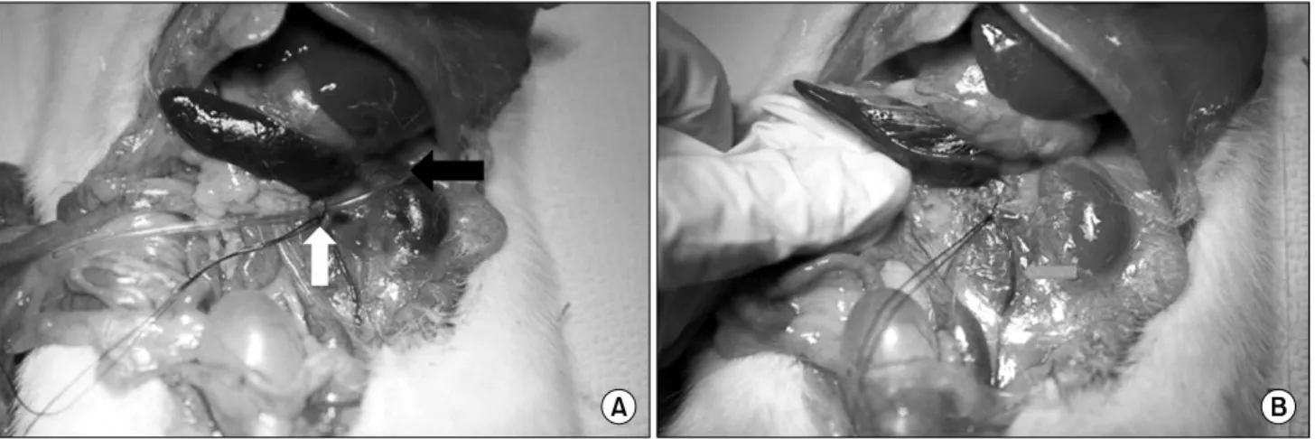
Fig. 1. (A) It shows ligation of proximal renal vein with plastic probe. Black arrow indicates plastic probe and white arrow indicates the engorgement of left gonadal vein and distal renal vein. (B) It shows partial obstruction of proximal renal vein after removal of plastic probe. Gray arrow indicates engorged left gonadal vein.
법을 이용하였다 (Fig. 1).
5. 고환 조직의 획득과 정모세포 밀도 측정 정계정맥류 유발 4주 후 안토시아닌 비투여 4주 군과 정계정맥류 유발 직후 안토시아닌 투여군을 희생 도살하였고 8주 후 안토시아닌 비투여 8주군 과 정계정맥류 유발 후 4주간의 관찰기간 이후에 안토시아닌 투여군을 희생 도살하였다. 각 군은 마 취 후 하복부 절개를 통해 양측 고환을 절제한 후 무게를 측정하고 조직을 획득하여 10% 중성 포르 말린에서 고정한 후 파라핀에 포매하여 절편을 얻 어 Hematoxyline-Eosin 염색을 시행하여 광학 현미 경으로 관찰하였다. 광학 현미경 400배 시야에서 비교적 원형에 가까운 정세관을 무작위 10곳을 선 정하여 정모 세포층의 두께와 정세관의 직경을 측 정하여 정모세포 밀도를 구하였다 (Fig. 2).
6. TUNEL (Terminal dexoynucleotidyl transferase mediated dUTP Nick End Labeling) assay 및 세 포자멸사의 평가
적출한 고환 조직에서 세포자멸사를 확인하기 위 하여 TUNEL assay를 시행하였다. TUNEL assay는 Apop Tag In Situ Apoptosis Detection Kits (Millipore Co., Massachusetts, US)을 사용하였고 고환 조직에 서 세포자멸사의 평가는 TUNEL assay에 양성을 보 이는 세포 수를 측정하여 각 군별 차이를 비교하였 다. TUNEL assay에 양성인 세포측정은 각 군의 슬 라이드에서 무작위 10부위를 선택하여 광학현미경
200배율에서 관찰되는 TUNEL assay에 양성인 세포 수를 측정하였다.
7. 산화 스트레스의 측정
고환 조직에서 산화 스트레스의 측정은 산화 변 형된 DNA를 반영하는 8-hydroxy-2'-deoxyguanosine (8-OHdG)를 정량적으로 측정하여 평가하였다.
DNeasy Blood & Tissue kit (Qiagen, Valencia, CA)를 이용하여 고환조직에서 DNA를 추출하고 DNA oxi- dation kit (Highly Sensitive 8-OHdG Check ELISA;
Japan Institute for the Control of Aging, Fukuroi, Japan)를 이용하여 8-OhdG을 측정하였다. 표준 8- OHdG (0.5∼40 ng/ml) 또는 고환조직으로부터 얻어 진 DNA를 8-OHdG로 미리 도포된 microtiter plate에 서 1시간 동안 8-OHdG에 대한 단일 클론 항체와 배양을 시키고 3,3',5,5'-tetramethylbenzidine을 첨가 하여 450 nm에서 흡광도를 측정하였다. 표준 곡선 과 수정 DNA 농도를 이용하여 조직 표본 농도를 계산하였다.
8. 통계분석방법
각 측정 수치를 평균±표준편차로 표기하였으며 통계는 SPSS for Microsoft Window 프로그램 (ver.
12.0)을 이용하였다. 통계학적 분석은 Kruskal-Wallis test, Mann-Witney U-test를 이용하여 분석하였으며 각 군 간의 비교는 Tukey 검정법을 시행하여 p값이 0.05 미만인 경우에 통계학적으로 유의한 것으로 판정하였다.
was no difference in spermato- genic cell density between (C) and (D) statistically (×400). (A) Group induced varicocele for 4 weeks without anthocyanin administration.
(B) Group received oral dose of anthocyanin (80 mg/kg) for 4 weeks right after varicocele induc- tion. (C) Group induced varico- cele for 8 weeks without antho- cyanin administration. (D) Group received oral dose of anthocyanin (80 mg/kg) for 4 weeks after 4 weeks observation following vari- cocele induction.
결 과
1. 고환의 무게
안토시아닌 비투여 4주군의 평균 우측 고환 무게 는 2.015±0.072 g이었고, 평균 좌측고환 무게는 1.480±0.035 g로 통계학적으로 유의하게 감소한 소 견을 보였다 (p<0.05). 한편 정계정맥류 유도 직후 부터 안토시아닌을 투여한 군의 평균 좌측 고환 무 게는 1.838±0.125 g으로 안토시아닌 비투여 4주군 에 비하여 통계학적으로 유의하게 증가한 소견을 보였다 (p<0.05). 한편 안토시아닌 비투여 8주군과 정계정맥류 유발 후 4주간의 관찰기간 이후에 안토 시아닌을 투여하는 군의 평균 좌측 고환무게는 1.320±0.065 g과 1.375±0.125 g으로 두 군 사이에 통 계학적으로 의미 있는 차이는 관찰되지 않았다 (p>
0.05) (Fig. 3).
2. 좌측 고환의 정모세포 밀도 (정모 세포층의 두 께/정세관의 직경)
안토시아닌 비투여 4주군에서 좌측 고환의 정모 세포 밀도는 0.272±0.026이었고 반면에 정계정맥류 유도 직후부터 안토시아닌을 투여한 군은 0.347±
0.029으로 나타나 안토시아닌 투여 군에서 정모세 포 밀도가 통계학적으로 의미 있게 증가되어 있었 다 (p<0.05). 한편 안토시아닌 비투여 8주군과 정계 정맥류 유발 후 4주간의 관찰기간 이후에 안토시아 닌을 투여한 군에서 좌측고환의 정모세포 밀도는 0.249± 0.031과 0.345±0.071으로 나타나 두 군 사이 에서 통계학적으로 유의한 차이는 관찰되지 않았다 (p>0.05) (Fig. 4).
3. 세포자멸사 확인 및 평가
세포자멸사가 발생한 세포는 세포고사체를 형성 하여 TUNEL assay에서 검정색 또는 짙은 갈색으로 관찰되었다 (Fig. 5). 안토시아닌 비투여 4주군, 정
Fig. 4. Spermatogenic cell density (germinal cell layer thickness/
diameter of seminiferous tubule). Spermatogenic cell density of group received anthocyanin right after varicocele induction is significantly higher than group induced varicocele varicocele for 4 weeks without anthocyanin administration statistically (p
<0.05). But there was no difference between group induced varicocele for 8 weeks without anthocyanin administration and group received anthocyanin after 4 weeks observation following varicocele induction statistically (p>0.05). *Anthocyanin 80 mg/kg: group received oral dose of anthocyanin (80 mg/kg) right after varicocele induction. **Anthocyanin 80 mg/kg:
group received oral dose of anthocyanin (80 mg/kg) after 4 weeks observation following varicocele induction.
Fig. 3. Mean weight of testes in each groups. Left testis weight of group received anthocyanin right after varicocele induction is significantly higher than group induced varicoele for 4 weeks without anthocyanin administration statistically (p<0.05). But there was no difference between group induced varicocele for 8 weeks without anthocyanin and group received anthocyanin after 4 weeks observation following varicocele induction statistically (p>0.05). *Anthocyanin 80 mg/kg: group received oral dose of anthocyanin (80 mg/kg) right after varicocele induction. **Anthocyanin 80 mg/kg: group received oral dose of anthocyanin (80 mg/kg) after 4 weeks observation following varicocele induction.
계정맥류 유도 직후부터 안토시아닌을 투여한 군, 안토시아닌 비투여 8주군 그리고 정계정맥류 유발 후 4주간의 관찰기간 이후에 안토시아닌을 투여한 군에서 TUNEL assay에 양성을 보이는 세포 수는 각 각 14.52±2.25, 6.30±1.10, 17.25±2.05, 그리고 15.15±
3.15으로 안토시아닌 비투여 4주군에 비하여 정계 정맥류 유발 후부터 안토시아닌을 투여한 군에서 TUNEL assay 양성 세포수가 통계학적으로 유의하 게 감소하였음을 확인하였다 (p<0.05) (Fig. 6). 하 지만 안토시아닌 비투여 8주군과 정계정맥류 유발 후 4주간의 관찰기간 이후에 안토시아닌을 투여한 군은 두 군 사이에 통계학적으로 유의한 차이는 보 이지 않았다 (p>0.05).
4. 고환 조직에서 산화스트레스의 측정
고환 조직에서 산화 스트레스는 고환 조직의 8- OHdG를 ELISA를 이용하여 정량적으로 측정하여 농도를 구하였다. 안토시아닌 비투여 4주군, 정계 정맥류 유도 후부터 안토시아닌을 투여한 군, 안토
시아닌 비투여 8주군 그리고 정계정맥류 유발 후 4주간의 관찰기간 이후에 안토시아닌을 투여한 군 에서 ELISA를 이용하여 측정한 8-OHdG의 농도는 각각 1.496±0.165, 0.582±0.105, 1.542±0.215, 그리고 1.516±0.114으로 안토시아닌 비투여 4주군에 비하 여 정계정맥류 유발 후부터 안토시아닌을 투여한 군에서 8-OHdG의 농도가 통계학적으로 유의하게 감소하였음을 확인하였다 (p<0.05) (Fig. 7). 하지만 안토시아닌 비투여 8주군과 정계정맥류 유발 후 4 주간의 관찰기간 이후에 안토시아닌을 투여한 군 은 두 군 사이에 통계학적으로 유의한 차이는 보이 지 않았다 (p>0.05).
고 찰
정계정맥류는 남성 불임 환자의 35%, 속발성 남 성 불임 환자의 81%를 차지하고 있어 비뇨기과 임
Fig. 5. Apoptotic bodies in TUNEL stain of left testis. Positive TUNEL stain cell called apoptotic body was showed in dark brown or black color in TUNEL stain (×200). (A) Counts of apoptotic bodies was more increased than that (B). (C) and (D) apoptotic bodies were more increased than that (A). But there was no difference in counts of apoptotic bodies between (C) and (D) statistically (×200). (A) Group induced varicocele for 4 weeks without anthocyanin administration. (B) Group received oral dose of anthocyanin (80 mg/kg) for 4 weeks right after varicocele induction. (C) Group induced varicocele for 8 weeks without anthocyanin administration. (D) Group received oral dose of anthocyanin (80 mg/kg) for 4 weeks after 4 weeks observation following varicocele induction.
상에서 주된 관심사 중의 하나이다. 현재까지 정계 정맥류 환자에서 확장된 덩굴 정맥 얼기를 개복7 및 복강경하8-10에서 수술적으로 결찰 하거나 혈관 조 영을 통하여 혈관을 폐쇄11시키는 수술 및 술기는 비정상 정액검사 소견을 가진 환자에게서 정액검 사 소견의 호전과 임신율 증가를 보이고 있어 현재 의 치료법을 적용할 환자의 선택이 임상에서 중요 하다고 할 수 있다. 하지만 세포 수준에서 정계정맥 류와 남성 불임의 명백한 관계는 아직 밝혀지지 않 았다. 성선 자극 호르몬 또는 남성 호르몬 분비의
감소,12,13 음낭 내 온도의 상승, 그리고 정맥혈 저류
에 의한 신정맥에서 정계정맥으로의 부신 및 신독
성 물질의 이동에 의한 저산소증 등의 가설들이 정 계정맥류 환자에서 정모세포 기능부전의 발생기전 으로 생각되고 있다.2 정확한 원인 및 발생기전을 밝히기 위하여 가임 남성에서 정계정맥류의 유무 에 따라 순차적 정액검사 및 고환 조직검사가 가장 이상적이지만 윤리적인 이유 등으로 본 연구진은 연 구에 필요한 충분한 조직을 얻을 수 있는 동물 실험 을 계획하였고 세포 수준에서의 결과를 얻었다.
본 연구에서 정계정맥류 유발 후 안토시아닌 비 투여 4주군은 우측 고환에 비하여 좌측 고환의 용 적이 유의하게 감소하였으며 좌측 고환에서의 정 모세포 밀도가 우측 고환의 정모세포 밀도에 비하
Fig. 6. Numbers of apoptotic bodies. In group received anthocyanin right after varicocele induction, count of apoptotic bodies is significantly lower than that of group induced varicocele for 4 weeks without anthocyanin administration statistically (p<0.05). But there was no difference between group induced varicocele for 8 weeks without anthocyanin.
administration and group received anthocyanin after 4 weeks observation following varicocele induction statistically (p>
0.05). *Anthocyanin 80 mg/kg: group received oral dose of anthocyanin (80 mg/kg) right after varicocele induction.
**Anthocyanin 80 mg/kg: group received oral dose of anthocyanin (80 mg/kg) after 4 weeks observation following varicocele induction
Fig. 7. Concentration of 8-OHdG (8-hydroxy-2'-deoxyguano- sine). In group received anthocyanin right after varicocele induction, concentration of 8-OHdG is significantly lower than that of group induced varicocele for 4 weeks without antho- cyanin administration statistically (p<0.05). But there was no difference between group induced varicocele for 8 weeks without anthocyanin administration and group received antho- cyanin after 4 weeks observation following varicocele induction statistically (p>0.05). *Anthocyanin 80 mg/kg: group received oral dose of anthocyanin (80 mg/kg) right after varicocele induction. **Anthocyanin 80 mg/kg: group received oral dose of anthocyanin (80 mg/kg) after 4 weeks observation following varicocele induction
여 통계학적으로 의미 있게 감소되었다. 이를 통하 여 Saypol 등6의 연구방법을 통한 정계정맥류 유발 은 실험적으로 정계정맥류 유발이 가능하며 본 연 구에 적합한 방법이라고 생각한다. 청소년기에 고 환용적의 감소는 정계정맥류와 밀접하게 연관되어 있음14은 이미 잘 알려져 있으며 Lyon 등15은 심한 경우 고환 위축과 정자생성 정체가 나타날 수 있다 고 보고하였다. Park 등16은 정계정맥류 유발 백서 에서 4주째 좌측 고환의 의미 있는 용적 감소와 정 세관의 퇴행성 변화 증가를 보고하였고 고환 용적 의 감소는 정계정맥류 환자에서 치료의 적응증에 해당되므로 본 연구모델에서 관찰된 고환 무게의 감소와 정모세포 밀도의 감소는 정계정맥류에 의 한 고환 손상의 결과라고 생각한다.
활성 산소 (Reactive oxygen species, ROS)와 불임 과의 연관성에 대한 연구가 시작 된 것은 그리 오래 되지 않았다 하지만 초기 연구자들은 ROS가 불임과 연관되어 있다고 추정하였다 Sharma와 Agarwal17과
Aitken 등18은 불임 환자에서 정액 내 ROS가 높게 측정됨을 보고하였고 특히 정계정맥류를 가진 환 자에서 활성 산소의 생성이 증가되어 있으며 이러 한 증가가 적어도 불임 발생과 관련되어 있다고 보 고하였다. 또한 정계정맥류 절제술을 받은 환자는 정계 정맥류가 없는 가임 남성과 동일한 수준의 산 소 자유 유리기를 보이고 있어 ROS가 정계정맥류 에 의한 불임에 관련되어 있음을 제시하였다.3 ROS는 정모 세포를 포함한 생존하고 있는 모든 세포의 정상적인 대사 과정에서 생성된다. 그러나 정자 자체,19,20 생체 이물의 합성21,22에 의해 또는 면 역 억제제23에 의하여 ROS가 과도하게 생성되는 경 우 독성 지질 과산화물 (toxic lipid peroxide)이 생성 된다. 또한 ROS가 축적되는 경우 세포는 다양한 항 산화 효소를 사용하여 방어기전을 보이는데 주된 해독 작용은 Catalase (CAT)와 Glutathione (GSH)에
(OH·)와 같은 ROS를 제거하는 안토시아닌의 항산 화 작용은 안토시아닌의 phenolic 구조에 기인하고 있음을 보고하였고 Shih 등29은 산화 스트레스를 받 는 과정에서 정상세포방어에 매우 중요한 anti- oxidant response element (ARE)-regulated Phase II en- zyme을 안토시아닌이 촉진시킴을 보고하였다. 현 재까지 안토시아닌의 생체 내 작용기전은 명확하 게 밝혀지지 않았지만 본 연구진은 안토시아닌의 항산화 작용과 다양한 생체 활성이 산화스트레스 와 관련한 정계정맥류에서도 효과가 있을 것으로 추정하고 연구를 진행하였다.
ROS는 고반응성이며 많은 세포내 분자들과 반응 할 수 있다. 특히 불포화 지방산 (인지질, 당지질, 글리세리드, 스테롤)과 산화될 수 있는 아미노산으 로 된 세포막 단백질의 산화는 세포막 투과성을 증 가시킨다. ROS는 과반응의 결과물로서 과산화물과 알코올 알데히드 및 지질 알데히드의 발생으로 자 가촉매 과정을 통하여 세포막 지질의 불포화 결합 을 공격할 수 있다. 그 결과로 세포 내 증가한 자유 유리기는 고도 불포화 지방산의 산화 파괴에 의하 여 지질 과산화물을 증가시킬 수 있다.30,31 특히 정 자의 경우 세포막에 불포화 지방산을 많이 함유되 어 있어 ROS의 공격을 보다 쉽게 받을 수 있으며 세포막 지질의 과산화와 세포막 투과성의 증가로 세포자멸사를 초래하며 활성 산소는 직접적으로 정자 DNA 손상을 일으키거나 세포 내 Ca2+의 농도 를 변화시켜 결국 세포자멸사를 유도하는 것으로 알려져 있다.32-34
본 연구에서 8-OHdG는 고환 조직 DNA의 산화 손 상을 반영하는 지표로서 정계정맥류 유발 후 안토시 아닌 비투여군의 좌측 고환에서 측정한 8-OHdG는 우측 고환에 비하여 통계학적으로 의미 있는 증가 소견을 보여주고 있으며 이는 정계정맥류에 의한
안토시아닌 투여군에서 세포고사체의 감소는 안토 시아닌의 항산화 작용에 의한 세포자멸사 예방의 결과라고 연구진은 생각한다.
본 연구에서 정계정맥류 유발 후 4주간의 관찰기 간을 가지고 안토시아닌을 투여한 군은 정계정맥 류 유발 직후 안토시아닌을 투여한 군과는 달리 고 환의 무게와 정모세포 밀도의 감소, 세포고사체의 숫자 증가, 그리고 고환조직 내 8-OHdG 농도 증가 를 보였고 이 결과는 8주 동안 정계정맥류를 유발 한 군의 결과와 유사 하였다. 정계정맥류로 인한 남 성 불임 환자에서 수술적 및 비수술적 치료로 정액 검사 소견의 호전과 임신율 증가가 관찰되는 점과 는 다르게 안토시아닌 투여에도 불구하고 상기의 결과가 관찰된 것에 대하여 본 연구진은 정계정맥 류로 인한 산화손상의 초기단계에서 항산화제인 안토시아닌은 산화손상으로부터 고환 조직의 정모 세포 손상을 예방하는데 효과적일 수 있으나 산화 손상으로 정모세포가 손상되고 또한 산화 스트레 스가 지속되는 상황에서 안토시아닌의 항산화 작 용 및 다양한 생리 활성은 정상적인 정자의 생성과 정에 제한적으로 작용할 것으로 추정하며 안토시 아닌의 투여만으로 정계정맥류로 인한 산화 스트 레스를 모두 예방할 수는 없을 것으로 생각한다.
결 론
정계정맥류 유발 백서 모델에서 정계정맥류 유 발 초기의 안토시아닌 투여는 ROS 생성 억제와 산 화 스트레스를 감소시켜 고환 용적과 정모세포 밀 도의 증가 그리고 고환 조직 내 8-OHdG와 세포고 사체 숫자의 감소를 유도하였다. 이 결과를 토대로 본 연구진은 정계정맥류 초기 환자에서 안토시아 닌은 건강한 정자를 형성하는데 효과적일 수 있다
고 생각한다. 한편 정계정맥류 유발 후 4주 관찰 기 간 이후에 안토시아닌의 투여는 안토시아닌 비투 여 8주군과 비교하여 의미 있는 차이가 없었다. 이 같은 결과를 명확하게 설명하기 위해서는 정계정 맥류와 안토시아닌에 대한 추가적인 실험이 필요 하며 이러한 실험 및 연구는 정계정맥류에 의한 남 성 불임 환자에서 안토시아닌의 임상적 적용을 가 능하게 할 것으로 연구진은 생각한다.
REFERENCES
1) Gorelick JI, Goldstein M. Loss of fertility in men with varicocele. Fertil Steril 1993;59:613-6
2) Naughton CK, Nangia AK, Agarwal A. Pathophysi- ology of varicoceles in male infertility. Hum Reprod Update 2001;7:473-81
3) Hendin BN, Kolettis PN, Sharma RK, Thomas AJ Jr, Agarwal A. Varicocele is associated with elevated spermatozoa reactive oxygen species production and diminished seminal plasma antioxidant capacity. J Urol 1999;161:1831-4
4) Fujisawa M, Hiramine C, Tanaka H, Okada H, Arakawa S, Kamidono S. Decrease in apoptosis of germ cells in the testes of infertile men with varicocele. World J Urol 1999;17:296-300
5) Jang H, Ha US, Kim SJ, Yoon BI, Han DS, Yuk SM, et al. Anthocyanin extracted from black soybean reduces prostate weight and promotes apoptosis in the prostatic hyperplasia-induced rat mode. J Agric Food Chem 2010;58:12686-91
6) Saypol DC, Howards SS, Turner TT, Miller ED Jr.
Influence of surgically induced varicocele in tes- ticular blood flow, temperature, and histology in adult rats and dogs. J Clin Invest 1981;68:39-45 7) Goldstein M. Surgical management of male in-
fertility and other scrotal disorders. In: Walsh PC, Retik AB, Vaughan ED, Wein AJ, editors.
Campbell's urology. 7th ed. Philadelphia: Saunders;
1998;1384-71
8) Hagood PG, Mehan DJ, Worischeck JH, Andrus GH, Parra RO. Laparoscopic varicocelectomy: prelimi- nary report of a new technique. J Urol 1992;147:
73-6
9) Ralph DJ, Timoney AG, Parker C, Pryor JP. Laparo- scopic varicocele ligation. Br J Urol 1993;72:230-3 10) Jarow JP, Assimos DG, Pittaway DE. Effectiveness
of laparoscopic varicocelectomy. Urology 1993;42:
544-7
11) Turek PJ, Lipshultz LI. The varicocele controversies II: diagnosis and management. AUA Update Series 1995;14:114-9
12) Cohen MS, Plaine L, Brown IS. The role of internal spermatic vein plasma catecholamine determination in sub-fertile men with varicoceles. Fertil Steril 1975;26:1243-9
13) Goldstein M, Eid JF. Elevation of intratesticular and scrotal temperature in men with varicocele. J Urol 1989;142:743-5
14) Shin JW, Kim SW, Paick JS. Effect of varicocele treatments in adolescents: changes of semen parame- ters after early varicocelectomy. Korean J Urol 2005;46:481-6
15) Lyon RP, Marshall S, Scott MP. Varicocele in child- hood and adolescence implication in adulthood in- fertility? Urology 1982;19:641-4
16) Park SP, Nam HJ, Park HJ, Park NC. Correlation between duration of varicocele and testicular damage in an experimental rat model. Korean J Androl 2009;27:18-24
17) Sharma RK, Agarwal A. Role of reactive oxygen species in male infertility. Urology 1996;48:835-50 18) Aitken RJ, Buckingham D, West K, Wu FC,
Zikopoulos K, Richardson DW. Differential con- tribution of leukocytes and spermatozoa to the gen- eration of reactive oxygen species in the ejaculates oligozoospermic patients and fertile donors. J Reprod Fertil 1992;94:451-62
19) de Lamirande E, Jiang H, Zini A, Kodama H, Gagnon C. Reactive oxygen species and sperm physiology.
Rev Reprod 1997;2:48-54
20) Aitken RJ, Clarkson JS. Cellular basis of defective sperm function and its association with the genesis of reactive oxygen species by human spermatozoa.
J Reprod Fertil 1987;81:459-69
21) Atessahin A, Karahan I, Türk G, Gür S, YIlmaz S, ÇeribasI AO. Protective role of lycopene on cispla- tin-induced changes in sperm characteristics, tes- ticular damage and oxidative stress in rats. Reprod Toxicol 2006;21:42-7
22) Ateşşahin A, Türk G, Karahan I, Yilmaz S, Ceribaşi AO, Bulmuş O. Lycopene prevents adriamycin-in- duced testicular toxicity in rats. Fertil Steril 2006;
85:1216-22
abetic patients on serum and on macrophages.
Atherosclerosis 2006;187:363-71
26) Sönmez M, Türk G, Yüce A. The effect of ascorbic acid supplementation on sperm quality, lipid perox- idation and testosterone levels of male Wistar rats.
Theriogenology 2005;63:2063-72
27) Sönmez M, Yüce A, Türk G. The protective effects of melatonin and vitamin E on antioxidant enzyme activities and epididymal sperm characteristics of ho- mocysteine treated male rats. Reprod Toxicol 2007;
23:226-31
28) Wang SY, Jiao H. Scavenging capacity of berry crops
Andrologia 2005;37:205-6
32) Agarwal A, Allamaneni SS. Sperm DNA damage as- sessment: a test whose time has come. Fertil Steril 2005;84:850-3
33) Saleh RA, Agarwal A, Sharma RK, Said TM, Sikka SC, Thomas AJ Jr. Evaluation of nuclear DNA dam- age in spermatozoa from infertile men with vari- cocele. Fertil Steril 2003;80:1431-6
34) Agarwal A, Said TM. Oxidative stress, DNA damage and apoptosis in male infertility: a clinical approach.
BJU Int 2005;95:503-7