ABSTRACT
Gastric volvulus (GV) is an uncommon pathology, with 10-20% of cases occurring in children, typically before one year of age. It often occurs in people with congenital diaphragmatic hernias, intestinal malrotation, eventration of the diaphragm, paraesophageal hernias, wandering spleens, asplenism, or intra-abdominal adhesions. We report a rare case of chronic GV after left hemihepatectomy for hepatoblastoma in a child. The patient was a 9-year-old boy who complained of upper abdominal pain and postprandial upper abdominal distension for one year. At the age of 4 months, he was diagnosed with hepatoblastoma and had undergone left hemihepatectomy. The upper gastrointestinal contrast study revealed chronic organoaxial gastric volvulus. After a surgical procedure involving adhesiolysis and an anterior wall gastropexy, the patient improved and the symptoms resolved. Although GV is a rare disease, it should be suspected in a patient with a previous abdominal surgical history who is complaining of abdominal distension and pain.
Keywords: Stomach volvulus; Hepatectomy
INTRODUCTION
Gastric volvulus (GV) is a rare pathology in pediatric patients. Notably, acute GV in newborns and infants may lead to life-threatening complications. The pathophysiology of GV usually involves a problem with the ligament that supports the gastric structure. Changes in the gastric anatomy or function, or problems with an adjacent organ may result in secondary GV.
Congenital diaphragmatic hernia, paraesophageal hernia, and wandering spleen are known secondary causes of GV [1-3]. Herein, we describe the rare case of a 9-year-old boy with chronic GV following left hemihepatectomy for hepatoblastoma.
Case Report
Received: Aug 18, 2018 Revised: Oct 4, 2018 Accepted: Oct 8, 2018 Correspondence to Taejin Park
Department of Surgery, Gyeongsang National University Changwon Hospital, Gyeongsang National University School of Medicine, 11 Samjeongja-ro, Seongsan-gu, Changwon 51472, Korea.
E-mail: [email protected]
Copyright © 2019 by The Korean Society of Pediatric Gastroenterology, Hepatology and Nutrition
This is an open-access article distributed under the terms of the Creative Commons Attribution Non-Commercial License (https://
creativecommons.org/licenses/by-nc/4.0/) which permits unrestricted non-commercial use, distribution, and reproduction in any medium, provided the original work is properly cited.
ORCID iDs Han Shin Lee
https://orcid.org/0000-0001-8928-0624 Eun Jung Jung
https://orcid.org/0000-0001-8413-613X Ji Sook Park
https://orcid.org/0000-0002-4704-2246 Taejin Park
https://orcid.org/0000-0002-8508-2353 Conflict of Interest
The authors have no financial conflicts of interest.
Han Shin Lee ,1 Eun Jung Jung ,1 Ji Sook Park ,2 and Taejin Park 1
1 Department of Surgery, Gyeongsang National University Changwon Hospital, Gyeongsang National University School of Medicine, Changwon, Korea
2 Department of Pediatrics, Gyeongsang National University Hospital, Gyeongsang National University School of Medicine, Jinju, Korea
Chronic Gastric Volvulus as a Late
Complication of Hepatectomy for
Hepatoblastoma in a Child: A Case
Report
CASE REPORT
A 9-year-old boy presented with recurrent abdominal pain, postprandial upper abdominal discomfort and bloating, and episodes of intermittent vomiting of 1 year duration.
At 4 months of age, he was diagnosed with hepatoblastoma and underwent open left hemihepatectomy. At the 5 year follow-up, there was no evidence of local recurrence and metastasis. On physical examination, his vital signs and weight were stable. The abdominal examination revealed a postprandially distended upper abdomen with no tenderness, guarding, or rebound tenderness. His bowel sounds were normal. The initial differential diagnosis was gastroesophageal reflux based on his symptoms. He received prokinetic medications for 3 months without improvement.
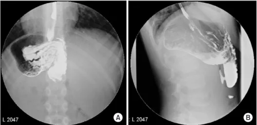
For a confirmatory diagnosis, an upper gastrointestinal contrast study was performed. The Gastrografin swallow study showed that the stomach was rotated along its longitudinal axis, causing delayed gastric emptying (Fig. 1). A final diagnosis of chronic GV was made.
Due to persistent abdominal bloating and discomfort, we decided to perform laparoscopic derotation and gastropexy.
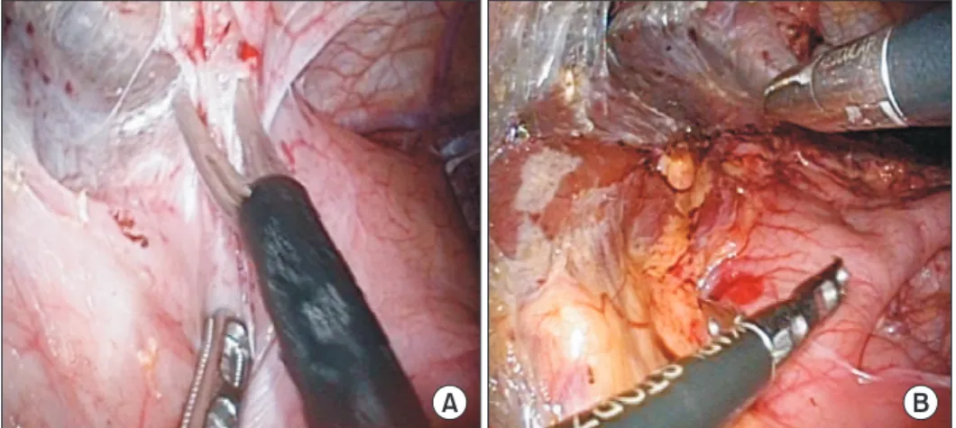
After obtaining the parent's informed consent, pneumoperitoneum was established by an open technique, followed by the placement of three 5 mm trocars. At the time of laparoscopy, the greater curvature of the stomach was attached to the abdominal wall and the left side of the liver. We gently separated the adhesions from the abdominal wall and liver using scissors and a harmonic scalpel (Ethicon Endo-Surgery Inc., Cincinnati, OH, USA) (Fig. 2). No strangulation or stricture of the stomach was observed. The anterior wall of the stomach was anchored to the abdominal wall by an absorbable multifilament suture (Fig. 3). We did not perform fundoplication.
The patient was discharged on postoperative day 10, and his postoperative course was uneventful.
He has not shown signs of postprandial abdominal distension 2 years postoperatively.
Chronic GV as a Late Complication of Hepatectomy for Hepatoblastoma in a Child
A B
Fig. 1. (A, B) Upper gastrointestinal study shows delayed contrast media with antral deformity.
DISCUSSION
The stomach has four ligaments (the gastrocolic, gastrohepatic, gastrophrenic, and gastrosplenic ligaments) that anchor it to the peritoneal cavity; any disruption in this anatomic environment may induce GV [4]. Depending on the axis of rotation of the stomach, GV can be classified into organoaxial, mesenteroaxial, or combined types [4]. GV can also be classified as acute or chronic and intra-abdominal or thoracic based on anatomic localization [4]. Acute GV is frequently associated with other anatomical abnormalities, such as a diaphragmatic or paraesophageal hernia and congenital asplenia [4]. Conversely, chronic GV is commonly idiopathic and rarely associated with anatomical anomalies of the stomach or adjacent organs [5].
The clinical symptoms of GV depend on the degree of rotation and subsequent obstruction [6]. Compared with acute GV, chronic GV can have more subtle manifestations with nonspecific symptoms, such as gastroesophageal reflux, respiratory infection, and recurrent abdominal pain varying from mild to severe [7]. The most common symptom of chronic GV is nonbilious vomiting. Gastric distension, difficulty in feeding, and failure to thrive are
A B
Fig. 2. (A) Operative finding shows multiple adhesions in the abdominal wall. (B) Operative finding shows greater curvature of stomach attached to the liver.
A B
Fig. 3. Intraoperative views. (A) The completion of adhesiolysis. (B) Anterior wall gastropexy was done with absorbable suture.
also more common with chronic GV than with acute GV [4]. Based on these symptoms, it is difficult to diagnosis chronic GV. In this case, the patient was asymptomatic for 7 years prior to the outpatient clinic visit. Postoperative anatomic defects are frequently associated with acute GV. Therefore, chronic GV was not initially suspected as a postoperative complication.
Consequently, the diagnosis of chronic GV requires a high level of clinical suspicion and an upper gastrointestinal contrast study. Treatment is categorized as medical or surgical according to the underlying pathology of GV. Acute GV requires emergency exploration due to the possibility of gastric perforation and gangrenous change. In chronic GV, the treatment plan is determined based on the patient's clinical condition, age, comorbidities, physical performance, and failure to thrive [5].
The medical treatment of chronic GV involves conservative management combined with body positioning (i.e., the head is kept slightly propped up in the prone position), prokinetic drugs, and antisecretory drugs [5]. Al-Salem [8] reported a successful outcome with conservative management in patients with chronic GV and concluded that mild to moderate symptoms of chronic GV should be treated conservatively. Persistent and severe symptoms are indications for surgical management.
Despite successful conservative management, there is a risk of chronic GV developing into acute GV [4]. The mortality rate of acute GV is more than twice that of chronic GV [4]. Thus, when determining the treatment options, conservative management may be considered for chronic GV. However, if the cause of chronic GV is suspected to be of secondary origin, surgical intervention must be considered as the primary treatment option.
GV due to postoperative adhesion in a child is very rare but should be considered in the differential diagnosis of postprandial abdominal bloating and vomiting after hepatectomy.
Early suspicion and appropriate treatment are necessary. The timing of the correction of chronic GV is still controversial; persistent or severe symptoms and GV with a secondary cause indicate that chronic GV should be treated surgically.
REFERENCES
1. McIntyre RC Jr, Bensard DD, Karrer FM, Hall RJ, Lilly JR. The pediatric diaphragm in acute gastric volvulus. J Am Coll Surg 1994;178:234-8.
PUBMED
2. Karande TP, Oak SN, Karmarkar SJ, Kulkarni BK, Deshmukh SS. Gastric volvulus in childhood. J Postgrad Med 1997;43:46-7.
PUBMED
3. Spector JM, Chappell J. Gastric volvulus associated with wandering spleen in a child. J Pediatr Surg 2000;35:641-2.
PUBMED | CROSSREF
4. Cribbs RK, Gow KW, Wulkan ML. Gastric volvulus in infants and children. Pediatrics 2008;122:e752-62.
PUBMED | CROSSREF
5. Porcaro F, Mattioli G, Romano C. Pediatric gastric volvulus: diagnostic and clinical approach. Case Rep Gastroenterol 2013;7:63-8.
PUBMED | CROSSREF
6. Darani A, Mendoza-Sagaon M, Reinberg O. Gastric volvulus in children. J Pediatr Surg 2005;40:855-8.
PUBMED | CROSSREF
7. Trecroci I, Morabito G, Romano C, Salamone I. Gastric volvulus in children--a diagnostic problem: two case reports. J Med Case Rep 2016;10:138.
PUBMED | CROSSREF
Chronic GV as a Late Complication of Hepatectomy for Hepatoblastoma in a Child
8. Al-Salem AH. Acute and chronic gastric volvulus in infants and children: who should be treated surgically? Pediatr Surg Int 2007;23:1095-9.
PUBMED | CROSSREF